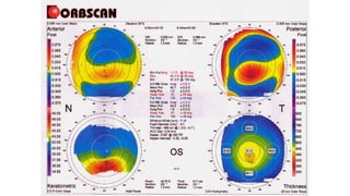
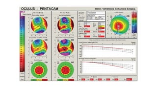
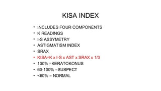
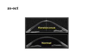
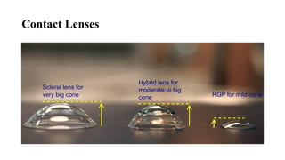
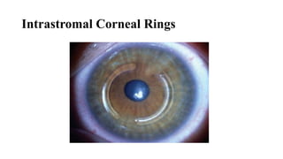
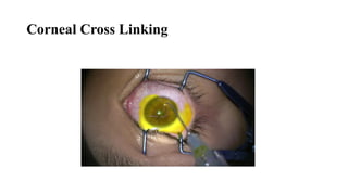
keratoconus
By Dr Priyanka Prasad
3rd
semester
Keratoconus
• term used for conical shape of cornea due to thinning and protrusion
• non inflammatory process
• usually bilateral but can be asymmetrical
• involves central two third of cornea just below the visual axis
• mild to marked visual impairment
Prevalence ,Distribution, And Course
• Estimates between 50 and 230 per 100000
• Asians and middle east > Caucasians
• No significant gender predominance
• Unilaterality 2-4%
• Onset at puberty 10-20 years gradual progression rate variable
Systemic Disorders
oAlagille Syndrome
oAtopic Dermatitis
oDowns Syndrome
oTurners Syndrome
oMarfans Syndrome
oEhlers Danlos
oCrouzon’s Syndrome
oBardet-biedl Syndrome
oNail Patella Syndrome
oGoltz- Gorlin Syndrome
Ocular Disorders
oVKC
oRP
oAniridia
oEctopia Lentis
oLeber’s Congenital Amaurosis
oBlue Sclera
Biochemical Changes
• High Levels Of Proteases, Cathepsins, Gelatinases
• Upregulation Of IL 1 Receptor Expression
• Downregulation Of Proteinase Inhibitor
• Decreased Levels Of Keratin Sulphate And Collagen
• Increased Fibroblastic Activity & Apoptosis
Pathology
• Epithelial changes – elongation of superficial cells at apex
tunel positive deeper epithelial cells signifying apoptosis
early degeneration of basal epithelial cells & iron deposition
disruption of basement membrane
• Bowman’s layer & ant stroma – z shaped interruptions
collagen fragmentation
loss of cohesion & interfibrillar spacing
fibroblastic activity
• Descemet’s membrane – rupture & fluid influx in hydrops
• Endothelium – usually uninvolved
pleomorphism and polymegathism are also documented
Types Of Cones In Keratoconus
• Round or nipple-shaped cones having ,</= 5 mm diameter , centre
lies more inferonasally
• Oval or sagging cone having > 5mm diameter lies more
inferotemporally
• Globus type in which ectasia involvs more than 75% of cornea
Diagnosis
• demographic profile
• clinical evaluations
• slit lamp biomicroscopy
• keratometry
• keratoscopy
• corneal topography
• anterior segment oct
Symptoms
• Progressive Blurring And/Or Distortion
• Photophobia
• Glare
• Monocular Diplopia
• Ocular Irritation
• Decrease contrast
Signs
• scissoring reflex in retinoscopy
• angulation of lower lid on down gaze also known as munson sign
• oil droplet sign (charleux sign)
• rizzuti phenomenon
Munson Sign
Oil Droplet Sign
Rizzuti Sign
Slit Lamp Biomicroscopy
• prominent corneal nerves
• vogt striae
• corneal scarring
• thinning of inferior or inferonasal corneal stroma
• corneal hydrops
• fleischer ring
Keratometry And Keratoscopy
• inferior keratometry in upward gaze will show steepening in inferior
cornea
Amsler Krumeich classification
Forme Fruste Keratoconus(FFKC)
• it is a subclinical disease not a variant of keratoconus in which
diagnosis is made with the help of topography unlike keratoconus in
which diagnosis is clinical.
• two ways to define
1. a normal cornea with fellow eye having keratoconus or family
history of keratoconus
2. an abnormal cornea in which corneal topography or corneal
hysteresis(ORA) or both are abnormal but there are no obvious
clinical signs of keratoconus
as-oct
Treatment
• spectacle correction
• contact lens
• intrastromal ring segments for mild to moderate disease
• corneal cross linking
• keratoplasty
1. PK
2. DALK
Contact Lenses
Intrastromal Corneal Rings
Corneal Cross Linking
Thank You

1.keratoconus 2.pptx...........................

  • 1.
    keratoconus By Dr PriyankaPrasad 3rd semester
  • 2.
    Keratoconus • term usedfor conical shape of cornea due to thinning and protrusion • non inflammatory process • usually bilateral but can be asymmetrical • involves central two third of cornea just below the visual axis • mild to marked visual impairment
  • 3.
    Prevalence ,Distribution, AndCourse • Estimates between 50 and 230 per 100000 • Asians and middle east > Caucasians • No significant gender predominance • Unilaterality 2-4% • Onset at puberty 10-20 years gradual progression rate variable
  • 4.
    Systemic Disorders oAlagille Syndrome oAtopicDermatitis oDowns Syndrome oTurners Syndrome oMarfans Syndrome oEhlers Danlos oCrouzon’s Syndrome oBardet-biedl Syndrome oNail Patella Syndrome oGoltz- Gorlin Syndrome Ocular Disorders oVKC oRP oAniridia oEctopia Lentis oLeber’s Congenital Amaurosis oBlue Sclera
  • 5.
    Biochemical Changes • HighLevels Of Proteases, Cathepsins, Gelatinases • Upregulation Of IL 1 Receptor Expression • Downregulation Of Proteinase Inhibitor • Decreased Levels Of Keratin Sulphate And Collagen • Increased Fibroblastic Activity & Apoptosis
  • 6.
    Pathology • Epithelial changes– elongation of superficial cells at apex tunel positive deeper epithelial cells signifying apoptosis early degeneration of basal epithelial cells & iron deposition disruption of basement membrane • Bowman’s layer & ant stroma – z shaped interruptions collagen fragmentation loss of cohesion & interfibrillar spacing fibroblastic activity • Descemet’s membrane – rupture & fluid influx in hydrops • Endothelium – usually uninvolved pleomorphism and polymegathism are also documented
  • 7.
    Types Of ConesIn Keratoconus • Round or nipple-shaped cones having ,</= 5 mm diameter , centre lies more inferonasally • Oval or sagging cone having > 5mm diameter lies more inferotemporally • Globus type in which ectasia involvs more than 75% of cornea
  • 8.
    Diagnosis • demographic profile •clinical evaluations • slit lamp biomicroscopy • keratometry • keratoscopy • corneal topography • anterior segment oct
  • 9.
    Symptoms • Progressive BlurringAnd/Or Distortion • Photophobia • Glare • Monocular Diplopia • Ocular Irritation • Decrease contrast
  • 10.
    Signs • scissoring reflexin retinoscopy • angulation of lower lid on down gaze also known as munson sign • oil droplet sign (charleux sign) • rizzuti phenomenon
  • 11.
  • 12.
  • 13.
  • 14.
    Slit Lamp Biomicroscopy •prominent corneal nerves • vogt striae • corneal scarring • thinning of inferior or inferonasal corneal stroma • corneal hydrops • fleischer ring
  • 15.
    Keratometry And Keratoscopy •inferior keratometry in upward gaze will show steepening in inferior cornea
  • 16.
  • 17.
    Forme Fruste Keratoconus(FFKC) •it is a subclinical disease not a variant of keratoconus in which diagnosis is made with the help of topography unlike keratoconus in which diagnosis is clinical. • two ways to define 1. a normal cornea with fellow eye having keratoconus or family history of keratoconus 2. an abnormal cornea in which corneal topography or corneal hysteresis(ORA) or both are abnormal but there are no obvious clinical signs of keratoconus
  • 21.
  • 22.
    Treatment • spectacle correction •contact lens • intrastromal ring segments for mild to moderate disease • corneal cross linking • keratoplasty 1. PK 2. DALK
  • 23.
  • 24.
  • 25.
  • 26.