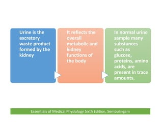
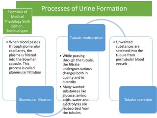
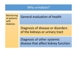
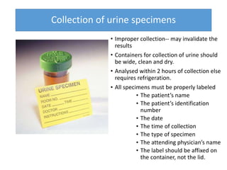
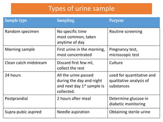
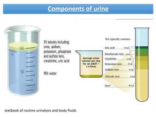
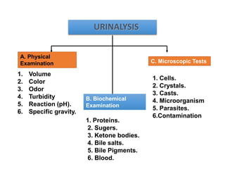
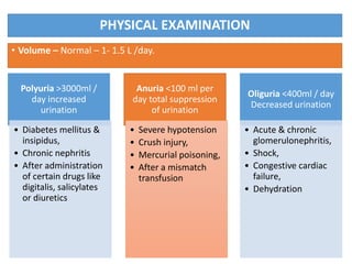
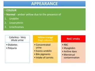
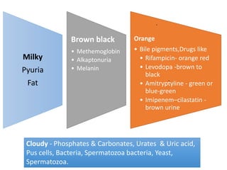
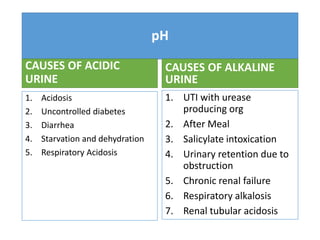
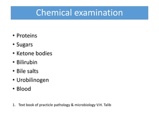
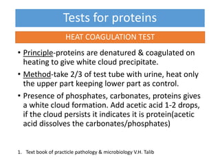
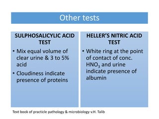
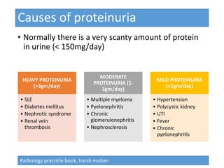
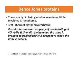
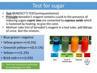
This document provides an overview of urine analysis in dentistry. It discusses the processes of urine formation, reasons for performing urinalysis, sample collection methods, and the components evaluated in a urinalysis. Physical examination assesses volume, color, odor, turbidity, pH, and specific gravity. Biochemical examination tests for proteins, sugars, ketone bodies, bile, and blood. Microscopic tests identify cells, crystals, casts, and microorganisms. Together these evaluations can help diagnose and monitor diseases affecting the kidneys, urinary tract, and other body systems.
It gives basic things regarding urinalysis and will be very useful for medical students, house surgeons, laboratory technicians and postgraduates in medicine.
This chapter is largely about the water and electrolytes ( salts )in your plasma and how the body manages to keep you from drying up and blowing away even if you are in the hot Texas sun and without liquid drink.
Indications for Urine examination include:
Suspected renal damage
Detection of UTI
Management of metabolic disorders
Diagnosis of jaundice
Management of Plasma cell dyscrasias
Diagnosis of pregnancy
Drug abuse
Physical Examination of Urine includes estimation of Appearance, Volume, Colour, Odour, reaction, Specific gravity and Osmolality.
The test measures the amount of sugar in a urine sample. Normal urine does not contain glucose. Microscopic Examination. A variety of normal and abnormal.
Microscopic examination of urine Casts • Urinary casts are cylindrical aggregations of particles that form in the distal nephron, dislodge
ARTIFICIAL INTELLIGENCE IN HEALTHCARE.pdfAnujkumaranit
Artificial intelligence (AI) refers to the simulation of human intelligence processes by machines, especially computer systems. It encompasses tasks such as learning, reasoning, problem-solving, perception, and language understanding. AI technologies are revolutionizing various fields, from healthcare to finance, by enabling machines to perform tasks that typically require human intelligence.
Report Back from SGO 2024: What’s the Latest in Cervical Cancer?bkling
Are you curious about what’s new in cervical cancer research or unsure what the findings mean? Join Dr. Emily Ko, a gynecologic oncologist at Penn Medicine, to learn about the latest updates from the Society of Gynecologic Oncology (SGO) 2024 Annual Meeting on Women’s Cancer. Dr. Ko will discuss what the research presented at the conference means for you and answer your questions about the new developments.
Title: Sense of Smell
Presenter: Dr. Faiza, Assistant Professor of Physiology
Qualifications:
MBBS (Best Graduate, AIMC Lahore)
FCPS Physiology
ICMT, CHPE, DHPE (STMU)
MPH (GC University, Faisalabad)
MBA (Virtual University of Pakistan)
Learning Objectives:
Describe the primary categories of smells and the concept of odor blindness.
Explain the structure and location of the olfactory membrane and mucosa, including the types and roles of cells involved in olfaction.
Describe the pathway and mechanisms of olfactory signal transmission from the olfactory receptors to the brain.
Illustrate the biochemical cascade triggered by odorant binding to olfactory receptors, including the role of G-proteins and second messengers in generating an action potential.
Identify different types of olfactory disorders such as anosmia, hyposmia, hyperosmia, and dysosmia, including their potential causes.
Key Topics:
Olfactory Genes:
3% of the human genome accounts for olfactory genes.
400 genes for odorant receptors.
Olfactory Membrane:
Located in the superior part of the nasal cavity.
Medially: Folds downward along the superior septum.
Laterally: Folds over the superior turbinate and upper surface of the middle turbinate.
Total surface area: 5-10 square centimeters.
Olfactory Mucosa:
Olfactory Cells: Bipolar nerve cells derived from the CNS (100 million), with 4-25 olfactory cilia per cell.
Sustentacular Cells: Produce mucus and maintain ionic and molecular environment.
Basal Cells: Replace worn-out olfactory cells with an average lifespan of 1-2 months.
Bowman’s Gland: Secretes mucus.
Stimulation of Olfactory Cells:
Odorant dissolves in mucus and attaches to receptors on olfactory cilia.
Involves a cascade effect through G-proteins and second messengers, leading to depolarization and action potential generation in the olfactory nerve.
Quality of a Good Odorant:
Small (3-20 Carbon atoms), volatile, water-soluble, and lipid-soluble.
Facilitated by odorant-binding proteins in mucus.
Membrane Potential and Action Potential:
Resting membrane potential: -55mV.
Action potential frequency in the olfactory nerve increases with odorant strength.
Adaptation Towards the Sense of Smell:
Rapid adaptation within the first second, with further slow adaptation.
Psychological adaptation greater than receptor adaptation, involving feedback inhibition from the central nervous system.
Primary Sensations of Smell:
Camphoraceous, Musky, Floral, Pepperminty, Ethereal, Pungent, Putrid.
Odor Detection Threshold:
Examples: Hydrogen sulfide (0.0005 ppm), Methyl-mercaptan (0.002 ppm).
Some toxic substances are odorless at lethal concentrations.
Characteristics of Smell:
Odor blindness for single substances due to lack of appropriate receptor protein.
Behavioral and emotional influences of smell.
Transmission of Olfactory Signals:
From olfactory cells to glomeruli in the olfactory bulb, involving lateral inhibition.
Primitive, less old, and new olfactory systems with different path
Factory Supply Best Quality Pmk Oil CAS 28578–16–7 PMK Powder in Stockrebeccabio
Factory Supply Best Quality Pmk Oil CAS 28578–16–7 PMK Powder in Stock
Telegram: bmksupplier
signal: +85264872720
threema: TUD4A6YC
You can contact me on Telegram or Threema
Communicate promptly and reply
Free of customs clearance, Double Clearance 100% pass delivery to USA, Canada, Spain, Germany, Netherland, Poland, Italy, Sweden, UK, Czech Republic, Australia, Mexico, Russia, Ukraine, Kazakhstan.Door to door service
Hot Selling Organic intermediates
Explore natural remedies for syphilis treatment in Singapore. Discover alternative therapies, herbal remedies, and lifestyle changes that may complement conventional treatments. Learn about holistic approaches to managing syphilis symptoms and supporting overall health.
Pulmonary Thromboembolism - etilogy, types, medical- Surgical and nursing man...VarunMahajani
Disruption of blood supply to lung alveoli due to blockage of one or more pulmonary blood vessels is called as Pulmonary thromboembolism. In this presentation we will discuss its causes, types and its management in depth.
Couples presenting to the infertility clinic- Do they really have infertility...Sujoy Dasgupta
Dr Sujoy Dasgupta presented the study on "Couples presenting to the infertility clinic- Do they really have infertility? – The unexplored stories of non-consummation" in the 13th Congress of the Asia Pacific Initiative on Reproduction (ASPIRE 2024) at Manila on 24 May, 2024.
Lung Cancer: Artificial Intelligence, Synergetics, Complex System Analysis, S...Oleg Kshivets
RESULTS: Overall life span (LS) was 2252.1±1742.5 days and cumulative 5-year survival (5YS) reached 73.2%, 10 years – 64.8%, 20 years – 42.5%. 513 LCP lived more than 5 years (LS=3124.6±1525.6 days), 148 LCP – more than 10 years (LS=5054.4±1504.1 days).199 LCP died because of LC (LS=562.7±374.5 days). 5YS of LCP after bi/lobectomies was significantly superior in comparison with LCP after pneumonectomies (78.1% vs.63.7%, P=0.00001 by log-rank test). AT significantly improved 5YS (66.3% vs. 34.8%) (P=0.00000 by log-rank test) only for LCP with N1-2. Cox modeling displayed that 5YS of LCP significantly depended on: phase transition (PT) early-invasive LC in terms of synergetics, PT N0—N12, cell ratio factors (ratio between cancer cells- CC and blood cells subpopulations), G1-3, histology, glucose, AT, blood cell circuit, prothrombin index, heparin tolerance, recalcification time (P=0.000-0.038). Neural networks, genetic algorithm selection and bootstrap simulation revealed relationships between 5YS and PT early-invasive LC (rank=1), PT N0—N12 (rank=2), thrombocytes/CC (3), erythrocytes/CC (4), eosinophils/CC (5), healthy cells/CC (6), lymphocytes/CC (7), segmented neutrophils/CC (8), stick neutrophils/CC (9), monocytes/CC (10); leucocytes/CC (11). Correct prediction of 5YS was 100% by neural networks computing (area under ROC curve=1.0; error=0.0).
CONCLUSIONS: 5YS of LCP after radical procedures significantly depended on: 1) PT early-invasive cancer; 2) PT N0--N12; 3) cell ratio factors; 4) blood cell circuit; 5) biochemical factors; 6) hemostasis system; 7) AT; 8) LC characteristics; 9) LC cell dynamics; 10) surgery type: lobectomy/pneumonectomy; 11) anthropometric data. Optimal diagnosis and treatment strategies for LC are: 1) screening and early detection of LC; 2) availability of experienced thoracic surgeons because of complexity of radical procedures; 3) aggressive en block surgery and adequate lymph node dissection for completeness; 4) precise prediction; 5) adjuvant chemoimmunoradiotherapy for LCP with unfavorable prognosis.
TEST BANK for Operations Management, 14th Edition by William J. Stevenson, Ve...kevinkariuki227
TEST BANK for Operations Management, 14th Edition by William J. Stevenson, Verified Chapters 1 - 19, Complete Newest Version.pdf
MANAGEMENT OF ATRIOVENTRICULAR CONDUCTION BLOCK.pdfJim Jacob Roy
Cardiac conduction defects can occur due to various causes.
Atrioventricular conduction blocks ( AV blocks ) are classified into 3 types.
This document describes the acute management of AV block.
The hemodynamic and autonomic determinants of elevated blood pressure in obes...
Urine analysis
1. URINE
ANALYSIS IN
DENTISTRY
• ASHISH RANGHANI
• PG PART 2
• GDCH, AHMEDABAD
UNDER GUIDANCE OF
DR. J.S SHAH
PROFESSOR AND HEAD
ORAL MEDICINE AND RADIOLOGY
GDCH
DATE- 28/07/2016
2. CONTENTS
1. Processes of Urine Formation
2. Why urinalysis?
3. Collection of urine specimens
4. Types of urine sample
5. Components of urine
6. Urinalysis
7. Physical Examination
Volume ,Color, Odor, Turbidity, Reaction (pH), Specific gravity
8. Biochemical Examination
Proteins, Sugers , Ketone bodies, Bile salts , Bile Pigments, Blood
9. Microscopic Tests
Cells, Crystals ,Casts, Microorganism
10. Urinary changes in Dental Diseases
3. Urine is the
excretory
waste product
formed by the
kidney
It reflects the
overall
metabolic and
kidney
functions of
the body
In normal urine
sample many
substances
such as
glucose,
proteins, amino
acids, are
present in trace
amounts.
Essentials of Medical Physiology Sixth Edition, Sembulingam
4. Processes of Urine Formation
• When blood passes
through glomerular
capillaries, the
plasma is filtered
into the Bowman
capsule. This
process is called
glomerular filtration
Glomerular filtration
• While passing
through the tubule,
the filtrate
undergoes various
changes both in
quality and in
quantity
• Many wanted
substances like
glucose, amino
acids, water and
electrolytes are
reabsorbed from
the tubules
Tubular reabsorption
• Unwanted
substances are
secreted into the
tubule from
peritubular blood
vessels
Tubular secretion
Essentials of
Medical
Physiology Sixth
Edition,
Sembulingam
6. Collection of urine specimens
• Improper collection-- may invalidate the
results
• Containers for collection of urine should
be wide, clean and dry.
• Analysed within 2 hours of collection else
requires refrigeration.
• All specimens must be properly labeled
• The patient’s name
• The patient’s identification
number
• The date
• The time of collection
• The type of specimen
• The attending physician’s name
• The label should be affixed on
the container, not the lid.
7. Types of urine sample
Sample type Sampling Purpose
Random specimen No specific time
most common, taken
anytime of day
Routine screening
Morning sample First urine in the morning,
most concentrated
Pregnancy test,
microscopic test
Clean catch midstream Discard first few ml,
collect the rest
Culture
24 hours All the urine passed
during the day and night
and next day 1st sample is
collected.
used for quantitative and
qualitative analysis of
substances
Postprandial 2 hours after meal Determine glucose in
diabetic monitoring
Supra-pubic aspired Needle aspiration Obtaining sterile urine
9. URINALYSIS
1. Volume
2. Color
3. Odor
4. Turbidity
5. Reaction (pH).
6. Specific gravity.
1. Proteins.
2. Sugers.
3. Ketone bodies.
4. Bile salts.
5. Bile Pigments.
6. Blood.
1. Cells.
2. Crystals.
3. Casts.
4. Microorganism
5. Parasites.
6.Contamination
A. Physical
Examination
B. Biochemical
Examination
C. Microscopic Tests
10. PHYSICAL EXAMINATION
• Volume – Normal – 1- 1.5 L /day.
Polyuria >3000ml /
day increased
urination
• Diabetes mellitus &
insipidus,
• Chronic nephritis
• After administration
of certain drugs like
digitalis, salicylates
or diuretics
Anuria <100 ml per
day total suppression
of urination
• Severe hypotension
• Crush injury,
• Mercurial poisoning,
• After a mismatch
transfusion
Oliguria <400ml / day
Decreased urination
• Acute & chronic
glomerulonephritis,
• Shock,
• Congestive cardiac
failure,
• Dehydration
11. APPEARANCE
• COLOUR
• Normal - amber yellow due to the presence of
1. Urobilin
2. Uroerythrin
3. Urochromes
Colorless - Very
dilute urine
• Diabetes
• Polyuria
Yellow orange
(high colored)
• Concentrated
urine
• Excess urobilin
• Bile pigments
• Intake of carrots
Red/ smoky
• RBC
• Myoglobin
• Aniline dyes
• Menstrual
contamination
12. Milky
Pyuria
Fat
Brown black
• Methemoglobin
• Alkaptonuria
• Melanin
Orange
• Bile pigments,Drugs like
• Rifampicin- orange red
• Levodopa -brown to
black
• Amitryptyline - green or
blue-green
• Imipenem–cilastatin -
brown urine
.
-
Cloudy - Phosphates & Carbonates, Urates & Uric acid,
Pus cells, Bacteria, Spermatozoa bacteria, Yeast,
Spermatozoa.
13. Specific Gravity
•It is directly proportional to the concentration
of solute & inversely proportional to the
volume
•Ranges between 1.003 to 1.030
14. LOW SPECIFIC
GRAVITY
HYPOSTHENURIA :indicates dilute
urine, which may be caused by
1. Diabetes insipidus ( can be
as low as 1.001)
2. Drinking excessive amounts
of liquid.
3. Pyelonephritis,
glomerulonephritis
4. Use of diuretics.
HYPERSTHENURIA : indicates very
concentrated urine, which may be
caused by
1. Dehydration
2. Diabetes mellitus
3. Adrenal insufficiency.
4. Toximea of pregnancy (protein
in the urine).
HIGH SPECIFIC
GRAVITY
16. ODOUR OF URINE
After prolonged standingAmmonia smell:
• Rancid : Tyrosinaemia.
Due to urinary infectionFecal smell:
• Mousy order : phenylketonuria
Ketone bodies is seen in diabetesFruity smell
• Maple syrup odour : MSUD
Normal odour Fresh urine has aromatic odor
17. pH
• Normal pH for urine ranges from 4.5 – 8.0 (average pH 6)
• Some foods (such as citrus fruits and dairy products) and
medications (such as antacids) can affect urine pH.
• In a diet high in protein the urine is more acidic, while a diet
high in vegetable material a urine that is more alkaline.
• Tested by:
• litmus paper
• pH paper
• dipsticks
18. pH
CAUSES OF ACIDIC
URINE
1. Acidosis
2. Uncontrolled diabetes
3. Diarrhea
4. Starvation and dehydration
5. Respiratory Acidosis
CAUSES OF ALKALINE
URINE
1. UTI with urease
producing org
2. After Meal
3. Salicylate intoxication
4. Urinary retention due to
obstruction
5. Chronic renal failure
6. Respiratory alkalosis
7. Renal tubular acidosis
19. Chemical examination
• Proteins
• Sugars
• Ketone bodies
• Bilirubin
• Bile salts
• Urobilinogen
• Blood
1. Text book of practicle pathology & microbiology V.H. Talib
20. Tests for proteins
• Principle-proteins are denatured & coagulated on
heating to give white cloud precipitate.
• Method-take 2/3 of test tube with urine, heat only
the upper part keeping lower part as control.
• Presence of phosphates, carbonates, proteins gives
a white cloud formation. Add acetic acid 1-2 drops,
if the cloud persists it indicates it is protein(acetic
acid dissolves the carbonates/phosphates)
HEAT COAGULATION TEST
1. Text book of practicle pathology & microbiology V.H. Talib
21. Other tests
SULPHOSALICYLIC ACID
TEST
• Mix equal volume of
clear urine & 3 to 5%
acid
• Cloudiness indicate
presence of proteins
HELLER’S NITRIC ACID
TEST
• White ring at the point
of contact of conc.
HNO3 and urine
indicate presence of
albumin
Text book of practicle pathology & microbiology v.H. Talib
22. Causes of proteinuria
• Normally there is a very scanty amount of protein
in urine (< 150mg/day)
HEAVY PROTEINURIA
(>3gm/day)
• SLE
• Diabetes mellitus
• Nephrotic syndrome
• Renal vein
thrombosis
MODERATE
PROTEINURIA (1-
3gm/day)
• Multiple myeloma
• Pyelonephritis
• Chronic
glomerulonephritis
• Nephrosclerosis
MILD PROTEINURIA
(<1gm/day)
• Hypertension
• Polycystic kidney
• UTI
• Fever
• Chronic
pyelonephritis
Pathology practicle book, harsh mohan
23. Bence Jones proteins
• These are light chain globulins seen in multiple
myeloma & lymphoma.
• Test- Thermal method(waterbath):
Proteins has unusual property of precipitating at
400 -600c & then dissolving when the urine is
brought to boiling(1000c) & reappears when the
urine is cooled.
1. Text book of practicle pathology & microbiology V.H. Talib
24. Test for sugar
• Blue-green= negative
• Yellow-green=+(<0.5%)
• Greenish yellow=++(0.5-1%)
• Yellow=+++(1-2%)
• Brick red=++++(>2%)
1. Text book of practicle pathology
& microbiology V.H. Talib
• Test-BENEDICT’S TEST(semiquantitative)
• Principle-benedict’s reagent contains cuso4.In the presence of
reducing sugars cupric ions are converted to cuprous oxide which
is hastened by heating, to give the color.
• Method- take 5ml of benedict’s reagent in a test tube, add 8drops
of urine. Boil the mixture.
25. Benedict’s test
• Detects all reducing substances like glucose,
fructose, & other reducing sustances.
• To confirm it is glucose, dipsticks can be used
(glucose oxidase)
26. Causes of glycosuria
• Glycosuria with hyperglycaemia-
1. Diabetes,
2. Acromegaly,
3. Cushing’s Disease,
4. Hyperthyroidism,
5. Drugs Like Corticosteroids.
• Glycosuria without hyperglycaemia-
Renal tubular dysfunction
Text book of practicle pathology & microbiology v.H. Talib
27. KETONE BODIES
• 3 types
Acetone
Acetoacetic acid
β-hydroxy butyric acid
They are products of fat metabolism
28. Rothera’s test
• Principle-acetone & acetoacetic
acid react with sodium
nitroprusside in the presence of
alkali to produce purple colour.
• Method- take 5ml of urine in a test
tube & saturate it with ammonium
sulphate. Then add one crystal of
sodium nitroprusside. Then gently
add 0.5ml of Strong ammonium
hydroxide along the sides of the
test tube.
• Appearance permanganate colored
ring at the junction of the two
fluids indicates a positive test
1. Text book of practicle pathology & microbiology V.H. Talib
29. Causes of ketonuria
• Diabetes
• Non-diabetic causes-
1. High Fever,
2. Starvation,
3. Severe Vomiting/Diarrhea
4. After General Anaesthesia
Text book of practicle pathology & microbiology v.H. Talib
30. Blood in urine
• Test- BENZIDINE TEST
• Method- mix 2ml of benzidine solution with 2ml of
hydrogen peroxide in a test tube. Take 2ml of urine &
add 2ml of above mixture. A blue color indicates +
reaction
Text book of practicle pathology & microbiology v.H. Talib
31. Causes of hematuria
• Acute & Chronic Glomerulonephritis,
• Chronic Passive Congestion Of The Kidney
• Renal TB,
• Leukaemias
• Severe UTI,
• Urinary Calculi
• Benign & Malignant Tumors Of The Kidney
& Urinary Tract
Text book of practicle pathology & microbiology v.H. Talib
32. BILE SALTS
Hay’s test
The test depends on the surface activity of bilirubin
as it lowers the surface tension of urine.
Sprinkle a little of precipitated sulfur powder on the
surface of 2 ml urine. If bilirubin is present, sulfur
powder will sink to the bottom of urine. If bile is
absent, sulfur will remain on the surface of urine.
Text book of practicle pathology & microbiology v.H. Talib
34. Urobilinogen
• Test- ehrlich test
• 5ml fresh urine + 0.5 ml Ehrilch's reagent, allow to
stand for 5 min →
• pink color on cold → normal trace.
• red color on cold → increased amounts.
• red color after heating → normal traces.
• Causes-hemolytic anemia's
Cause- obstruction to bile flow (obstructive jaundice)
35. Microscopic examination of urine
• A sample of well-mixed urine
(usually 10-15 ml) is
centrifuged in a test tube at
relatively low speed (about
2000-3,000 rpm) for 5-10
minutes which produces a
concentration of sediment
(cellular matter) at the
bottom of the tube.
• A drop of sediment is poured
onto a glass slide, a coverslip
is place over it & observed
under microscope
Urinalysis: a comprehensive review,
36. A variety of normal and abnormal cellular
elements may be seen in urine sediment such as
1. Red blood cells
2. White blood cells
3. Mucus
4. Various epithelial cells
5. Various crystals
6. Bacteria
7. Casts
37. Hematuria is the presence of
abnormal numbers of red cells
in urine due to any of several
possible causes
• Renal stone
• Kidney tumors
• kidney trauma,
• Upper and lower
urinary tract infections,
• Polycystic kidney
WBC in high numbers indicate
inflammation or infection
somewhere along the urinary or
genital tract
• UTI
• Prostatitis
• Chronic pyelonephritis
• Renal stone
• Renal tumours
• Cystitis
38. • The most common type of cast- hyaline casts
• Seen in fever, exercise, damage to the glomerular capillary.
• Red blood cells may stick together and form red blood cell
casts. Such casts are indicative of glomerulonephritis, with
leakage of RBC's from glomeruli, or severe tubular damage
• White blood cell casts
• Their presence indicates inflammation of the kidney.
TYPES OF CAST
Acellular cast
• Hyaline casts
• Granular casts
• Waxy casts
• Fatty casts
• Pigment casts
• Crystal casts
Cellular cast
• Red cell casts
• White cell casts,
• Epithelial cell cast
40. URINE ANALYSIS IN DIABETES
• Diabetes mellitus (DM) also known as a group of
metabolic diseases in which there are high blood sugar
levels over a prolonged period.
• This high blood sugar produces the symptoms of
frequent urination, increased thirst, and increased
hunger.
• Untreated, diabetes can cause many complications.
Acute complications include diabetic ketoacidosis and
nonketotic hyper osmolar coma.
• Serious long-term complications include heart disease,
stroke, kidney failure, foot ulcers and damage to the
eyes
A study on abnormal constituents of urine in diabetic patients
41. • In diabetes mellitus mainly glucose and ketone
bodies are elevated
• Glucosuria occurs in mainly during diabetis mellitus
and renal diabetes.
• These ketone bodies are present in the urine this
may be due to diabetic ketoacidosis
• It occurs when the body cannot use sugar (glucose)
as a fuel source because there is little or no insulin.
Fat is used for fuel instead
42. Diagnosis of Multiple
Myeloma
Two of the 4 following criteria are
generally required for diagnosis of
multiple myeloma:
1. Radiographic evidence of osteolytic
bone lesions
2. >20% plasma cells in bone marrow
aspirates or biopsy specimens.
3. Demonstration of monoclonal or
biclonal gammopathy with serum
electrophoresis
4. Demonstration of Bence-Jones
proteinuria
Systemic Lupus
Erythematosus
• Heavy proteinuria (>
3gm/day)
Pathology practicle book, harsh mohan
43. Mercury concentrations in urine
• Urine levels of mercury less than 20 ng/mL are
considered safe.
• The mercury body burden of dental personnel is
normally higher than in the general population.
• This increased body burden is attributed to dental
personnel mixing and applying dental amalgam and
removing amalgam restorations
44. References
1. Text book of practicle pathology & microbiology v.H. Talib
2. Pathology practicle book, harsh mohan
3. Urinalysis in clinical practice, sekhar chakraborty
4. Graff’s textbook of routine urinalysis and body fluids
5. Salma mahaboob, madan mohan rao m, a study on abnormal constituents
of urine in diabetic patients, ujmds 2014, page 64-67
6. Urinalysis: a comprehensive review, jeff Simerville, m.D., Georgetown
university school of medicine, washington, d.C
7. Essentials of Medical Physiology Sixth Edition, Sembulingam