
Recommended
Recommended
More Related Content
What's hot
What's hot (20)
Similar to Urine crystal
Similar to Urine crystal (20)
Recently uploaded
Recently uploaded (20)
Urine crystal
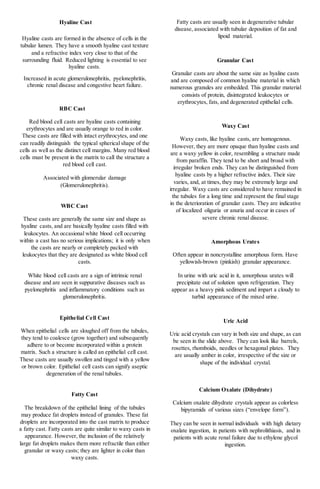
- 1. Hyaline Cast Hyaline casts are formed in the absence of cells in the tubular lumen. They have a smooth hyaline cast texture and a refractive index very close to that of the surrounding fluid. Reduced lighting is essential to see hyaline casts. Increased in acute glomerulonephritis, pyelonephritis, chronic renal disease and congestive heart failure. RBC Cast Red blood cell casts are hyaline casts containing erythrocytes and are usually orange to red in color. These casts are filled with intact erythrocytes, and one can readily distinguish the typical spherical shape of the cells as well as the distinct cell margins. Many red blood cells must be present in the matrix to call the structure a red blood cell cast. Associated with glomerular damage (Glomerulonephritis). WBC Cast These casts are generally the same size and shape as hyaline casts, and are basically hyaline casts filled with leukocytes. An occasional white blood cell occurring within a cast has no serious implications; it is only when the casts are nearly or completely packed with leukocytes that they are designated as white blood cell casts. White blood cell casts are a sign of intrinsic renal disease and are seen in suppurative diseases such as pyelonephritis and inflammatory conditions such as glomerulonephritis. Epithelial Cell Cast When epithelial cells are sloughed off from the tubules, they tend to coalesce (grow together) and subsequently adhere to or become incorporated within a protein matrix. Such a structure is called an epithelial cell cast. These casts are usually swollen and tinged with a yellow or brown color. Epithelial cell casts can signify aseptic degeneration of the renal tubules. Fatty Cast The breakdown of the epithelial lining of the tubules may produce fat droplets instead of granules. These fat droplets are incorporated into the cast matrix to produce a fatty cast. Fatty casts are quite similar to waxy casts in appearance. However, the inclusion of the relatively large fat droplets makes them more refractile than either granular or waxy casts; they are lighter in color than waxy casts. Fatty casts are usually seen in degenerative tubular disease, associated with tubular deposition of fat and lipoid material. Granular Cast Granular casts are about the same size as hyaline casts and are composed of common hyaline material in which numerous granules are embedded. This granular material consists of protein, disintegrated leukocytes or erythrocytes, fats, and degenerated epithelial cells. Waxy Cast Waxy casts, like hyaline casts, are homogenous. However, they are more opaque than hyaline casts and are a waxy yellow in color, resembling a structure made from paraffin. They tend to be short and broad with irregular broken ends. They can be distinguished from hyaline casts by a higher refractive index. Their size varies, and, at times, they may be extremely large and irregular. Waxy casts are considered to have remained in the tubules for a long time and represent the final stage in the deterioration of granular casts. They are indicative of localized oliguria or anuria and occur in cases of severe chronic renal disease. Amorphous Urates Often appear in noncrystalline amorphous form. Have yellowish-brown (pinkish) granular appearance. In urine with uric acid in it, amorphous urates will precipitate out of solution upon refrigeration. They appear as a heavy pink sediment and impart a cloudy to turbid appearance of the mixed urine. Uric Acid Uric acid crystals can vary in both size and shape, as can be seen in the slide above. They can look like barrels, rosettes, rhomboids, needles or hexagonal plates. They are usually amber in color, irrespective of the size or shape of the individual crystal. Calcium Oxalate (Dihydrate) Calcium oxalate dihydrate crystals appear as colorless bipyramids of various sizes (“envelope form”). They can be seen in normal individuals with high dietary oxalate ingestion, in patients with nephrolithiasis, and in patients with acute renal failure due to ethylene glycol ingestion.
- 2. Calcium Oxalate (Monohydrate) Calcium oxalate monohydrate crystals are colorless and can assume several shapes, including ovoids, biconcave disks, rods and dumbbells. This form of crystals is seen ethylene glycol poisoning. Amorphous Phosphate Similar to amorphous urates except usually colorless. In urine with phosphate in it, amorphous phosphates will precipitate out of solution upon refrigeration. They appear as a heavy white sediment and impart a cloudy to turbid appearance of the mixed urine.’ Calcium Phosphate Large flat-shaped plates or wedge-shaped prisms. The prisms often appear in rosettes. Single prisms are usually blunt on one end and pointed on the other end. Although considered normal they may also be associated with kidney stone formation. Triple Phosphate Typically appear in "coffin-lid" form. May also appear as "fern-leaf" shape if freshly formed. Although considered normal they may also be associated with kidney stone formation Ammonium Biurate Yellowish-brown, can be seen in a "thorn apple" shape (round with thorny projections) or in spherical form. Ammonium biruate crystals can be seen in normal urine. However, the presence of ammonium biurate crystals especially in combination with a urine pH 9.0 or higher usually indicates an old or poorly preserved specimen. Best practice is to NOT report any urinalysis results on the sample as it has been compromised. A recollect should be requested. Calcium Carbonate Appearance: Small, colorless granules or dumbbells. Not clinically significant but can be confused with other elements. A unique feature of calcium carbonate is that the crystals effervesce with hydrochloric acid or acetic acid. This can help to confirm the presence of calcium carbonate in the urine. Cystine Appearance: Colorless, thin, hexagonal plates Cystine crystals are found in the inherited condition, cystinuria. Cystine crystals are the most frequent cause of kidney stones in children. The presence of cystine crystals should be confirmed by cyanide-nitroprusside test (turns red-purple). Cholesterol Appearance: clear, flat plates with notched corners. The appearance of cholesterol is associated with the Nephrotic Syndrome. Cholesterol crystals are accompanied by a positive biochemical test for protein. They usually appear after the urine sample has been refrigerated and may be accompanied by oval fat bodies, fatty casts, and free fat droplets in the sediment. Leucine Appearance: yellow-brown spheroids with concentric rings around the outer edge and radial striations in the center. Leucine crystals may be seen in liver disorders in which amino acid metabolism is impaired. The presence of leucine crystals is often accompanied by a positive biochemical test for bilirubin and is often accompanied by tyrosine crystals in the same sediment. Tyrosine Appearance: colorless to yellow-brown single needles. Also seen as sheaves or rosettes. Tyrosine crystals may be seen in tyrosinemia and in certain liver disorders in which amino acid metabolism is impaired. The presence of tyrosine crystals is usually accompanied by a positive biochemical test for bilirubin and are often accompanied by the presence of leucine crystals in the sediment. Bilirubin Appearance: Yellow-brown needles or granules. They are frequently attached to the surface of cells. Bilirubin crystals are seen in several hepatic disorders. The appearance of bilirubin crystals should be accompanied by a positive biochemical test for bilirubin (reagent test pad and Ictotest).
- 3. Sulfonamides Appearance: flat needles, sheaves of small needles or as spheroids. Often brown in color. The presence of sulfanomide crystals usually indicates administration of the drug and not necessarily a pathological condition. However, their presence is also associated with kidney stone formation. Radiographic Dyes Appearance: flat needles or sheaves accompanied by round globules but are variable in form. When the presence of radiopaque dye crystals is suspected, the ordering location should be consulted to confirm administration of contrast media. The presence of these crystals in the urine is associated with very high specfic gravity results by refractometry (>1.035). Specific gravity by the reagent test pad method is not affected by the presence of these crystals. Ampicillin Ampicillin crystal appear as colorless needles that tend to form bundles following refrigeration. These crystals are present in urine due to massive doses of penicillin compounds without adequate hydration. Starch Starch granule contamination may occur when cornstarch is the powder used in powdered gloves. The granules are highly refractile spheres, usually with a dimpled center. They resemble fat droplets when polarized, producing a maltese cross formation. Oil Droplets Highly refractile and may resemble RBCs. Oil droplets may result from contamination by immersion oil or lotions and creamsand may be seen with fecal contamination. Air Bubbles Air bubbles are also highly refractile. It occurs when the specimen is placed under a coverslip. Pollen Grains Seasonal contaminants that appear as spheres with a cell wall and occasional concentric circles. Fiber Cotton, plant, and paper fibers maybe confused for urinary casts. Care in sample collection and handling will minimize the presence of such material.