Classification of Joints
Ajoint, also called an articulation, is any place where adjacent bones or bone and cartilage
come together (articulate with each other) to form a connection. Joints are classified both
structurally and functionally.
1.Structural Classification of Joints
1. Fibrous Joint is where the adjacent bones are united by fibrous connective tissue.
2. Cartilaginous Joint, the bones are joined by hyaline cartilage or fibrocartilage.
3. Synovial Joint, the articulating surfaces of the bones are not directly connected, but
instead come into contact with each other within a joint cavity that is filled with a
lubricating fluid.
1. Fibrous Joints
At a fibrous joint, the adjacent bones are directly connected to each other by fibrous connective
tissue, and thus the bones do not have a joint cavity between them
Fibrous Joints. Fibrous joints form strong connections between bones.
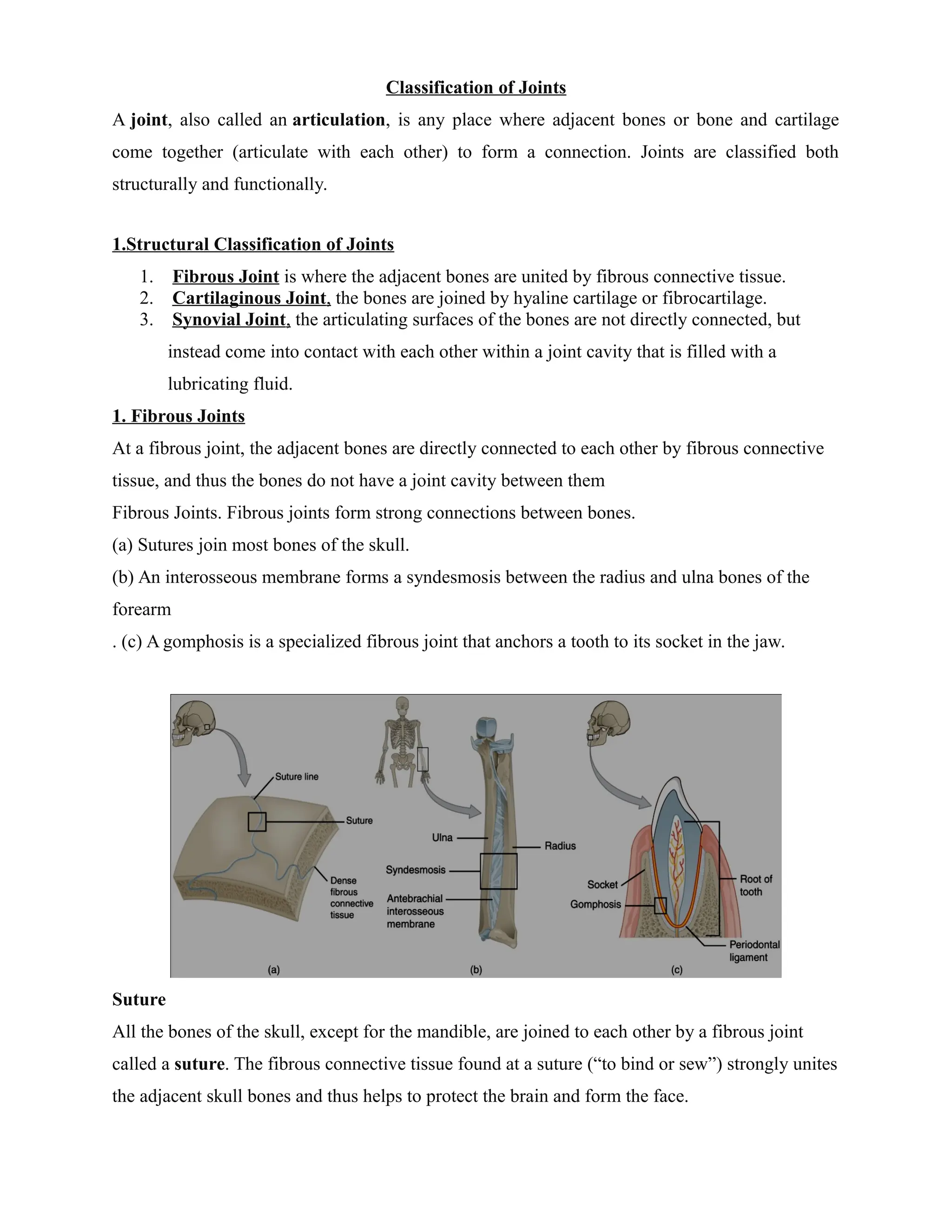
(a) Sutures join most bones of the skull.
(b) An interosseous membrane forms a syndesmosis between the radius and ulna bones of the
forearm
. (c) A gomphosis is a specialized fibrous joint that anchors a tooth to its socket in the jaw.
Suture
All the bones of the skull, except for the mandible, are joined to each other by a fibrous joint
called a suture. The fibrous connective tissue found at a suture (“to bind or sew”) strongly unites
the adjacent skull bones and thus helps to protect the brain and form the face.
2.
Syndesmosis
A syndesmosis (“fastenedwith a band”) is a type of fibrous joint in which two parallel bones are
united to each other by fibrous connective tissue.
The syndesmoses found in the forearm and leg serve to unite parallel bones and prevent
their separation.
Syndesmoses are found between the bones of the forearm (radius and ulna) and the leg
(tibia and fibula).
Gomphosis
A gomphosis (“fastened with bolts”) is the specialized fibrous joint that anchors the root of a
tooth into its bony socket within the maxillary bone (upper jaw) or mandible bone (lower jaw) of
the skull.
Due to the immobility of a gomphosis, this type of joint is functionally classified as a
synarthrosis.
2-Cartilaginous Joints
As the name indicates, at a cartilaginous joint, the adjacent bones are united by cartilage, a tough
but flexible type of connective tissue. These types of joints lack a joint cavity and involve bones
that are joined together by either hyaline cartilage or fibrocartilage.
There are two types of cartilaginous joints.
1. Synchondrosis is a cartilaginous joint where the bones are joined by hyaline cartilage. Also
classified as a synchondrosis are places where bone is united to a cartilage structure, such as
between the anterior end of a ribs and the costal cartilage of the thoracic cage.
2. Symphysis, where the bones are joined by fibrocartilage.
Examples in which the gap between the bones is narrow include the pubic symphysis and
the manubriosternal joint. At the pubic symphysis, the pubic portions of the right and left
hip bones of the pelvis are joined together by fibrocartilage across a narrow gap.
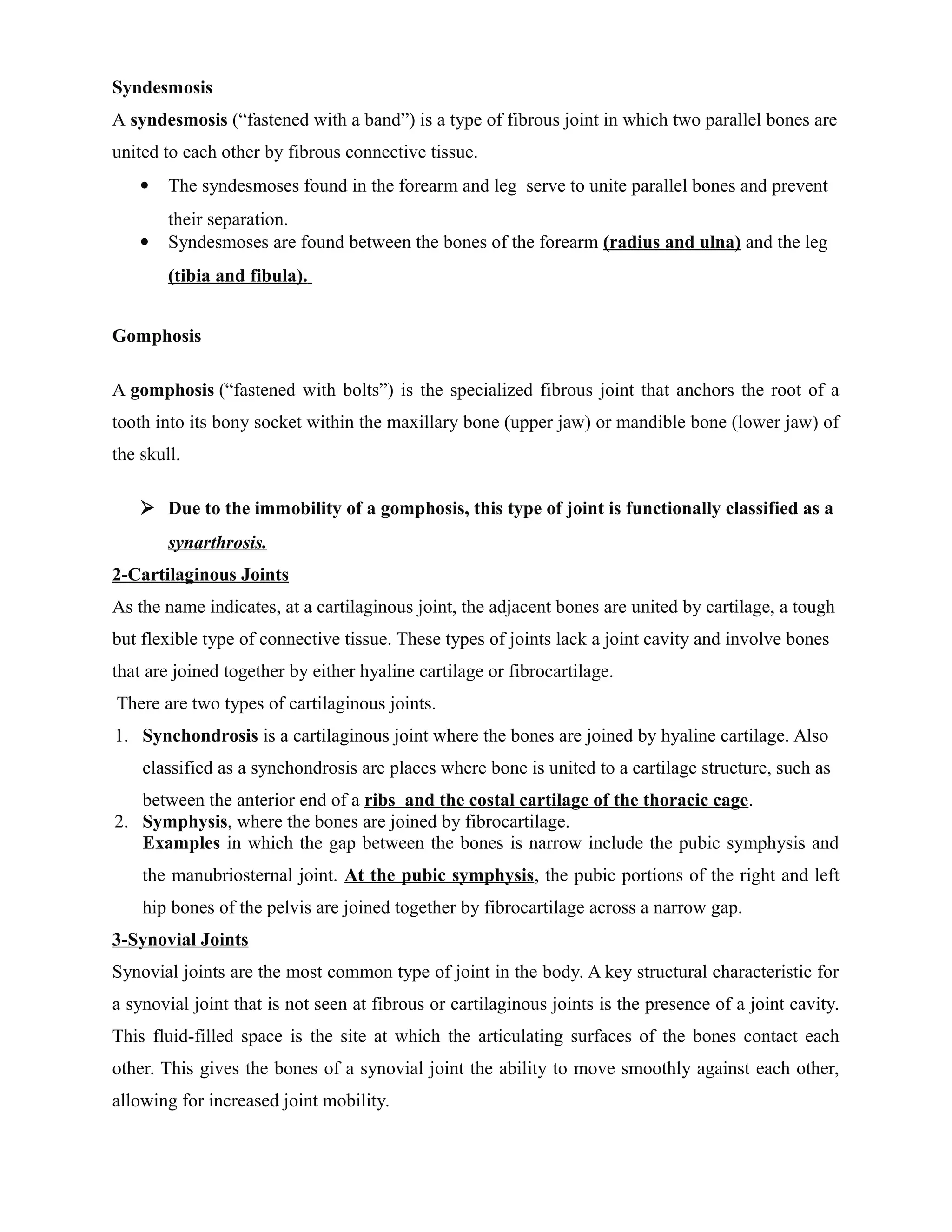
3-Synovial Joints
Synovial joints are the most common type of joint in the body. A key structural characteristic for
a synovial joint that is not seen at fibrous or cartilaginous joints is the presence of a joint cavity.
This fluid-filled space is the site at which the articulating surfaces of the bones contact each
other. This gives the bones of a synovial joint the ability to move smoothly against each other,
allowing for increased joint mobility.
3.
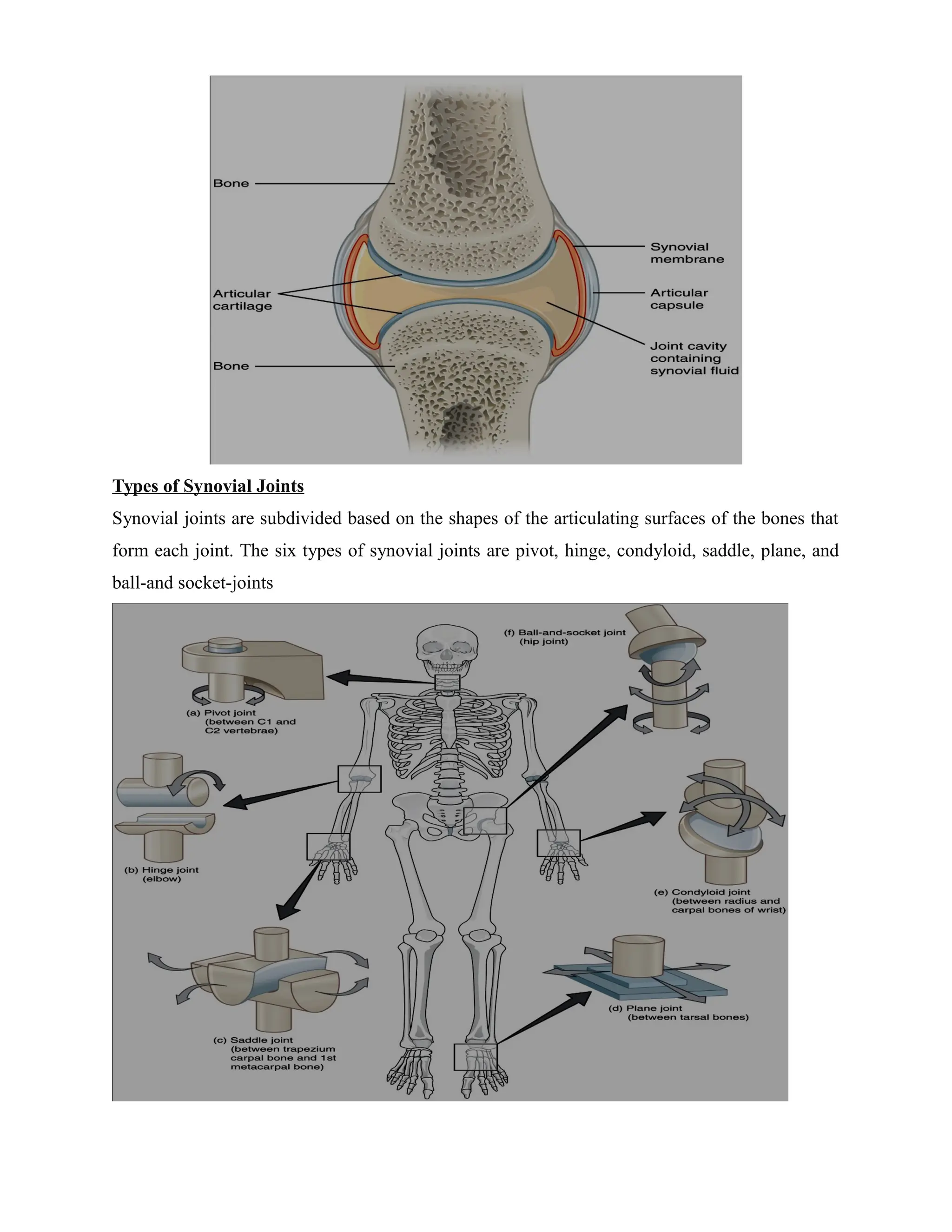
Types of SynovialJoints
Synovial joints are subdivided based on the shapes of the articulating surfaces of the bones that
form each joint. The six types of synovial joints are pivot, hinge, condyloid, saddle, plane, and
ball-and socket-joints
4.
1. Pivot joint-At a pivot joint, a rounded portion of a bone is enclosed within a ring
formed partially by the articulation with another bone and partially by a ligament. The
bone rotates within this ring.
Example are articulation between the C1 (atlas) and the dens of the C2 (axis)
vertebrae, which provides the side-to-side rotation of the head, or at the proximal
radioulnar joint between the head of the radius and the radial notch of the ulna.
2. Hinge Joint-In a hinge joint, the convex end of one bone articulates
with the concave end of the adjoining bone
A good example is the elbow joint, with the articulation between the
trochlea of the humerus and the trochlear notch of the ulna.
3. Condyloid Joint-At a condyloid joint (ellipsoid joint), the shallow
depression at the end of one bone articulates with a rounded structure
from an adjacent bone or bones
Example - the radiocarpal joint of the wrist, between the shallow depression at the distal
end of the radius bone.
4. Saddle Joint-At a saddle joint, both of the articulating surfaces for the
bones have a saddle shape, which is concave in one direction and
convex in the other.
Example- the thumb that ability to move away from the palm of the
hand along two planes.
5. Plane Joint-At a plane joint (gliding joint), the articulating surfaces of
the bones are flat or slightly curved and of approximately the same
size, which allows the bones to slide against each other
Example - the carpal bones (intercarpal joints) of the wrist or tarsal
bones (intertarsal joints) of the foot, between the clavicle and
acromion of the scapula.
5.
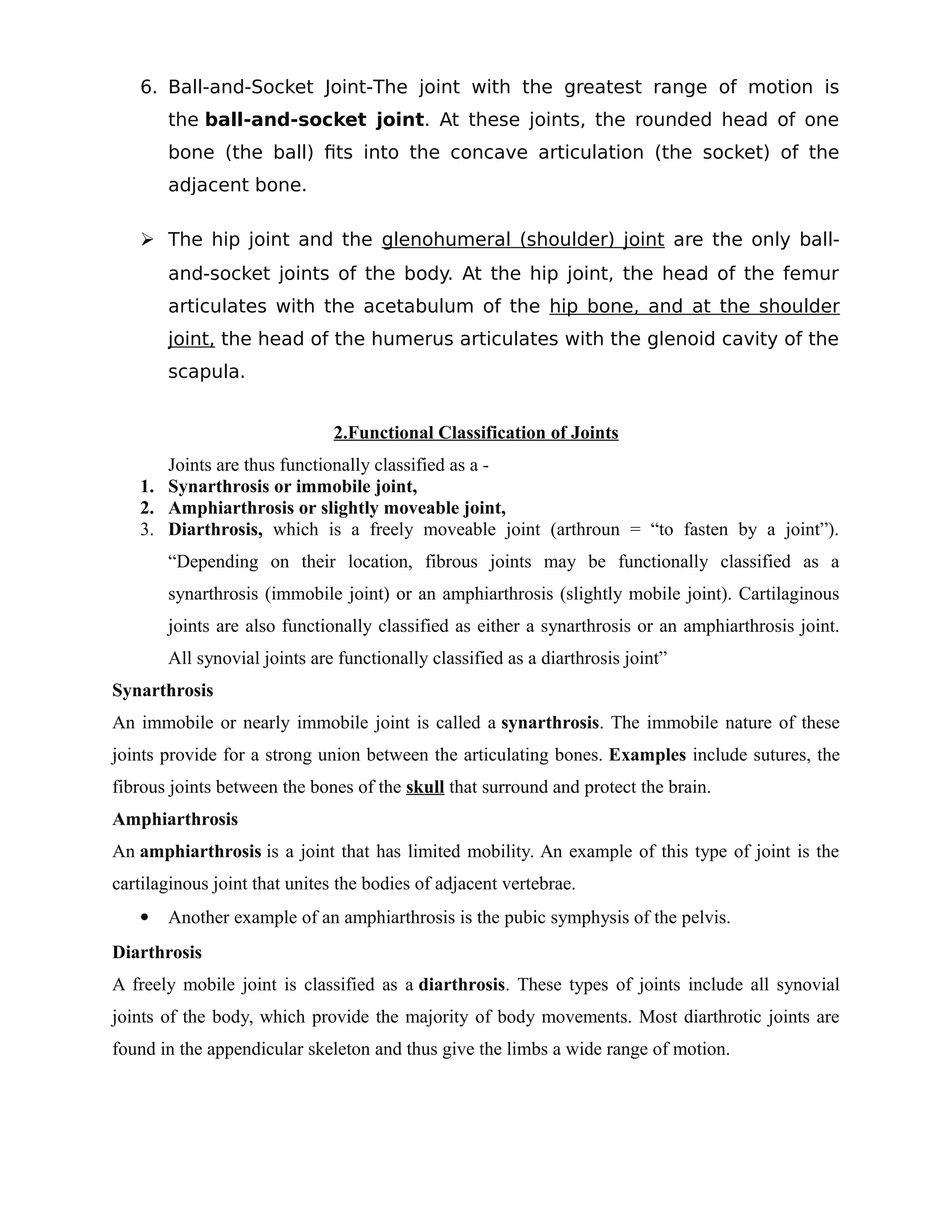
6. Ball-and-Socket Joint-Thejoint with the greatest range of motion is
the ball-and-socket joint. At these joints, the rounded head of one
bone (the ball) fits into the concave articulation (the socket) of the
adjacent bone.
The hip joint and the glenohumeral (shoulder) joint are the only ball-
and-socket joints of the body. At the hip joint, the head of the femur
articulates with the acetabulum of the hip bone, and at the shoulder
joint, the head of the humerus articulates with the glenoid cavity of the
scapula.
2.Functional Classification of Joints
Joints are thus functionally classified as a -
1. Synarthrosis or immobile joint,
2. Amphiarthrosis or slightly moveable joint,
3. Diarthrosis, which is a freely moveable joint (arthroun = “to fasten by a joint”).
“Depending on their location, fibrous joints may be functionally classified as a
synarthrosis (immobile joint) or an amphiarthrosis (slightly mobile joint). Cartilaginous
joints are also functionally classified as either a synarthrosis or an amphiarthrosis joint.
All synovial joints are functionally classified as a diarthrosis joint”
Synarthrosis
An immobile or nearly immobile joint is called a synarthrosis. The immobile nature of these
joints provide for a strong union between the articulating bones. Examples include sutures, the
fibrous joints between the bones of the skull that surround and protect the brain.
Amphiarthrosis
An amphiarthrosis is a joint that has limited mobility. An example of this type of joint is the
cartilaginous joint that unites the bodies of adjacent vertebrae.
Another example of an amphiarthrosis is the pubic symphysis of the pelvis.
Diarthrosis
A freely mobile joint is classified as a diarthrosis. These types of joints include all synovial
joints of the body, which provide the majority of body movements. Most diarthrotic joints are
found in the appendicular skeleton and thus give the limbs a wide range of motion.
6.
Types of BodyMovements
Synovial joints allow the body a tremendous range of movements. Each movement at a synovial
joint results from the contraction or relaxation of the muscles that are attached to the bones on
either side of the articulation. The type of movement that can be produced at a synovial joint is
determined by its structural type. While the ball-and-socket joint gives the greatest range of
movement at an individual joint, in other regions of the body, several joints may work together to
produce a particular movement.
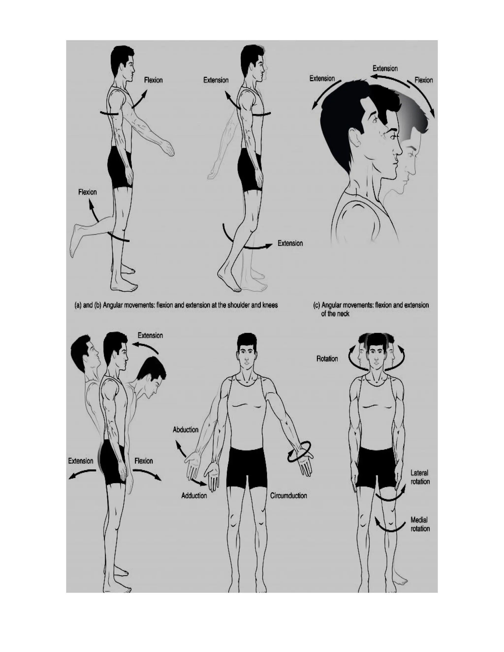
In the Figure below-
1. Synovial joints give the body many ways in which to move.
(a) – (b) Flexion and extension motions are in the sagittal (anterior–posterior) plane of motion.
These movements take place at the shoulder, hip, elbow, knee, wrist, metacarpophalangeal,
metatarsophalangeal, and interphalangeal joints.
(c)–(d) Anterior bending of the head or vertebral column is flexion, while any posterior-going
movement is extension.
(e) Abduction and adduction are motions of the limbs, hand, fingers, or toes in the coronal
(medial–lateral) plane of movement.
8.
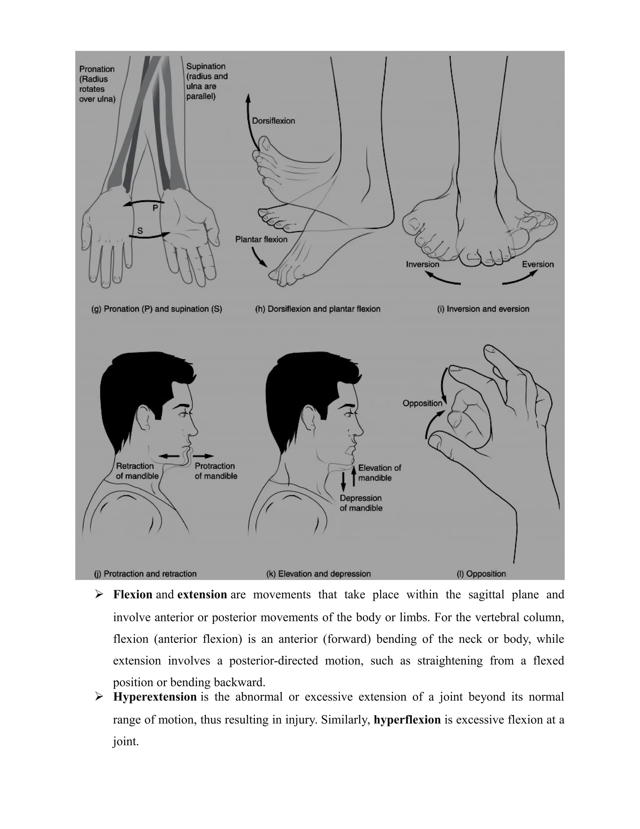
Flexion andextension are movements that take place within the sagittal plane and
involve anterior or posterior movements of the body or limbs. For the vertebral column,
flexion (anterior flexion) is an anterior (forward) bending of the neck or body, while
extension involves a posterior-directed motion, such as straightening from a flexed
position or bending backward.
Hyperextension is the abnormal or excessive extension of a joint beyond its normal
range of motion, thus resulting in injury. Similarly, hyperflexion is excessive flexion at a
joint.
9.
Abduction andadduction motions occur within the coronal plane and involve medial-
lateral motions of the limbs, fingers, toes, or thumb. Abduction moves the limb laterally
away from the midline of the body, while adduction is the opposing movement that
brings the limb toward the body or across the midline.
Circumduction is the movement of a body region in a circular manner, in which one end
of the body region being moved stays relatively stationary while the other end describes a
circle.
Rotation can occur within the vertebral column, at a pivot joint, or at a ball-and-socket
joint. Rotation of the neck or body is the twisting movement produced by the summation
of the small rotational movements available between adjacent vertebrae.
Supination and pronation are movements of the forearm. In the anatomical position, the
upper limb is held next to the body with the palm facing forward.
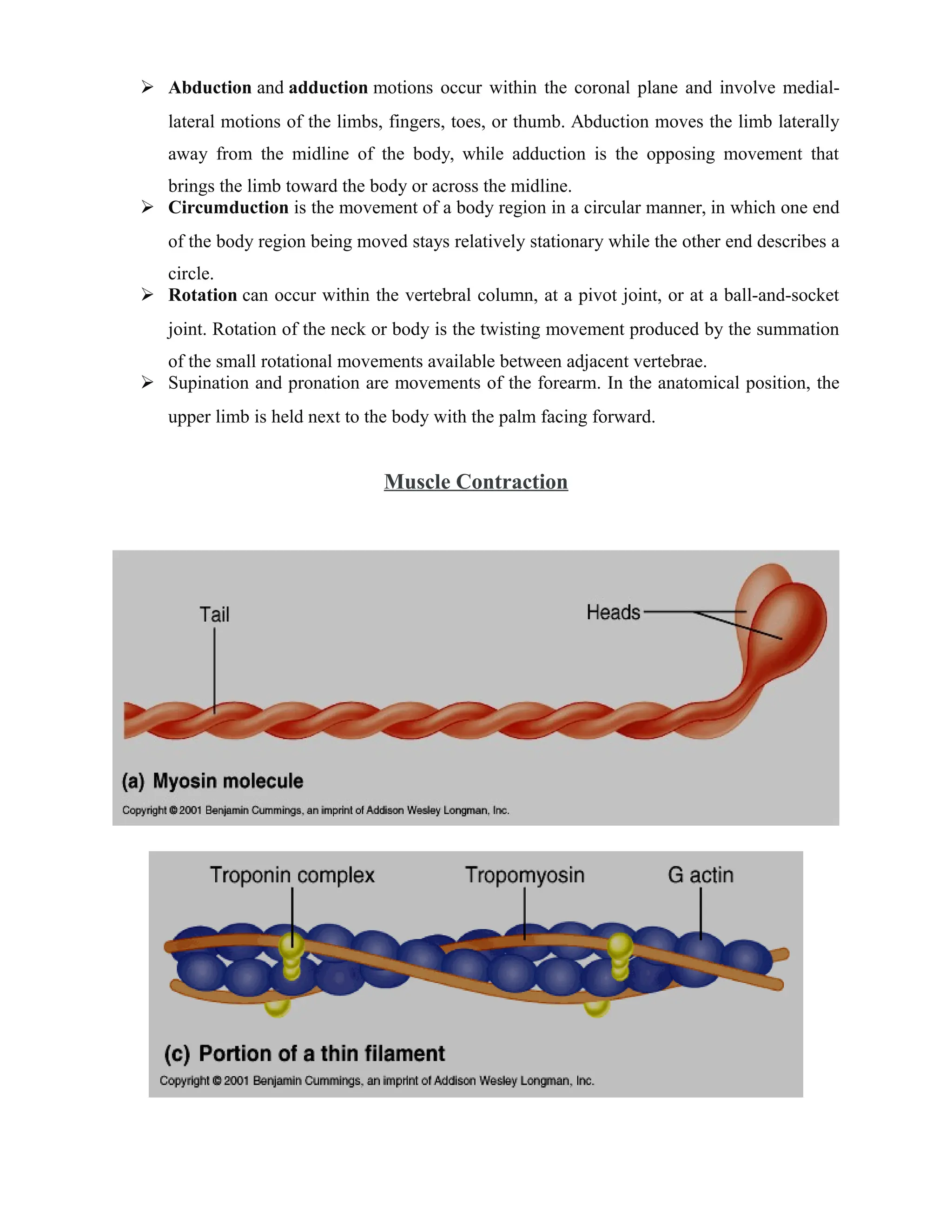
Muscle Contraction
10.
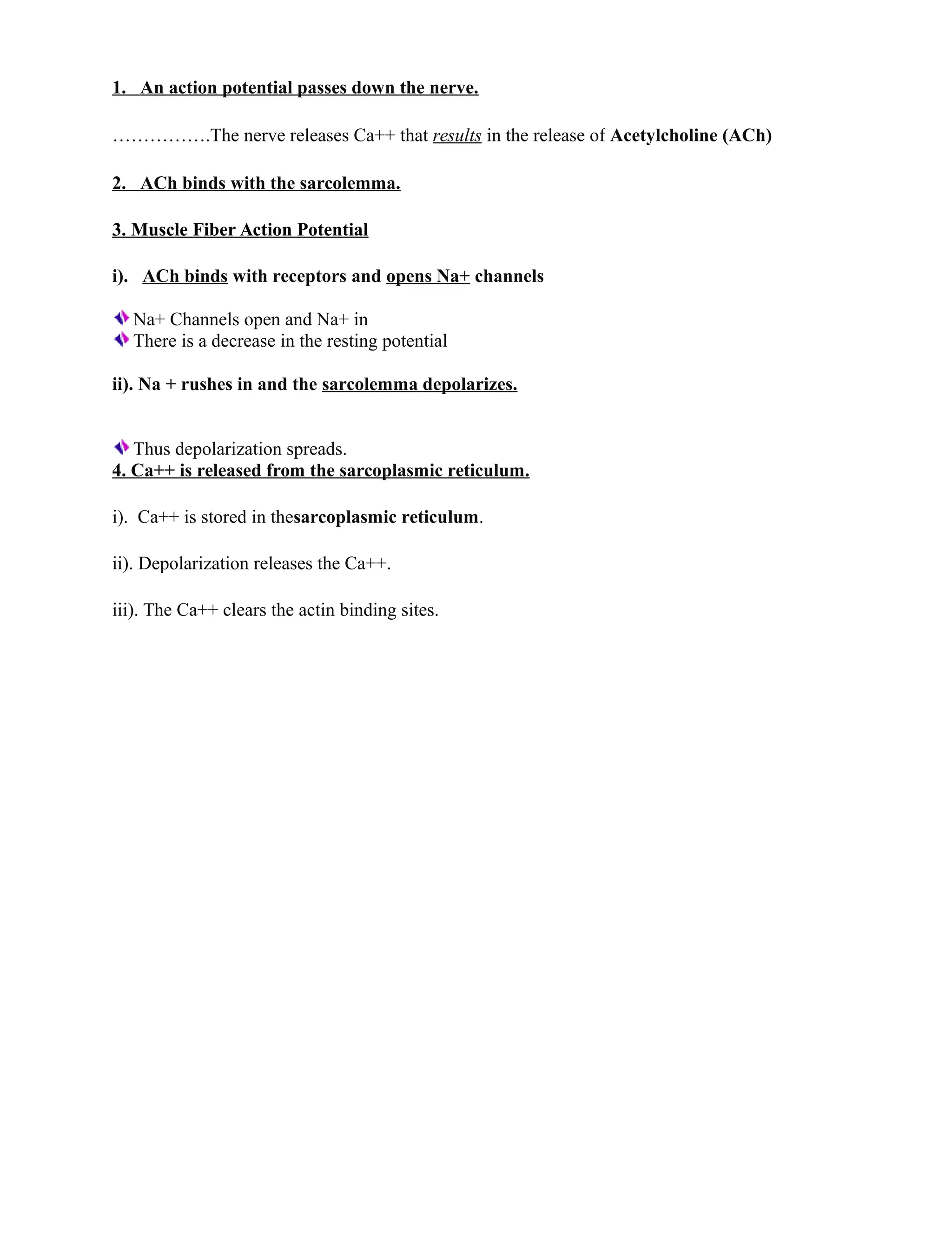
1. An actionpotential passes down the nerve.
…………….The nerve releases Ca++ that results in the release of Acetylcholine (ACh)
2. ACh binds with the sarcolemma.
3. Muscle Fiber Action Potential
i). ACh binds with receptors and opens Na+ channels
Na+ Channels open and Na+ in
There is a decrease in the resting potential
ii). Na + rushes in and the sarcolemma depolarizes.
Thus depolarization spreads.
4. Ca++ is released from the sarcoplasmic reticulum.
i). Ca++ is stored in thesarcoplasmic reticulum.
ii). Depolarization releases the Ca++.
iii). The Ca++ clears the actin binding sites.
11.
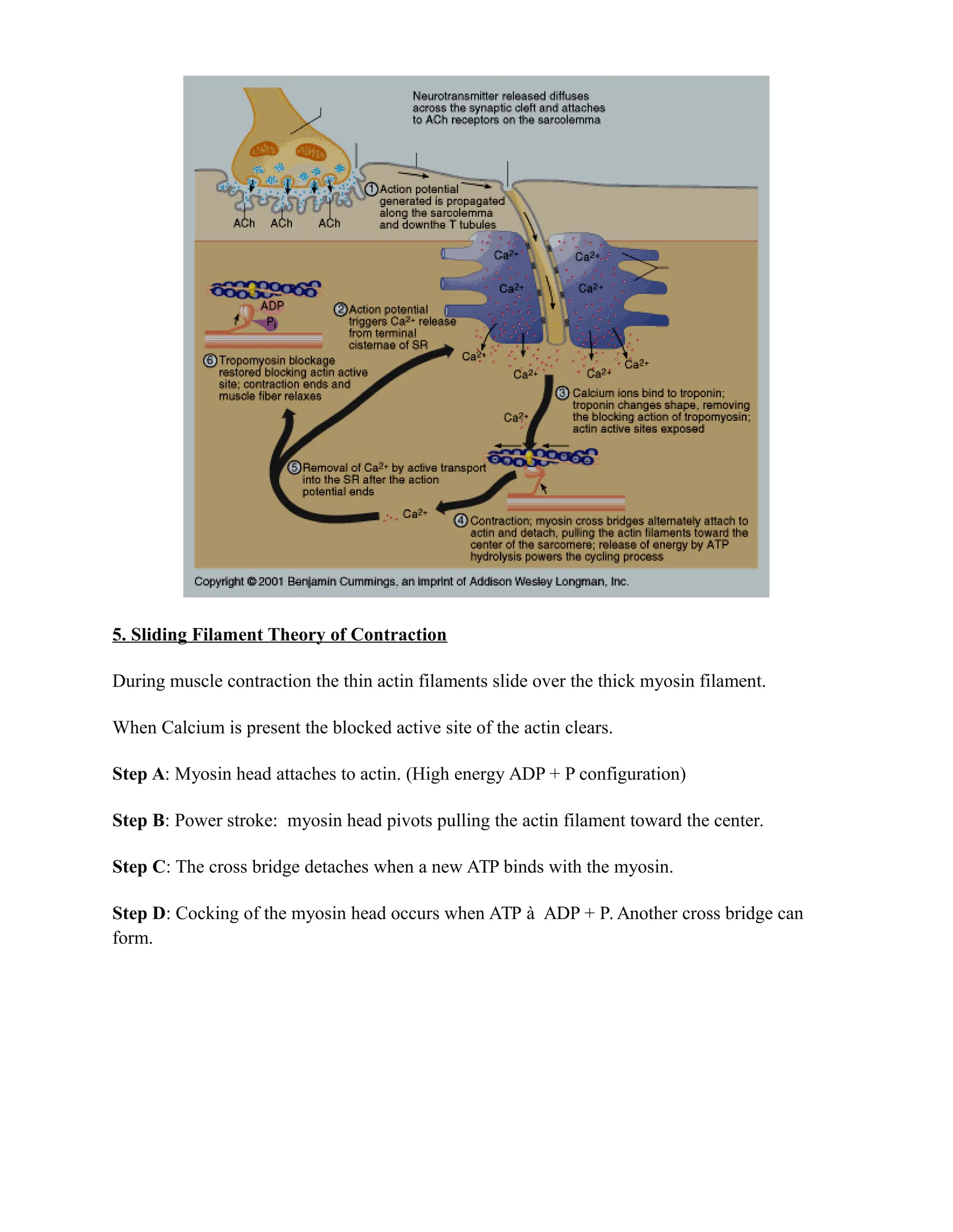
5. Sliding FilamentTheory of Contraction
During muscle contraction the thin actin filaments slide over the thick myosin filament.
When Calcium is present the blocked active site of the actin clears.
Step A: Myosin head attaches to actin. (High energy ADP + P configuration)
Step B: Power stroke: myosin head pivots pulling the actin filament toward the center.
Step C: The cross bridge detaches when a new ATP binds with the myosin.
Step D: Cocking of the myosin head occurs when ATP à ADP + P. Another cross bridge can
form.
12.
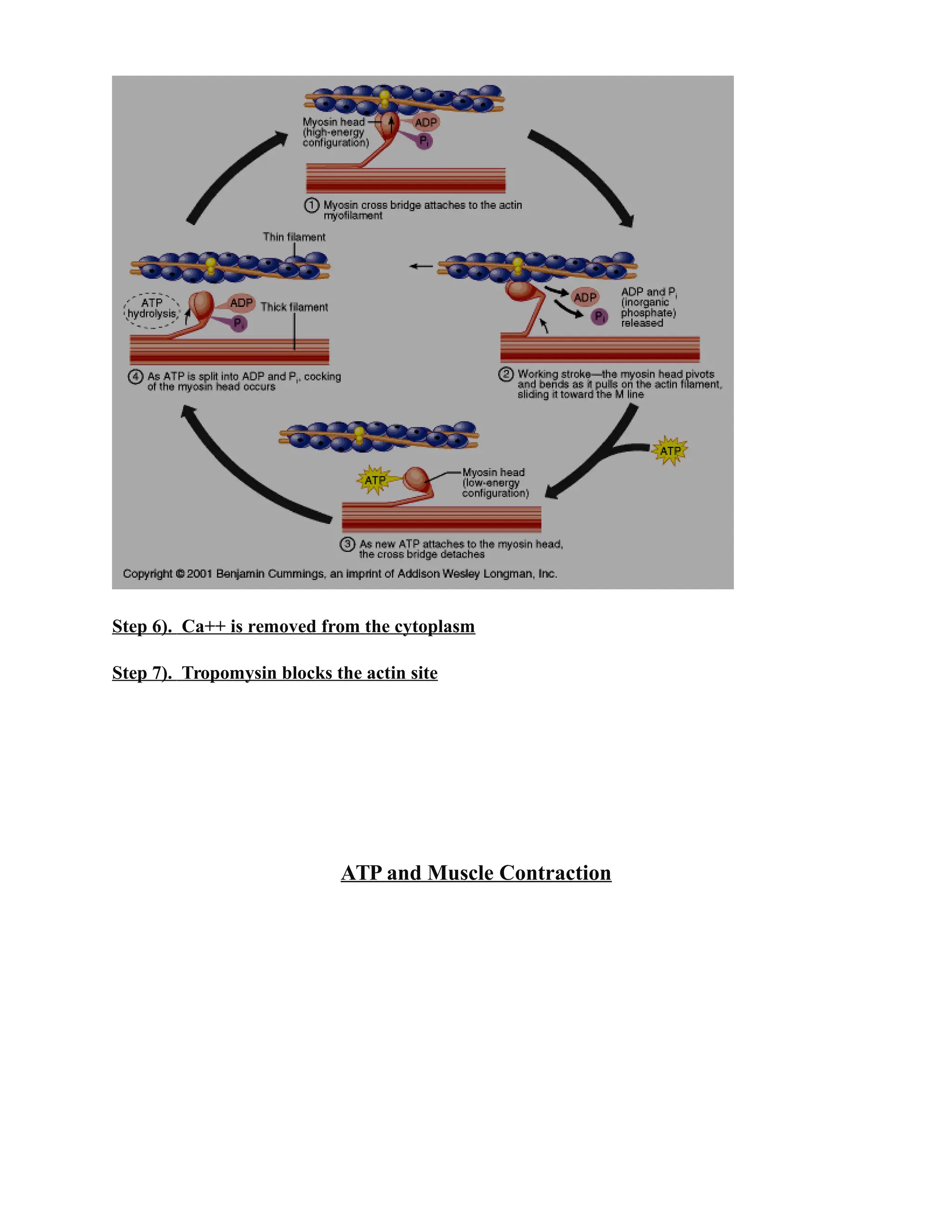
Step 6). Ca++is removed from the cytoplasm
Step 7). Tropomysin blocks the actin site
ATP and Muscle Contraction
13.
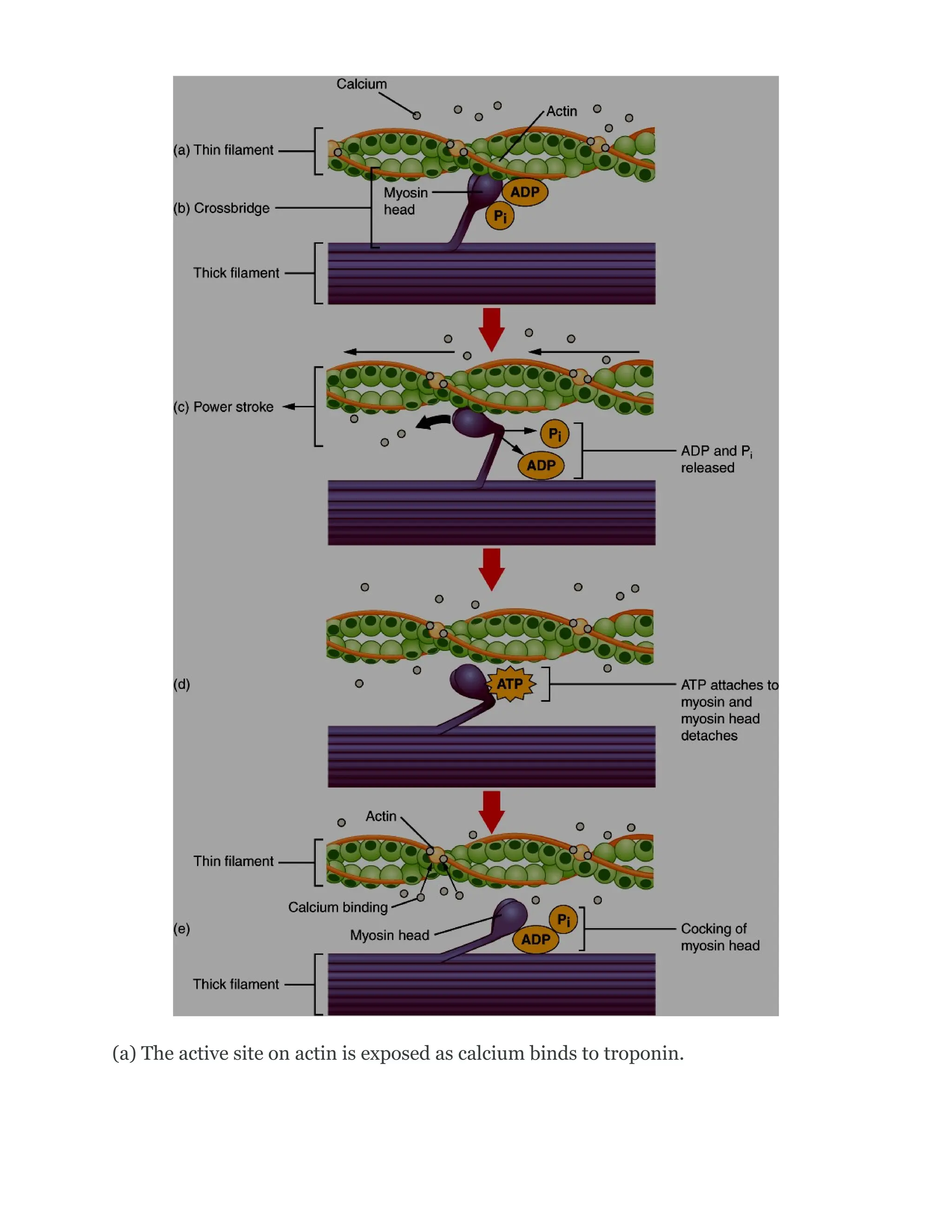
(a) The activesite on actin is exposed as calcium binds to troponin.
14.
(b) The myosinhead is attracted to actin, and myosin binds actin at its actin-binding
site, forming the cross-bridge.
(c) During the power stroke, the phosphate generated in the previous contraction
cycle is released.
(d) A new molecule of ATP attaches to the myosin head, causing the cross-bridge to
detach. (e) The myosin head hydrolyzes ATP to ADP and phosphate, which returns
the myosin to the cocked position.
Creatine phosphate is a molecule that can store energy in its phosphate bonds. In a
resting muscle, excess ATP transfers its energy to creatine, producing ADP and creatine
phosphate. This acts as an energy reserve that can be used to quickly create more ATP.
When the muscle starts to contract and needs energy, creatine phosphate transfers its
phosphate back to ADP to form ATP and creatine. This reaction is catalyzed by the
enzyme creatine kinase and occurs very quickly;
What are “T-tubules” and what is their role?
ANS- Transverse tubules (T-tubules) are extensions of the sarcolemma (muscle cell
membrane) that penetrate into the centre of skeletal and cardiac muscle cells.
T-TUBULES is to conduct impulses from the surface of the cell (SARCOLEMMA)
down into the cell and, specifically, to another structure in the cell called the
SARCOPLASMIC RETICULUM.
The T-tubules are inward extensions of the sarcolemma that trigger the release of Ca+
+
from SR during an Action Potential. (b) Ca++
binds to tropomyosin, and this slides the
tropomyosin rods away from the binding sites.
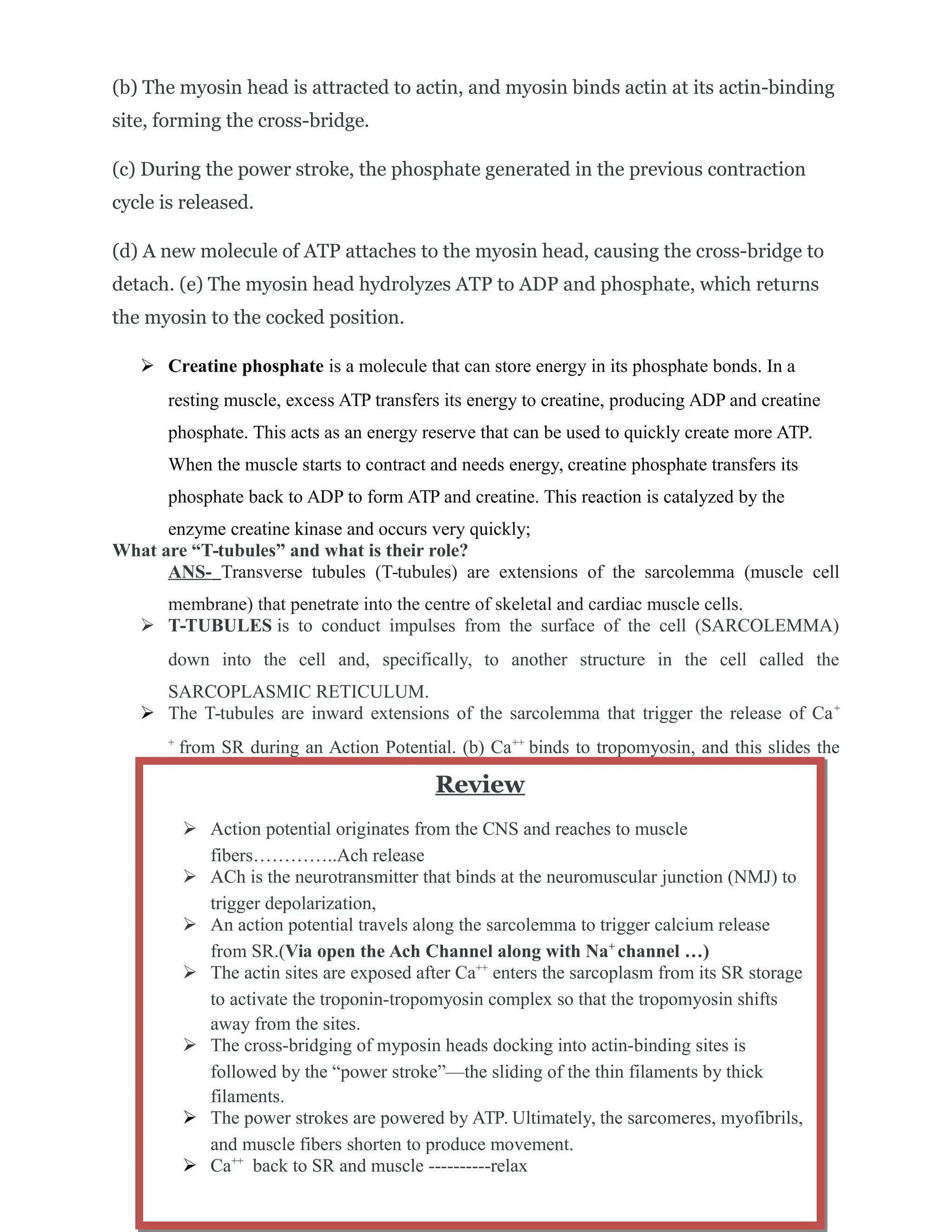
Review
Action potential originates from the CNS and reaches to muscle
fibers…………..Ach release
ACh is the neurotransmitter that binds at the neuromuscular junction (NMJ) to
trigger depolarization,
An action potential travels along the sarcolemma to trigger calcium release
from SR.(Via open the Ach Channel along with Na+
channel …)
The actin sites are exposed after Ca++
enters the sarcoplasm from its SR storage
to activate the troponin-tropomyosin complex so that the tropomyosin shifts
away from the sites.
The cross-bridging of myposin heads docking into actin-binding sites is
followed by the “power stroke”—the sliding of the thin filaments by thick
filaments.
The power strokes are powered by ATP. Ultimately, the sarcomeres, myofibrils,
and muscle fibers shorten to produce movement.
Ca++
back to SR and muscle ----------relax
15.
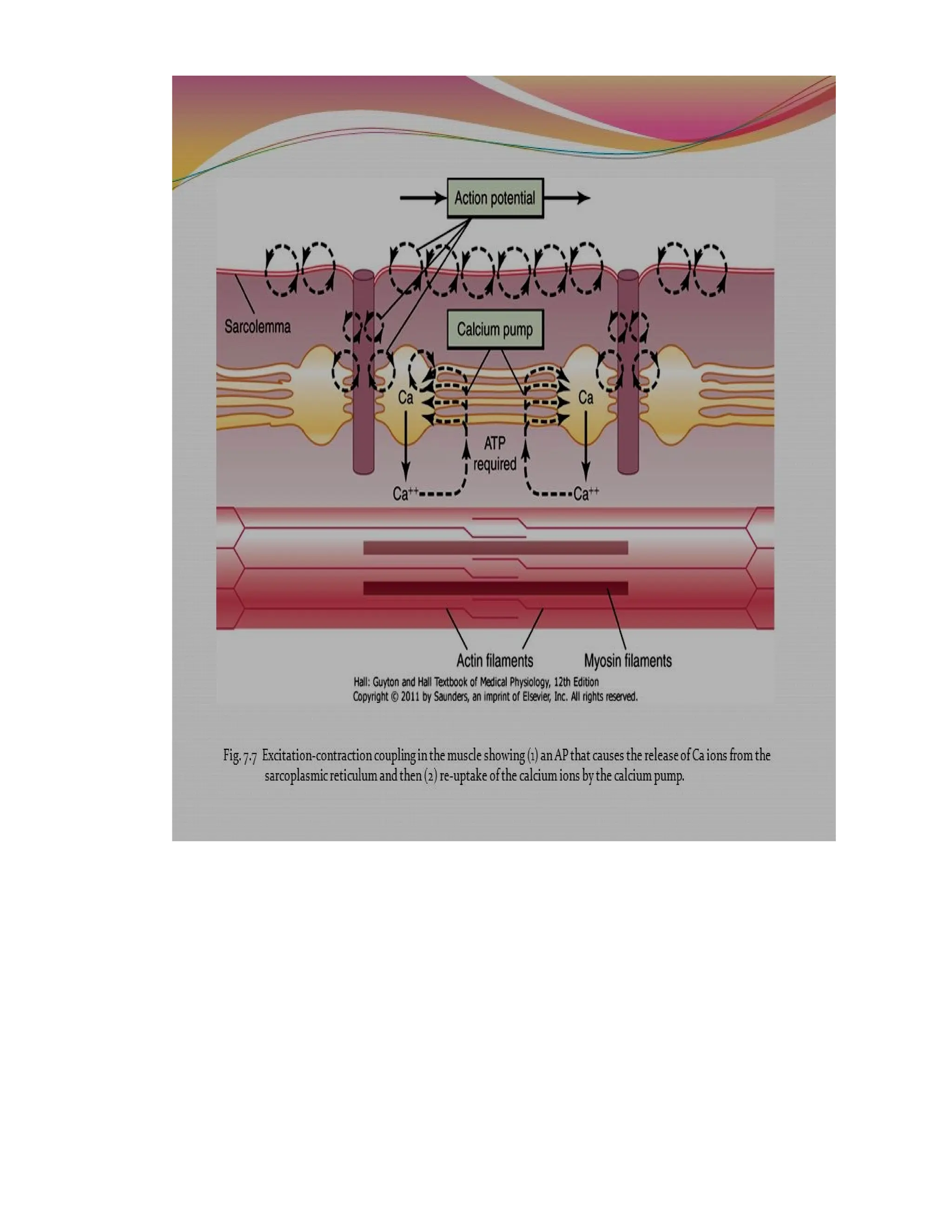
EXCITATION-CONTRACTION COUPLING
Both neuronsand skeletal muscle cells are electrically excitable, meaning that they are able to
generate action potentials. An action potential is a special type of electrical signal that can travel
along a cell membrane as a wave. This allows a signal to be transmitted quickly and faithfully
over long distances.
Note- Although the term excitation-contraction coupling confuses or scares some students, it
comes down to this: for a skeletal muscle fiber to contract, its membrane must first be
“excited”—in other words, it must be stimulated to fire an action potential. The muscle fiber
action potential, which sweeps along the sarcolemma as a wave, is “coupled” to the actual
contraction through the release of calcium ions (Ca++
) from the SR. Once released, the Ca+
+
interacts with the shielding proteins, forcing them to move aside so that the actin-binding sites
are available for attachment by myosin heads. The myosin then pulls the actin filaments toward
the center, shortening the muscle fiber.