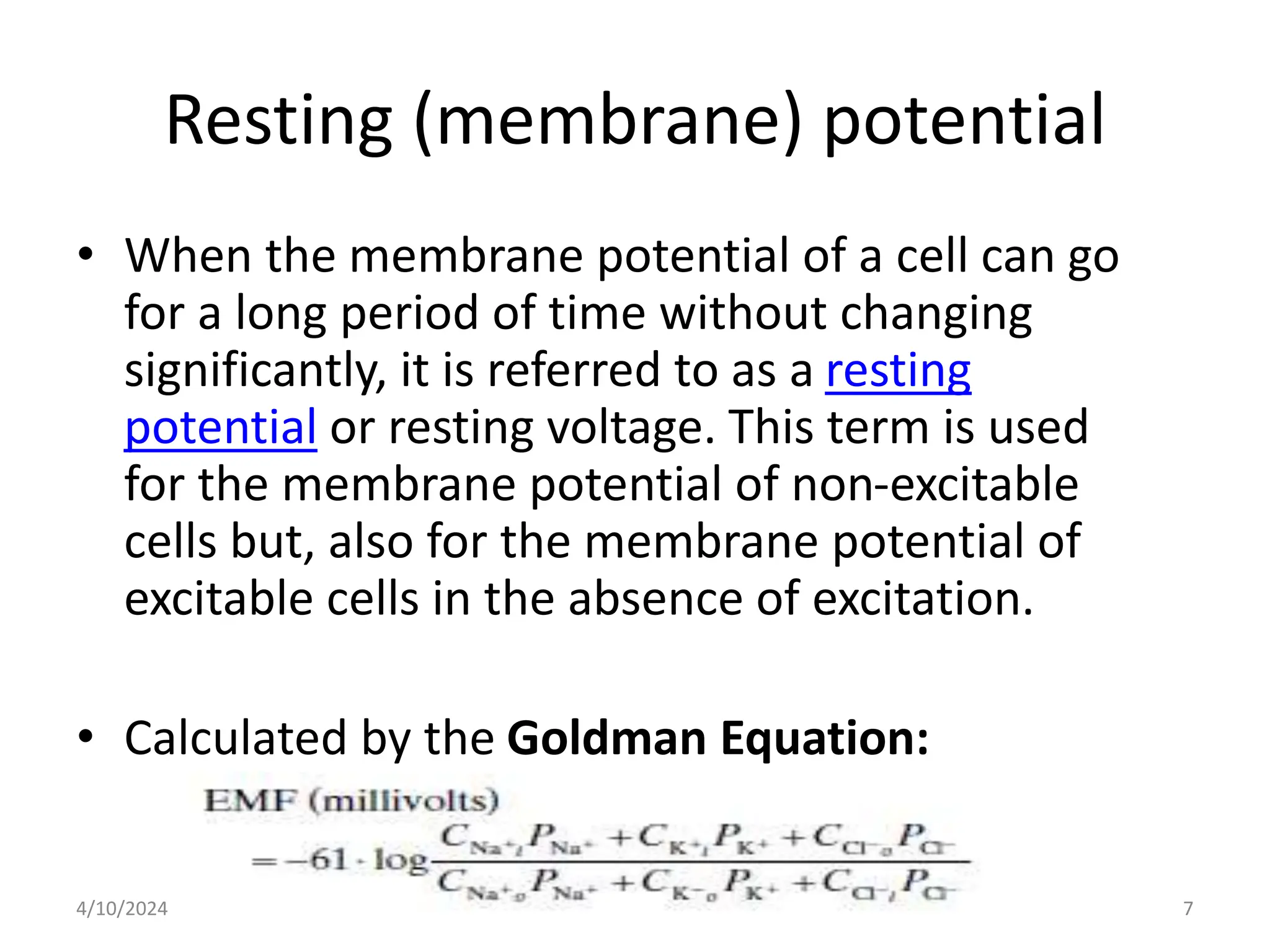
IONIC BASIS OF RESTING MEMBRANE
AND ACTION POTENTIALS
Dr. Hidaayah O.
Jimoh-Abdulghaffaar
4/10/2024 1
Outline
• Introduction
• Resting membrane potential
– Ionic basis
• Action potential
– History
– Ionic basis
– Initiation
– Components
– Stages
• Clinical correlates
4/10/2024 2
Introduction
• The membranes of almost all cells in the body
have electrical potentials.
• The membrane of some cells (nerve and muscle)
can generate rapidly-changing electrochemical
impulses. These impulses are used to transmit
signals.
• The functions of some cells (glandular cells,
ciliated cells and macrophages) are activated by
local changes in membrane potentials.
4/10/2024 3
Introduction Cont’d
• Membrane potentials are due to the diffusion
of ions down their concentration gradients,
the electric charge of the ion, and any
membrane pumps for that ion.
• Influx is the net movement of ions into the cell
from the ECF.
• Efflux is the net movement of ions out of the
cell to the ECF.
• Flux (the movement of charges) is always
measured in millivolts (mV).
4/10/2024 4
Introduction Cont’d
4/10/2024 5
Resting Membrane
Potential
4/10/2024 6
Resting (membrane) potential
• When the membrane potential of a cell can go
for a long period of time without changing
significantly, it is referred to as a resting
potential or resting voltage. This term is used
for the membrane potential of non-excitable
cells but, also for the membrane potential of
excitable cells in the absence of excitation.
• Calculated by the Goldman Equation:
4/10/2024 7
4/10/2024 8
Fig. 1: Resting Membrane Potential
Ionic Basis
• Potassium concentration is great inside a nerve fiber
membrane but, very low outside the membrane.
• Assuming the membrane is permeable to K+ only, K+
diffuse outward through the membrane, making the
outside more electropositive and leaving the inside
electronegative.
• This leads to the development of a potential difference
between the inside and outside - diffusion potential.
• Diffusion potential is 94mV, negative inside the fiber.
4/10/2024 9
Fig. 2: Equilibrium Potential of Potassium
4/10/2024 10
Ionic Basis Cont’d
• There is a high concentration of Na+ outside the
membrane and a low concentration in side the cell.
• The membrane is highly permeable to Na+ but
impermeable to other ions.
• Na+ diffuse inside the cell, making the inside more
electropositive and the outside, electronegative.
• Diffusion potential is 61mV positive inside the cell.
4/10/2024 11
Fig. 3: Equilibrium Potential of Sodium
4/10/2024 12
4/10/2024 13
Fig. 4: Resting Membrane Potential
Fig. 5: Diffusion Potentials of Sodium and Potassium
4/10/2024 14
Fig. 6: The Sodium-Potassium ATPase Pump
4/10/2024 15
RMP
• In nerves, -70mV when not transmitting nerve
signals.
• Cardiac muscle?
• Skeletal muscle?
• Salivary gland?
• Red blood cell?
• Other cells/tissues??
4/10/2024 16
Action Potential
4/10/2024 17
Fig. 7: The Squid Giant Axon
4/10/2024 18
4/10/2024 19
4/10/2024 20
4/10/2024 21
Action Potential – history Cont’d
4/10/2024 22
Fig. 8: Graphical Representation of Action Potential
4/10/2024 23
Ionic Basis of Action Potential
• An action potential occurs when there is a reversal of
the normal resting potential, going from negative to
positive - depolarization.
• Depolarization occurs when a stimulus causes the
voltage-gated Na+ channels to open, allowing Na+ to
rapidly influx down its concentration gradient.
• The sudden in-rush of positive sodium ions reverses the
membrane potential for a few milliseconds.
• Then the voltage-gated K+ channels open, allowing K+
to rapidly efflux due to its concentration gradient. This
brings the membrane back to negative inside and is
called repolarization.
4/10/2024 24
Resting stage
• RMP before action potential begins.
• Membrane is said to be polarized at this stage.
4/10/2024 25
Depolarization stage
• Membrane suddenly becomes permeable to Na+…..
• This rapidly neutralizes the normal “polarized state” of
-90mV.
• In large nerve fibers, the great excess of Na+ causes the
membrane potential to actually overshoot beyond the zero
level.
• However, this doesn’t occur in smaller fibers.
• Activated by the voltage-gated Na+ channel.
4/10/2024 26
Repolarization stage
• After being highly permeable to Na+, within
milliseconds, the membrane Na+ channels begin
to close and the K+ channels open more than
normal.
• There is rapid diffusion of K+ to the exterior.
• This helps to re-establish the normal RMP.
• Activated by the voltage-gated K+ channel.
4/10/2024 27
Ionic Basis of Action Potential
4/10/2024 28
Sodium channels have 2 gates,
a normal voltage (activation)
gate (which is closed at rest)
and an inactivation gate (which
is open at rest). The rapid
opening of the voltage gate
lets Na+ rush in and
depolarizes the cell. This is
immediately followed by
closing of the inactivation gate
which stops the Na+ influx. At
the same time the K+ gate
opens letting K+ efflux
(repolarization).
4/10/2024 29
Ionic Basis of Action Potentials
0 mV
-80 mV
PK>>PNa
PNa>>PK
PK>>PNa
ENa
EK
Time 
1. At rest only K+
leak channels
open, PK>>PNa
2. With stimulus,
voltage-gated Na
channels open,
PNa>>PK
Na+ flows into
the cell carrying
positive charge
3. Delayed opening
of voltage-gated
K channels,
PK>>PNa
K+ flows out of
cell removing
positive charge
4/10/2024 30
Summary
4/10/2024 31
4/10/2024 32
Action Potential
0 mV
-80 mV
Rising phase or
depolarization
Falling phase or
Repolarization
Resting membrane
potential
Threshold Potential
Undershoot or after-
hyperpolarization
Overshoot
4/10/2024 33
4/10/2024 34
4/10/2024 35
4/10/2024 36
Clinical Correlates
• Local anesthetics e.g. procaine, lidocaine
– Prevent generation of action potential by blocking the
opening of voltage-gated Na+ channels thereby
preventing depolarization and transmission of pain
sensation to the brain.
• Tetrodotoxin
– Potent toxin produced by puffer fish which bind to
voltage-gated Na+ channels.
– Ingestion of improperly-prepared fish diet even in
small quantities can cause death.
4/10/2024 37

IONIC BASIS OF RESTING MEMBRANE AND ACTION POTENTIALS.pptx

  • 1.
    IONIC BASIS OFRESTING MEMBRANE AND ACTION POTENTIALS Dr. Hidaayah O. Jimoh-Abdulghaffaar 4/10/2024 1
  • 2.
    Outline • Introduction • Restingmembrane potential – Ionic basis • Action potential – History – Ionic basis – Initiation – Components – Stages • Clinical correlates 4/10/2024 2
  • 3.
    Introduction • The membranesof almost all cells in the body have electrical potentials. • The membrane of some cells (nerve and muscle) can generate rapidly-changing electrochemical impulses. These impulses are used to transmit signals. • The functions of some cells (glandular cells, ciliated cells and macrophages) are activated by local changes in membrane potentials. 4/10/2024 3
  • 4.
    Introduction Cont’d • Membranepotentials are due to the diffusion of ions down their concentration gradients, the electric charge of the ion, and any membrane pumps for that ion. • Influx is the net movement of ions into the cell from the ECF. • Efflux is the net movement of ions out of the cell to the ECF. • Flux (the movement of charges) is always measured in millivolts (mV). 4/10/2024 4
  • 5.
  • 6.
  • 7.
    Resting (membrane) potential •When the membrane potential of a cell can go for a long period of time without changing significantly, it is referred to as a resting potential or resting voltage. This term is used for the membrane potential of non-excitable cells but, also for the membrane potential of excitable cells in the absence of excitation. • Calculated by the Goldman Equation: 4/10/2024 7
  • 8.
    4/10/2024 8 Fig. 1:Resting Membrane Potential
  • 9.
    Ionic Basis • Potassiumconcentration is great inside a nerve fiber membrane but, very low outside the membrane. • Assuming the membrane is permeable to K+ only, K+ diffuse outward through the membrane, making the outside more electropositive and leaving the inside electronegative. • This leads to the development of a potential difference between the inside and outside - diffusion potential. • Diffusion potential is 94mV, negative inside the fiber. 4/10/2024 9
  • 10.
    Fig. 2: EquilibriumPotential of Potassium 4/10/2024 10
  • 11.
    Ionic Basis Cont’d •There is a high concentration of Na+ outside the membrane and a low concentration in side the cell. • The membrane is highly permeable to Na+ but impermeable to other ions. • Na+ diffuse inside the cell, making the inside more electropositive and the outside, electronegative. • Diffusion potential is 61mV positive inside the cell. 4/10/2024 11
  • 12.
    Fig. 3: EquilibriumPotential of Sodium 4/10/2024 12
  • 13.
    4/10/2024 13 Fig. 4:Resting Membrane Potential
  • 14.
    Fig. 5: DiffusionPotentials of Sodium and Potassium 4/10/2024 14
  • 15.
    Fig. 6: TheSodium-Potassium ATPase Pump 4/10/2024 15
  • 16.
    RMP • In nerves,-70mV when not transmitting nerve signals. • Cardiac muscle? • Skeletal muscle? • Salivary gland? • Red blood cell? • Other cells/tissues?? 4/10/2024 16
  • 17.
  • 18.
    Fig. 7: TheSquid Giant Axon 4/10/2024 18
  • 19.
  • 20.
  • 21.
  • 22.
    Action Potential –history Cont’d 4/10/2024 22
  • 23.
    Fig. 8: GraphicalRepresentation of Action Potential 4/10/2024 23
  • 24.
    Ionic Basis ofAction Potential • An action potential occurs when there is a reversal of the normal resting potential, going from negative to positive - depolarization. • Depolarization occurs when a stimulus causes the voltage-gated Na+ channels to open, allowing Na+ to rapidly influx down its concentration gradient. • The sudden in-rush of positive sodium ions reverses the membrane potential for a few milliseconds. • Then the voltage-gated K+ channels open, allowing K+ to rapidly efflux due to its concentration gradient. This brings the membrane back to negative inside and is called repolarization. 4/10/2024 24
  • 25.
    Resting stage • RMPbefore action potential begins. • Membrane is said to be polarized at this stage. 4/10/2024 25
  • 26.
    Depolarization stage • Membranesuddenly becomes permeable to Na+….. • This rapidly neutralizes the normal “polarized state” of -90mV. • In large nerve fibers, the great excess of Na+ causes the membrane potential to actually overshoot beyond the zero level. • However, this doesn’t occur in smaller fibers. • Activated by the voltage-gated Na+ channel. 4/10/2024 26
  • 27.
    Repolarization stage • Afterbeing highly permeable to Na+, within milliseconds, the membrane Na+ channels begin to close and the K+ channels open more than normal. • There is rapid diffusion of K+ to the exterior. • This helps to re-establish the normal RMP. • Activated by the voltage-gated K+ channel. 4/10/2024 27
  • 28.
    Ionic Basis ofAction Potential 4/10/2024 28 Sodium channels have 2 gates, a normal voltage (activation) gate (which is closed at rest) and an inactivation gate (which is open at rest). The rapid opening of the voltage gate lets Na+ rush in and depolarizes the cell. This is immediately followed by closing of the inactivation gate which stops the Na+ influx. At the same time the K+ gate opens letting K+ efflux (repolarization).
  • 29.
  • 30.
    Ionic Basis ofAction Potentials 0 mV -80 mV PK>>PNa PNa>>PK PK>>PNa ENa EK Time  1. At rest only K+ leak channels open, PK>>PNa 2. With stimulus, voltage-gated Na channels open, PNa>>PK Na+ flows into the cell carrying positive charge 3. Delayed opening of voltage-gated K channels, PK>>PNa K+ flows out of cell removing positive charge 4/10/2024 30
  • 31.
  • 32.
  • 33.
    Action Potential 0 mV -80mV Rising phase or depolarization Falling phase or Repolarization Resting membrane potential Threshold Potential Undershoot or after- hyperpolarization Overshoot 4/10/2024 33
  • 34.
  • 35.
  • 36.
  • 37.
    Clinical Correlates • Localanesthetics e.g. procaine, lidocaine – Prevent generation of action potential by blocking the opening of voltage-gated Na+ channels thereby preventing depolarization and transmission of pain sensation to the brain. • Tetrodotoxin – Potent toxin produced by puffer fish which bind to voltage-gated Na+ channels. – Ingestion of improperly-prepared fish diet even in small quantities can cause death. 4/10/2024 37