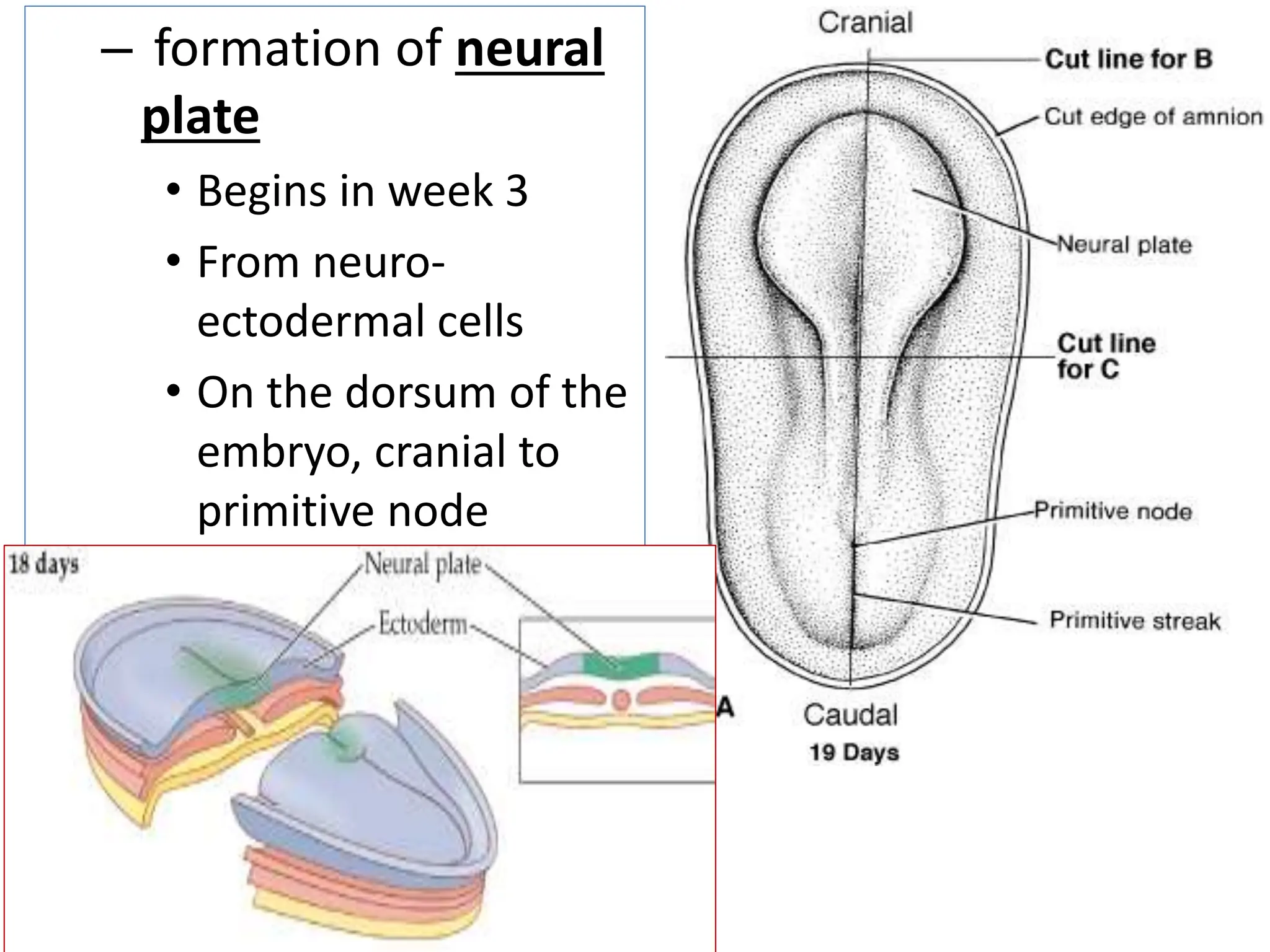
Neurulation
• Formation of the
neural tube
• Inductive factors:
– Inactivation of BMP by
chordin, noggin &
follistatin
• produced by notochord &
primitive node
– Expression of WNT-3A
&FGF; & production of
retinoic acid
• (WNT-3A = wingless-type
MMTV integration site
family, member 3A) 2
– formation of neural
plate
• Begins in week 3
• From neuro-
ectodermal cells
• On the dorsum of the
embryo, cranial to
primitive node
• Formation of neural
folds & neural groove
• Formation of the neural
crest
4
• Formation of the neural tube
– Forms as neural folds touch and
fuse
• beginning in the cervical region (level of
5th somites)
– Two neuropores are then formed:
• Rostral (anterior) neuropore
• Caudal (posterior) neuropore
5
• Fusion of the neural folds progress
craniocaudally, leaving two
neuropores:
– Cranial neuropore
• closes by 25th day (18-20 somite stage)
– Caudal neuropore
• closes by 27th day (25 somite stage)
Summary:
Stages in
neural tube
formation
Derivatives of the neural tube
• Cranial ⅔ forms the brain vesicles
• Caudal ⅓ forms the spinal cord
8
Clinical correlates of neurulation
• Neural tube defects
– Anencephaly (with cranioschisis)
– Cranial meningocoele
– Meningoencephalocoele
– Meningohydroencephalocoele
– Spina bifida:
• spina bifida occulta
• spina bifida cystica:
– Meningocoele
– Meningomyelocoele
• Rachischisis (myeloschisis)
9
Cranial meningocoele
Anencephaly:
associated with cranioschisis
Spina bifida occulta
Spina bifida with
meningocoele
Spina bifida with meningomyelocoele
Rachischisis
Formation of somites
• See ‘’Period of Organogenesis’’

8_Nembryologydeveneurulation_Somites.pptx

  • 2.
    Neurulation • Formation ofthe neural tube • Inductive factors: – Inactivation of BMP by chordin, noggin & follistatin • produced by notochord & primitive node – Expression of WNT-3A &FGF; & production of retinoic acid • (WNT-3A = wingless-type MMTV integration site family, member 3A) 2
  • 3.
    – formation ofneural plate • Begins in week 3 • From neuro- ectodermal cells • On the dorsum of the embryo, cranial to primitive node
  • 4.
    • Formation ofneural folds & neural groove • Formation of the neural crest 4
  • 5.
    • Formation ofthe neural tube – Forms as neural folds touch and fuse • beginning in the cervical region (level of 5th somites) – Two neuropores are then formed: • Rostral (anterior) neuropore • Caudal (posterior) neuropore 5
  • 6.
    • Fusion ofthe neural folds progress craniocaudally, leaving two neuropores: – Cranial neuropore • closes by 25th day (18-20 somite stage) – Caudal neuropore • closes by 27th day (25 somite stage)
  • 7.
  • 8.
    Derivatives of theneural tube • Cranial ⅔ forms the brain vesicles • Caudal ⅓ forms the spinal cord 8
  • 9.
    Clinical correlates ofneurulation • Neural tube defects – Anencephaly (with cranioschisis) – Cranial meningocoele – Meningoencephalocoele – Meningohydroencephalocoele – Spina bifida: • spina bifida occulta • spina bifida cystica: – Meningocoele – Meningomyelocoele • Rachischisis (myeloschisis) 9
  • 10.
  • 11.
  • 12.
  • 13.
  • 14.
    Spina bifida withmeningomyelocoele
  • 15.
  • 16.
    Formation of somites •See ‘’Period of Organogenesis’’