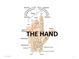
2. • The hand is the region of the upper limb distal
to the wrist joint.
• It is subdivided into three parts:
• the wrist;
• the metacarpus;
• and the digits (five fingers including the
thumb).
• The hand has an anterior surface (palm) and a
dorsal surface (dorsum of hand).
3/21/2024 2
3. BONES OF HAND
• There are three groups of bones in the hand:
the eight carpal bones are the bones of the
wrist;
the five metacarpals (I to V) are the bones of
the metacarpus.
the phalanges are the bones of the digits-the
thumb has only two, the rest of the digits have
three.
3/21/2024 3
4. • The carpal bones and metacarpals of the
index, middle, ring, and little fingers
(metacarpals II to V) tend to function as a unit
and form much of the bony framework of the
palm.
• The metacarpal bone of the thumb functions
independently and has increased flexibility at
the carpometacarpal joint to provide
opposition of the thumb to the fingers.
3/21/2024 4
5. CARPAL BONES;
• The small carpal bones of the wrist are arranged
in two rows, a proximal and a distal row, each
consisting of four bones
Proximal row
• From lateral to medial and when viewed from
anteriorly, the proximal row of bones consists of:
the boat-shaped scaphoid;
the lunate, which has a 'crescent shape';
the three-sided triquetrum bone;
the pea-shaped pisiform
3/21/2024 5
8. • The pisiform is a sesamoid bone in the tendon
of flexor carpi ulnaris and articulates with the
anterior surface of the triquetrum.
• The scaphoid has a prominent tubercle on its
lateral palmar surface that is directed
anteriorly.
3/21/2024 8
10. Distal row
• From lateral to medial and when viewed from
anteriorly, the distal row of carpal bones
consists of:
the irregular four-sided trapezium bone;
the four-sided trapezoid;
the capitate, which has a head;
the hamate, which has a hook.
3/21/2024 10
11. • The trapezium articulates with the metacarpal
bone of the thumb and has a distinct tubercle
on its palmar surface that projects anteriorly.
• The largest of the carpal bones, the capitate,
articulates with the base of the metacarpal III.
• The hamate, which is positioned just lateral
and distal to the pisiform, has a prominent
hook (hook of hamate) on its palmar surface
that projects anteriorly.
3/21/2024 11
13. METACARPALS
• Each of the five metacarpal bones is related to
one digit:
metacarpal I is related to the thumb;
metacarpals II to V are related to the index,
middle, ring, and little fingers, respectively.
3/21/2024 13
15. • Each metacarpal consists of a base, a shaft
(body), and distally, a head.
• All of the bases of the metacarpals articulate
with the carpal bones; in addition, the bases
of the metacarpal bones of the fingers
articulate with each other.
• All of the heads of the metacarpal bones
articulate with the proximal phalanges of the
digits. The heads form the knuckles on the
dorsal surface of the hand when the fingers
are flexed.
3/21/2024 15
17. PHALANGES
• The phalanges are the bones of the digits:
• the thumb has two-a proximal and a distal phalanx;
• the rest of the digits have three-a proximal, a middle,
and a distal phalanx.
• Each phalanx has a base, a shaft (body), and distally, a
head.
• The base of each proximal phalanx articulates with the
head of the related metacarpal bone.
• The head of each distal phalanx is nonarticular and
flattened into a crescent-shaped palmar tuberosity,
which lies under the palmar pad at the end of the digit.
3/21/2024 17
20. WRIST JOINT
• The wrist joint is a synovial joint between the
distal end of the radius and the articular disc
overlying the distal end of the ulna, and the
scaphoid, lunate, and triquetrum.
• Together, the articular surfaces of the carpals
form an oval shape with a convex contour,
which articulates with the corresponding
concave surface of the radius and articular
disc.
3/21/2024 20
21. Reinforcement of the wrist joint is by palmar
radiocarpal, palmar ulnocarpal, and dorsal
radiocarpal ligaments.
In addition, radial and ulnar collateral
ligaments of the wrist joint span the distance
between the styloid processes of the radius
and ulna and the adjacent carpal bones.
These ligaments reinforce the medial and
lateral sides of the wrist joint and support
them during flexion and extension.
3/21/2024 21
22. Movements around the wrist joint include;
abduction, adduction, flexion, and extension.
Because the radial styloid process extends
further distally than does the ulnar styloid
process, the hand can be adducted to a
greater degree than it can be abducted.
3/21/2024 22
24. CARPAL JOINTS
• Is a synovial joint between the carpal bones
which share a common articular cavity.
• The joint capsule is reinforced by numerous
ligaments.
• Although movement at the carpal joints
(intercarpal joints) is limited, they do
contribute to the positioning of the hand in
abduction, adduction, flexion, and,
particularly, extension.
3/21/2024 24
26. CARPOMETACARPAL JOINTS
• There are five carpometacarpal joints between
the metacarpals and the related distal row of
carpal bones.
• The saddle joint, between metacarpal I and the
trapezium, imparts a wide range of mobility to
the thumb that is not a feature of the rest of the
digits.
• Movements at this carpometacarpal joint are
flexion, extension, abduction, adduction,
rotation, and circumduction.
3/21/2024 26
28. • The carpometacarpal joints between
metacarpals II to V and the carpal bones are
much less mobile than the carpometacarpal
joint of the thumb, allowing only limited
gliding movements.
• Movement of the joints increases medially so
metacarpal V slides to the greatest degree.
3/21/2024 28
30. METACARPOPHALANGEAL JOINTS
• The joints between the distal heads of the
metacarpals and the proximal phalanges of the
digits are condylar type of synovial joint, which
allow flexion, extension, abduction, adduction,
circumduction, and limited rotation.
• The capsule of each joint is reinforced by the
palmar ligament and by medial and lateral
collateral ligaments.
• In addition to the above ligaments, there is the
deep transverse metacarpal ligaments.
3/21/2024 30
32. • The three deep transverse metacarpal ligaments
are thick bands of connective tissue connecting
the palmar ligaments of the
metacarpophalangeal joints of the fingers to each
other.
• They are important because, by linking the heads
of the metacarpal bones together, they restrict
the movement of these bones relative to each
other.
• As a result, they help form a unified skeletal
framework for the palm of the hand.
3/21/2024 32
33. INTERPHALANGEAL JOINTS OF HAND
• The interphalangeal joints of the hand are
hinge joints that allow mainly flexion and
extension.
• They are reinforced by medial and lateral
collateral ligaments and palmar ligaments.
3/21/2024 33
34. CLINICAL
• The commonest carpal injury is a fracture across the
waist of the scaphoid bone.
• It is uncommon to see other injuries.
• In approximately 10% of individuals, the scaphoid bone
has a sole blood supply from the radial artery, which
enters through the distal portion of the bone to supply
the proximal portion.
• When a fracture occurs across the waist of the
scaphoid, the proximal portion therefore undergoes
avascular necrosis.
• It is impossible to predict which patients have this
blood supply.
3/21/2024 34
38. The carpal tunnel is formed anteriorly at the
wrist by a deep arch formed by the carpal bones
and the flexor retinaculum .
The base of the carpal arch is formed medially
by the pisiform and the hook of the hamate and
laterally by the tubercles of the scaphoid and
trapezium.
The flexor retinaculum is a thick connective
tissue ligament that bridges the space between
the medial and lateral sides of the base of the
arch and converts the carpal arch into the carpal
tunnel.
3/21/2024 38
39. • The four tendons of the flexor digitorum
profundus, the four tendons of the flexor
digitorum superficialis, and the tendon of the
flexor pollicis longus pass through the carpal
tunnel, as does the median nerve.
3/21/2024 39
40. • Free movement of the tendons in the carpal
tunnel is facilitated by synovial sheaths, which
surround the tendons.
• All the tendons of the flexor digitorum profundus
and flexor digitorum superficialis are surrounded
by a single synovial sheath; a separate sheath
surrounds the tendon of the flexor pollicis longus.
• The median nerve lies anterior to the tendons in
the carpal tunnel.
3/21/2024 40
41. NOTE:
• That although the ulnar artery, ulnar nerve,
and tendon of palmaris longus pass into the
hand, they do so anterior to the flexor
retinaculum and do not pass through the
carpal tunnel.
• The tendon of palmaris longus is not
surrounded by a synovial sheath.
3/21/2024 41
43. Carpal tunnel syndrome
• Carpal tunnel syndrome is an entrapment syndrome
caused by pressure on the median nerve within the
carpal tunnel.
• This gives rise to loss of sensations and weakness of
the muscles of the thenar eminence.
• Aetiology: due to involvement of the bone (arthritis,
dislocation of the lunate, fracture of the wrist)
• Soft tissue pathology (tenosynovitis, acromegaly,
obesity)
• Carpal tunnel syndrome is more common in female
• Occurs between 40-70 years of age
• NZUBE READ THIS WELL AND GOGGLE IT.
3/21/2024 43
44. • Patients typically report pain and pins-and-
needles in the distribution of the median nerve.
Weakness and loss of muscle bulk of the thenar
muscles may also occur. Gently tapping over the
median nerve (in the region of the flexor
retinaculum) readily produces these symptoms
(Tinel's sign).
• Initial treatment is aimed at reducing the
inflammation and removing any repetitive insults
that produce the symptoms. If this does not lead
to improvement nerve conduction studies will be
necessary to confirm nerve entrapment, which
may require surgical decompression of the flexor
retinaculum.
3/21/2024 44
45. PALMAR APONEUROSIS
• The palmar aponeurosis is a triangular-shaped
condensation of deep fascia that covers the palm
and is anchored to the skin in distal regions.
• The apex of the triangle is continuous with the
palmaris longus tendon, when present;
otherwise, it is anchored to the flexor
retinaculum.
• From this point, fibers radiate to extensions at
the base of the digits that project into each of the
index, middle, ring, and little fingers and, to a
lesser extent, the thumb.
3/21/2024 45
46. • Transverse fibers interconnect the more
longitudinally arranged bundles that continue
into the digits.
• Vessels, nerves, and long flexor tendons lie
deep to the palmar aponeurosis in the palm.
3/21/2024 46
48. ANATOMICAL SNUFFBOX
• The 'anatomical snuffbox' is a term given to
the triangular depression formed on the
posterolateral side of the wrist and
metacarpal I by the extensor tendons passing
into the thumb.
• The base of the triangle is at the wrist and the
apex is directed into the thumb.
• The impression is most apparent when the
thumb is extended
3/21/2024 48
49. the lateral border is formed by the tendons of
the abductor pollicis longus and extensor
pollicis brevis;
the medial border is formed by the tendon of
the extensor pollicis longus;
the floor of the impression is formed by the
scaphoid and trapezium, and distal ends of the
tendons of the extensor carpi radialis longus
and extensor carpi radialis brevis.
3/21/2024 49
52. • The radial artery passes obliquely through the
anatomical snuffbox, deep to the extensor
tendons of the thumb and lies adjacent to the
scaphoid and trapezium.
• Terminal parts of the superficial branch of the
radial nerve pass subcutaneously over the
snuffbox as does the origin of the cephalic
vein from the dorsal venous arch of the hand.
3/21/2024 52
54. • The intrinsic muscles of the hand are the
adductor pollicis, interossei, thenar,
hypothenar, palmaris brevis, and lumbrical
muscles.
• Unlike the extrinsic muscles that originate in
the forearm, insert in the hand, and function
in forcefully gripping ('power grip') with the
hand, the intrinsic muscles occur entirely in
the hand and mainly execute precision
movements ('precision grip') with the fingers
and thumb.
3/21/2024 54
55. • All of the intrinsic muscles of the hand are
innervated by the deep branch of the ulnar
nerve except for the three thenar and two
lateral lumbrical muscles, which are
innervated by the median nerve.
• The intrinsic muscles are predominantly
innervated by spinal cord segment T1 with a
contribution from C8.
3/21/2024 55
56. Intrinsic muscles of the hand
MUSCLE ORIGIN INSERTION INNERVATION ACTION
Palmaris
brevis
Palmar
aponeurosis and
flexor
retinaculum
Dermis of skin
on the medial
margin of the
hand
Superficial
branch of the
ulnar nerve
[C8,T1]
Improves grip
Dorsal
interossei
(four muscles)
Adjacent sides
of metacarpals
Extensor hood
and base of
proximal
phalanges of
index, middle,
and ring fingers
Deep branch of
ulnar nerve
[C8,T1]
Abduction of
index, middle,
and ring fingers
at the
metacarpophala
ngeal joints
3/21/2024 56
57. Intrinsic muscles of the hand
MUSCLE ORIGIN INSERTION INNERVATION ACTION
Palmar
interossei
(four muscles)
Sides of
metacarpals
Extensor hoods
of the thumb,
index, ring, and
little fingers and
the proximal
phalanx of
thumb
Deep branch of
ulnar nerve
[C8,T1]
Adduction of
the thumb,
index, ring, and
little fingers at
the
metacarpophala
ngeal joints
Adductor
pollicis
Transverse
head-
metacarpal III;
oblique head-
capitate and
bases of
metacarpals II
and III
Base of proximal
phalanx and
extensor hood
of thumb
Deep branch of
ulnar nerve
[C8,T1]
Adducts thumb
3/21/2024 57
58. Intrinsic muscles of the hand
MUSCLE ORIGIN INSERTION INNERVATION ACTION
Lumbricals
(four
muscles)
Tendons of
flexor digitorum
profundus
Extensor hoods
of index, ring,
middle, and
little fingers
Medial two by
the deep branch
of the ulnar
nerve; lateral
two by digital
branches of the
median nerve
Flex
metacarpophala
ngeal joints
while extending
interphalangeal
joints
3/21/2024 58
59. Thenar muscles of the hand
MUSCLE ORIGIN INSERTION INNERVATION ACTION
Opponens
pollicis
Tubercle of
trapezium and
flexor
retinaculum
Lateral margin
and adjacent
palmar surface
of metacarpal I
Recurrent
branch of
median nerve
[C8,T1]
Medially rotates
thumb
Abductor
pollicis
brevis
Tubercles of
scaphoid and
trapezium and
adjacent flexor
Proximal
phalanx and
extensor hood
of thumb
Recurrent
branch of
median nerve
[C8,T1]
Abducts thumb
at
metacarpophala
ngeal joint
3/21/2024 59
60. Thenar muscles of the hand
MUSCLE ORIGIN INSERTION INNERVATION ACTION
Flexor
pollicis
brevis
Tubercle of the
trapezium and
flexor
retinaculum
Proximal
phalanx of the
thumb
Recurrent
branch of
median nerve
[C8,T1]
pophalangeal
joint
3/21/2024 60
61. Hypothenar muscles of the hand
MUSCLE ORIGIN INSERTION INNERVATION ACTION
Opponens
digiti
minimi
Hook of hamate
and flexor
retinaculum
Medial aspect of
metacarpal V
Deep branch of
ulnar nerve
[C8,T1]
Laterally rotates
metacarpal V
Abductor
digiti
minimi
Pisiform, the
pisohamate
ligament, and
tendon of flexor
carpi ulnaris
Proximal
phalanx of little
finger
Deep branch of
ulnar nerve
[C8,T1]
Abducts little
finger at
metacarpophala
ngeal joint
3/21/2024 61
62. Hypothenar muscles of the hand
MUSCLE ORIGIN INSERTION INNERVATION ACTION
Flexor digiti
minimi
brevis
Hook of the
hamate and
flexor
retinaculum
Proximal
phalanx of little
finger
Deep branch of
ulnar nerve
[C8,T1]
Flexes little
finger at
metacarpophala
ngeal joint
3/21/2024 62
64. • Blood supply to the hand is majorly by the radial
and ulnar arteries, which form two
interconnected vascular arches (superficial and
deep) in the palm.
• Vessels to the digits, muscles, and joints originate
from the two arches and the parent arteries:
the radial artery contributes substantially to the
supply of the thumb and the lateral side of the
index finger;
the remaining digits and the medial side of the
index finger are supplied mainly by the ulnar
artery.
3/21/2024 64
66. Ulnar artery and superficial palmar arch
• The ulnar artery and ulnar nerve enter the hand
on the medial side of the wrist.
• It lies between the palmaris brevis and the flexor
retinaculum and is lateral to the ulnar nerve and
the pisiform bone.
• Distally, it forms the superficial palmar arch,
which is superficial to the long flexor tendons of
the digits.
• On the lateral side of the palm, the arch
communicates with a palmar branch of the radial
artery.
3/21/2024 66
67. • One branch of the ulnar artery in the hand is
the deep palmar branch, which arises from
the medial aspect of the ulnar artery,
penetrates the origin of the hypothenar
muscles.
• It anastomose with the deep palmar arch
derived from the radial artery.
3/21/2024 67
69. • Branches from the superficial palmar arch
include:
1. A palmar digital artery to the medial side of the
little finger;
2. three large, common palmar digital arteries,
which ultimately provide the principal blood
supply to the lateral side of the little finger, both
sides of the ring and middle fingers, and the
medial side of the index finger-they are joined
by palmar metacarpal arteries from the deep
palmar arch before bifurcating into the proper
palmar digital arteries, which enter the fingers.
3/21/2024 69
71. Radial artery and deep palmar arch
• The radial artery curves around the lateral
side of the wrist, passes over the floor of the
anatomical snuffbox and into the deep plane
of the palm by penetrating anteriorly through
the back of the hand.
• It access the deep plane of the palm and form
the deep palmar arch.
3/21/2024 71
72. • The deep palmar arch passes medially
through the palm between the metacarpal
bones and the long flexor tendons of the
digits.
• On the medial side of the palm, it
communicates with the deep palmar branch
of the ulnar artery.
3/21/2024 72
74. • Before penetrating the back of the hand, the
radial artery gives rise to two vessels:
1. a dorsal carpal branch, which passes
medially as the dorsal carpal arch, across the
wrist and gives rise to dorsal metacarpal
arteries, which subsequently divide to
become small dorsal digital arteries, which
enter the fingers;
2. the first dorsal metacarpal artery, which
supplies adjacent sides of the index finger
and thumb.
3/21/2024 74
75. • Two vessels, the princeps pollicis artery and
the radialis indicis artery, arise from the radial
artery.
• The princeps pollicis artery is the major blood
supply to the thumb,
• and the radialis indicis artery supplies the
lateral side of the index finger.
3/21/2024 75
76. • The deep palmar arch gives rise to:
• three palmar metacarpal arteries, which join
the common palmar digital arteries from the
superficial palmar arch of ulnar; and
• three perforating branches, which
anastomose with the dorsal metacarpal
arteries from the dorsal carpal arch.
3/21/2024 76
78. VEINS
• As generally found in the upper limb, the hand
contains interconnected networks of deep and
superficial veins.
• The deep veins follow the arteries; the
superficial veins drain into a dorsal venous
network on the back of the hand over the
metacarpal bones
3/21/2024 78
79. • The cephalic vein originates from the lateral
side of the dorsal venous network and passes
over the anatomical snuffbox into the
forearm.
• The basilic vein originates from the medial
side of the dorsal venous network and passes
into the dorsomedial aspect of the forearm.
3/21/2024 79
Articular surfaces;
The carpal bones have numerous articular surfaces. All of them articulate with each other, and the carpal bones in the distal row articulate with the metacarpals of the digits. With the exception of the metacarpal of the thumb, all movements of the metacarpal bones on the carpal bones are limited.
The expansive proximal surfaces of the scaphoid and lunate articulate with the radius to form the wrist joint.
Significantly, a deep transverse metacarpal ligament does not occur between the palmar ligament of the metacarpophalangeal joint of the thumb and the palmar ligament of the index finger. The absence of this ligament, and the presence of a saddle joint between metacarpal I and the trapezium, are responsible for the increased mobility of the thumb relative to the rest of the digits of the hand.