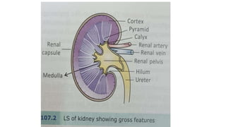
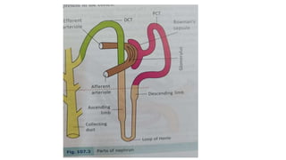
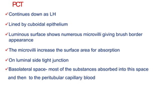
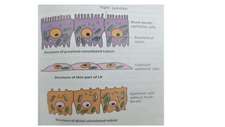
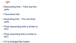
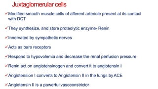
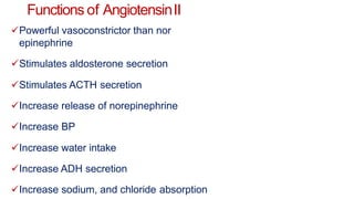
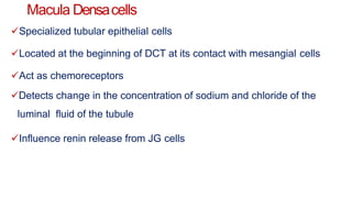
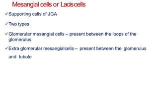
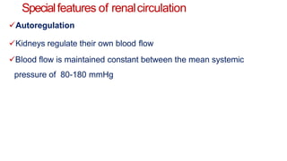
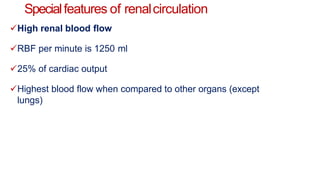
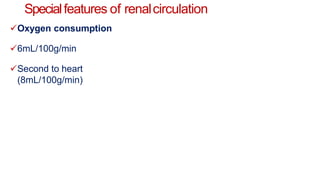
Structure and function
of kidney &
Renal circulation
Thestudent should be ableto
Describe the structure and function of the kidney
Describe the structure and function of the Juxta Glomerular apparatus
Explainthe physiological and clinical significance of
the renin-angiotensin system
Explain special features of renal circulation
Introduction
Metabolic waste products are generated continuously in the
body.
They have to be excreted out to prevent their accumulation.
Urinary system is responsible for
1. Formation
2. Storage
3. Expulsion of urine through which water-soluble wastes are
excreted.
Urinary system
The urinary system consists of
1. Kidney
2. Ureters
3. Urinary bladder
4. Urethra
Kidney is the primary excretory
organ
Functions of kidney
Excretory functions
1. Excreteswaste water-
soluble
metabolic products like urea, uric
acid,
creatinine, ammonium chloride, urobilinogen, etc.
2. Some drugs and environmental toxins consumed are also excreted
into the urine
3. In Diabetes mellitus, glucose is excreted in the urine
Functions of kidney
Non-excretory functions
1. Regulation of blood electrolytes
2. Regulates levels of several electrolytes and
ions like sodium, potassium, calcium, chloride and
phosphates.
Functions of kidney
Non-excretory functions
1. Regulation of blood pH
2. Acid-base balance
3. Excretes variable amount of hydrogen into the
urine
4. Conserves bicarbonate ions
Functions of kidney
Non-excretory functions
1. Regulation of blood volume
2. By conserving or eliminating water in the
urine.
3. Changes in blood volume affects blood
pressure.
Functions of kidney
Non-excretory functions
1. Regulation of blood pressure
2. Adjust the level of blood volume which in turn influences
B.P.
3. Secretes renin
4. Renin activates renin-angiotensin-aldosterone system
5. This system increases blood pressure
Functions of kidney
Non-excretory functions
1. Maintain blood osmolality
2. Regulates levels of solutes in the blood
3. Regulates the water content of the blood
4. Kidney maintains blood osmolality of around 290
milliosmoles
Functions of kidney
Non-excretory functions
1. Kidney produces hormones
2. 1,25(OH) cholecalciferol (calcitriol) – regulates the blood
calcium level
3. Erythropoietin- regulates the production of RBC
4. Renin- activates renin-angiotensin-aldosterone system
5. Bradykinin and prostaglandins- regulate B.P
6. Thrombopoietin- regulates platelets production
Functions of kidney
Non-excretory functions
1. Detoxification
2. Detoxifies toxic
substances
3. Excretes them in urine
If 75%of renal tissueremoved??
After the loss of one kidney, the left-out kidney produces a renal growth
factor which increases the size of glomeruli and length of the nephron
causing the increase in the size of the kidney.
Structure of kidney
Two kidneys are situated in the abdominal cavity.
Each kidney weighs about 150 grams.
Bean shaped
Left kidney is placed slightly higher level than the right kidney
Longitudinal slit in the middle third of medial border- hilum/hilus
Renal artery and nerves enter, renal vein, lymphatics and ureter leave the
kidney through the hilum.
Hilum leads to a wide space called the renal pelvis.
Pelvis continues as ureter.
Structure of kidney
Pelvis divides into the calyceal system
Each kidney contains 8-10 calyces
The space between two calyces is called a pyramid
Pyramid contains numerous pores- papillae
Ducts of Bellini drain the urine through these pores into the calyx and
then into the pelvis
The ducts of Bellini are formed by collecting ducts
In the LS section, the kidney shows an outer cortex and inner medulla
Nephron
Functional and anatomical unit of the kidney
Each kidney has 1-1.5 million nephrons
The length of the nephron varies from 45-
65mm
Parts of the nephron
1. Renal of malphigian corpuscles
2. Proximal convoluted tubule (PCT)
3. Loop of Henle (LH)
4. Distal convoluted tubule (DCT)
5. Collecting duct (CD)
Renalor malphigiancorpuscle
Consist of Bowman’s capsule, Glomerulus
Present in the cortex
Blind cup like the beginning of the nephron is called Bowman’s capsule
It consists of squamous epithelial cells
The parietal layer of BC is continuous with PCT
The visceral layer epithelium is in close contact with the glomerulus
The whole membrane consists of capillary endothelial layer, basal
lamina, an epithelial cell layer
Renalor malphigiancorpuscle
There are pores in between endothelial cells (70-90
nm)
The epithelial cell layer has pores (25 nm)
The basal lamina prevents filtration of proteins
Renalor malphigiancorpuscle
Glomerulus
Afferent arteriole divides and forms a tuft of capillaries called the
glomerulus
These occupy the space of Bowman’s capsule
The capillaries join and form the efferent arterioles
Glomerular capillaries are 50 times more permeable than
capillaries in the skeletal muscle
PCT
Continues down as LH
Lined by cuboidal epithelium
Luminous surface shows numerous microvilli giving brush border
appearance
The microvilli increase the surface area for absorption
On luminal side tight junction
Basolateral space- most of the substances absorbed into this space
and then to the peritubular capillary blood
LH
Descending limb – Thick and thin
parts
Transverse limb
Ascending limb – Thin and thick
parts
Thick descending limb is similar to
PCT
Thick ascending limb is similar to
DCT
LH is arranged like hairpin
DCT
Lined by cuboidal epithelial
cells
Resembles like cells of PCT
But few microvilli
No brush border
Two parts
1. Early DCT/ diluting segment
2. Late DCT
EarlyDCT
Sodium, chloride
absorbed
Water not absorbed
Causes dilution of fluid
EarlyDCT
Sodium, chloride
absorbed
Water not absorbed
Causes dilution of fluid
LateDCT& Cortical collectingduct
Both have similar functional characteristics
Made up of two types of cells
Principalcells(P cells)– absorptionof sodium and water and secretionof
potassium
Intercalated cells (I Cells)- H+ secretion and HCO3- and K+ absorption
Juxtaglomerular apparatus
JGApparatusis present at the point of contact of DCTwith afferent
and efferent arteriole of the same NEPHRON
Three types of cells
Juxtaglomerular cells (JG cells)
Macula densa cells (MD)
Mesangial or Lacis cells
Juxtaglomerular apparatus
Juxtaglomerular cells
Modified smooth muscle cells of afferent arteriole present at its contact
with DCT
They synthesize, and store proteolytic enzyme- Renin
Innervated by sympathetic nerves
Acts as baro receptors
Respond to hypovolemia and decrease the renal perfusion pressure
Renin act on angiotensinogen and convert it to angiotensin I
Angiotension I converts to Angiotensin II in the lungs byACE
Angiotensin II is a powerful vasoconstrictor
Functions of AngiotensinII
Powerful vasoconstrictor than nor
epinephrine
Stimulates aldosterone secretion
Stimulates ACTH secretion
Increase release of norepinephrine
Increase BP
Increase water intake
Increase ADH secretion
Increase sodium, and chloride absorption
Macula Densacells
Specialized tubular epithelial cells
Located at the beginning of DCT at its contact with mesangial cells
Act as chemoreceptors
Detects change in the concentration of sodium and chloride of the
luminal fluid of the tubule
Influence renin release from JG cells
Mesangial cells or Laciscells
Supporting cells of JGA
Two types
Glomerular mesangial cells – present between the loops of the
glomerulus
Extra glomerular mesangialcells – present between the glomerulus
and tubule
AppliedPhysiology
Renin-Angiotensin system is implicated in the genesis of hypertension
Drugs that block the action of RAS are used in the treatment of
hypertension (ACE inhibitors)
Twotypes of nephrons
1. Cortical nephrons
2. Juxtamedullarynephrons
Renalblood flow
Arterial blood supply
Each kidney is supplied by a renal artery
It is a branch of the abdominal aorta
On entering the kidney, it divides into branchesand passes between
the pyramids – interlobar arteries
The interlobar arteries finally give rise to the afferent arterioles
Afferent arterioles divide into a tuft of capillaries – glomerulus
These in turn join and form efferent arteriole
EA in cortical nephrons forms- peritubular capillary plexus
EA in juxta medullary nephrons gives vasarecta
Renalblood flow
Venous
drainage
Stellate veins
Interlobular
veins
Arcuate veins
Interlobar vein
Renal vein
Inferior vena
Specialfeatures of renalcirculation
Portal Circulation
Vein is interposed between capillaries
Venous portal system
In the kidney it is an arterial portal system
Arteriole is interposed between the capillaries
Glomerulus – EA- Peritubular capillaries/ vasa
recta
Specialfeatures of renalcirculation
High pressure in the glomerulus
Renal artery is a direct branch of the abdominal aorta
The pressure in the glomerular capillaries is 45 mmHg
Much higher compared to systemic capillary pressure 30mmHg
This high pressure and high permeability is responsible for the
formation of filtrate
Specialfeatures of renalcirculation
High permeability
Glomerulus is 50 times more permeable than capillaries in skeletal
muscle
Specialfeatures of renalcirculation
Three types of capillaries
Glomerulus – Tuft of capillaries present in the Bowman’s
capsule.
Peritubular capillary plexus
Vasa recta
Specialfeatures of renalcirculation
Autoregulation
Kidneys regulate their own blood flow
Blood flow is maintained constant between the mean systemic
pressure of 80-180 mmHg
Specialfeatures of renalcirculation
High renal blood flow
RBF per minute is 1250 ml
25% of cardiac output
Highest blood flow when compared to other organs (except
lungs)
Specialfeatures of renalcirculation
Oxygen consumption
6mL/100g/min
Second to heart
(8mL/100g/min)
structure and function of kidney.pptx

structure and function of kidney.pptx

  • 1.
    Structure and function ofkidney & Renal circulation
  • 2.
    Thestudent should beableto Describe the structure and function of the kidney Describe the structure and function of the Juxta Glomerular apparatus Explainthe physiological and clinical significance of the renin-angiotensin system Explain special features of renal circulation
  • 3.
    Introduction Metabolic waste productsare generated continuously in the body. They have to be excreted out to prevent their accumulation. Urinary system is responsible for 1. Formation 2. Storage 3. Expulsion of urine through which water-soluble wastes are excreted.
  • 4.
    Urinary system The urinarysystem consists of 1. Kidney 2. Ureters 3. Urinary bladder 4. Urethra Kidney is the primary excretory organ
  • 6.
    Functions of kidney Excretoryfunctions 1. Excreteswaste water- soluble metabolic products like urea, uric acid, creatinine, ammonium chloride, urobilinogen, etc. 2. Some drugs and environmental toxins consumed are also excreted into the urine 3. In Diabetes mellitus, glucose is excreted in the urine
  • 7.
    Functions of kidney Non-excretoryfunctions 1. Regulation of blood electrolytes 2. Regulates levels of several electrolytes and ions like sodium, potassium, calcium, chloride and phosphates.
  • 8.
    Functions of kidney Non-excretoryfunctions 1. Regulation of blood pH 2. Acid-base balance 3. Excretes variable amount of hydrogen into the urine 4. Conserves bicarbonate ions
  • 9.
    Functions of kidney Non-excretoryfunctions 1. Regulation of blood volume 2. By conserving or eliminating water in the urine. 3. Changes in blood volume affects blood pressure.
  • 10.
    Functions of kidney Non-excretoryfunctions 1. Regulation of blood pressure 2. Adjust the level of blood volume which in turn influences B.P. 3. Secretes renin 4. Renin activates renin-angiotensin-aldosterone system 5. This system increases blood pressure
  • 11.
    Functions of kidney Non-excretoryfunctions 1. Maintain blood osmolality 2. Regulates levels of solutes in the blood 3. Regulates the water content of the blood 4. Kidney maintains blood osmolality of around 290 milliosmoles
  • 12.
    Functions of kidney Non-excretoryfunctions 1. Kidney produces hormones 2. 1,25(OH) cholecalciferol (calcitriol) – regulates the blood calcium level 3. Erythropoietin- regulates the production of RBC 4. Renin- activates renin-angiotensin-aldosterone system 5. Bradykinin and prostaglandins- regulate B.P 6. Thrombopoietin- regulates platelets production
  • 13.
    Functions of kidney Non-excretoryfunctions 1. Detoxification 2. Detoxifies toxic substances 3. Excretes them in urine
  • 14.
    If 75%of renaltissueremoved?? After the loss of one kidney, the left-out kidney produces a renal growth factor which increases the size of glomeruli and length of the nephron causing the increase in the size of the kidney.
  • 15.
    Structure of kidney Twokidneys are situated in the abdominal cavity. Each kidney weighs about 150 grams. Bean shaped Left kidney is placed slightly higher level than the right kidney Longitudinal slit in the middle third of medial border- hilum/hilus Renal artery and nerves enter, renal vein, lymphatics and ureter leave the kidney through the hilum. Hilum leads to a wide space called the renal pelvis. Pelvis continues as ureter.
  • 17.
    Structure of kidney Pelvisdivides into the calyceal system Each kidney contains 8-10 calyces The space between two calyces is called a pyramid Pyramid contains numerous pores- papillae Ducts of Bellini drain the urine through these pores into the calyx and then into the pelvis The ducts of Bellini are formed by collecting ducts In the LS section, the kidney shows an outer cortex and inner medulla
  • 18.
    Nephron Functional and anatomicalunit of the kidney Each kidney has 1-1.5 million nephrons The length of the nephron varies from 45- 65mm Parts of the nephron 1. Renal of malphigian corpuscles 2. Proximal convoluted tubule (PCT) 3. Loop of Henle (LH) 4. Distal convoluted tubule (DCT) 5. Collecting duct (CD)
  • 20.
    Renalor malphigiancorpuscle Consist ofBowman’s capsule, Glomerulus Present in the cortex Blind cup like the beginning of the nephron is called Bowman’s capsule It consists of squamous epithelial cells The parietal layer of BC is continuous with PCT The visceral layer epithelium is in close contact with the glomerulus The whole membrane consists of capillary endothelial layer, basal lamina, an epithelial cell layer
  • 21.
    Renalor malphigiancorpuscle There arepores in between endothelial cells (70-90 nm) The epithelial cell layer has pores (25 nm) The basal lamina prevents filtration of proteins
  • 22.
    Renalor malphigiancorpuscle Glomerulus Afferent arterioledivides and forms a tuft of capillaries called the glomerulus These occupy the space of Bowman’s capsule The capillaries join and form the efferent arterioles Glomerular capillaries are 50 times more permeable than capillaries in the skeletal muscle
  • 24.
    PCT Continues down asLH Lined by cuboidal epithelium Luminous surface shows numerous microvilli giving brush border appearance The microvilli increase the surface area for absorption On luminal side tight junction Basolateral space- most of the substances absorbed into this space and then to the peritubular capillary blood
  • 26.
    LH Descending limb –Thick and thin parts Transverse limb Ascending limb – Thin and thick parts Thick descending limb is similar to PCT Thick ascending limb is similar to DCT LH is arranged like hairpin
  • 27.
    DCT Lined by cuboidalepithelial cells Resembles like cells of PCT But few microvilli No brush border Two parts 1. Early DCT/ diluting segment 2. Late DCT
  • 28.
    EarlyDCT Sodium, chloride absorbed Water notabsorbed Causes dilution of fluid
  • 29.
    EarlyDCT Sodium, chloride absorbed Water notabsorbed Causes dilution of fluid
  • 30.
    LateDCT& Cortical collectingduct Bothhave similar functional characteristics Made up of two types of cells Principalcells(P cells)– absorptionof sodium and water and secretionof potassium Intercalated cells (I Cells)- H+ secretion and HCO3- and K+ absorption
  • 32.
    Juxtaglomerular apparatus JGApparatusis presentat the point of contact of DCTwith afferent and efferent arteriole of the same NEPHRON Three types of cells Juxtaglomerular cells (JG cells) Macula densa cells (MD) Mesangial or Lacis cells
  • 33.
  • 34.
    Juxtaglomerular cells Modified smoothmuscle cells of afferent arteriole present at its contact with DCT They synthesize, and store proteolytic enzyme- Renin Innervated by sympathetic nerves Acts as baro receptors Respond to hypovolemia and decrease the renal perfusion pressure Renin act on angiotensinogen and convert it to angiotensin I Angiotension I converts to Angiotensin II in the lungs byACE Angiotensin II is a powerful vasoconstrictor
  • 35.
    Functions of AngiotensinII Powerfulvasoconstrictor than nor epinephrine Stimulates aldosterone secretion Stimulates ACTH secretion Increase release of norepinephrine Increase BP Increase water intake Increase ADH secretion Increase sodium, and chloride absorption
  • 36.
    Macula Densacells Specialized tubularepithelial cells Located at the beginning of DCT at its contact with mesangial cells Act as chemoreceptors Detects change in the concentration of sodium and chloride of the luminal fluid of the tubule Influence renin release from JG cells
  • 37.
    Mesangial cells orLaciscells Supporting cells of JGA Two types Glomerular mesangial cells – present between the loops of the glomerulus Extra glomerular mesangialcells – present between the glomerulus and tubule
  • 38.
    AppliedPhysiology Renin-Angiotensin system isimplicated in the genesis of hypertension Drugs that block the action of RAS are used in the treatment of hypertension (ACE inhibitors) Twotypes of nephrons 1. Cortical nephrons 2. Juxtamedullarynephrons
  • 40.
    Renalblood flow Arterial bloodsupply Each kidney is supplied by a renal artery It is a branch of the abdominal aorta On entering the kidney, it divides into branchesand passes between the pyramids – interlobar arteries The interlobar arteries finally give rise to the afferent arterioles Afferent arterioles divide into a tuft of capillaries – glomerulus These in turn join and form efferent arteriole EA in cortical nephrons forms- peritubular capillary plexus EA in juxta medullary nephrons gives vasarecta
  • 41.
  • 42.
    Specialfeatures of renalcirculation PortalCirculation Vein is interposed between capillaries Venous portal system In the kidney it is an arterial portal system Arteriole is interposed between the capillaries Glomerulus – EA- Peritubular capillaries/ vasa recta
  • 43.
    Specialfeatures of renalcirculation Highpressure in the glomerulus Renal artery is a direct branch of the abdominal aorta The pressure in the glomerular capillaries is 45 mmHg Much higher compared to systemic capillary pressure 30mmHg This high pressure and high permeability is responsible for the formation of filtrate
  • 44.
    Specialfeatures of renalcirculation Highpermeability Glomerulus is 50 times more permeable than capillaries in skeletal muscle
  • 45.
    Specialfeatures of renalcirculation Threetypes of capillaries Glomerulus – Tuft of capillaries present in the Bowman’s capsule. Peritubular capillary plexus Vasa recta
  • 46.
    Specialfeatures of renalcirculation Autoregulation Kidneysregulate their own blood flow Blood flow is maintained constant between the mean systemic pressure of 80-180 mmHg
  • 47.
    Specialfeatures of renalcirculation Highrenal blood flow RBF per minute is 1250 ml 25% of cardiac output Highest blood flow when compared to other organs (except lungs)
  • 48.
    Specialfeatures of renalcirculation Oxygenconsumption 6mL/100g/min Second to heart (8mL/100g/min)