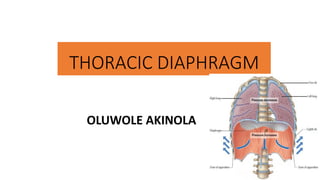
2. The Diaphragm
• Is a dome-
shaped,
musculotendino
us partition
• between the
thoracic and
abdominal
cavities
• Surfaces:
• Has a concave
inferior surface
and
• a convex
superior surface
4. Domes of the Diaphragm
•2 domes (right and left)
• Separated by an
aponeurotic central
tendon
•Fibrous pericardium is
apposed to, and partly
fused with this tendon
•The right dome is higher
up than the left
• owing to the presence of
the liver beneath this
dome.
5. Domes of the Diaphragm
•During expiration,
• right dome reaches as high
up as the 5th rib
• left dome reaches the 5th
intercostal space (in the
midclavicular line)
•The position of the dome
varies with respiration,
posture and the state of
abdominal organs
6. Importance of the Diaphragm
•Is the chief muscle of
inspiration
•During inspiration, the
domes of the diaphragm
descend
• towards the abdominal
cavity
• thereby increasing
intrathoracic volume
7. Parts of the Diaphragm
Based on peripheral attachment of
its fibres, the diaphragm may be
divided into the following parts:
• Sternal part,
• consists of two muscular slips
attached to the xiphoid
process
• Costal part,
• consists of slips that arise from
the lower six costal cartilages
and their ribs
• Its fibres form the domes of
the diaphragm
• Lumbar part
• consists of fibres that arise
from the arcuate ligaments
and crura of the diaphragm
8. Peripheral Attachment of
the Diaphragm
Diaphragm is attached
peripherally to the ff:
1. Posterior surface of the
xiphoid process
• This gives rise to fibres of the
sternal part of the diaphragm
2. Lower six costal cartilage and
their ribs
• These give rise to the costal part
3. Median, medial and lateral
arcuate ligaments.
• These give rise to some posterior
fibres of the diaphragm
4. Right and left crura,
• give rise to some posterior fibres
of the diaphragm
9. • Right crus of the diaphragm
• attached to the upper 3 lumbar vertebrae
• Left crus
• attached to the upper 2 lumbar vertebrae
• Median arcuate ligament
• a fibrous arc that links the right and left crura across the
midline
• It lies anterior to aortic hiatus, and thus, to the
descending aorta
• Medial and lateral arcuate ligaments
• fibrous thickening of the fascia of psoas major and
quadratus lumborum, respectively
Peripheral Attachment of
the Diaphragm
• Medial and lateral arcuate
ligaments
• fibrous thickening of the
fascia of psoas major and
quadratus lumborum,
respectively
10. Central Attachment of the Diaphragm
• From the peripheral sites of
attachment, fibres of the diaphragm
converge on the central tendon
• Central tendon
• is trifoliate,
• lies near the centre of the diaphragm
• is connected to the fibrous
pericardium by pericardiacophrenic
ligaments; but
• has no bony attachment
12. Arterial Supply of the Diaphragm
•Superior phrenic arteries
• from the thoracic aorta
•Musculophrenic and
pericardiacophrenic
arteries
• from internal thoracic
arteries
•Inferior phrenic arteries
• from abdominal aorta
13. Venous Drainage of the Diaphragm
• Musculophrenic veins
• tributaries of internal thoracic veins
• Pericardiacophrenic veins
• tributaries of internal thoracic veins
• Superior phrenic vein (right side only)
• tributary in inferior vena cava [IVC]
• Inferior phrenic veins
• right vein drains into the IVC, while
• left one drains into the IVC and left
suprarenal vein
14. Lymphatic Drainage
of the Diaphragm
Lymph vessels from the
diaphragm drain into the
following nodes:
• Diaphragmatic nodes
• From these nodes, lymph
drains into phrenic,
parasternal and posterior
mediastinal nodes
• Upper lumbar nodes
15. Apertures of the Diaphragm
• Diaphragm has openings via which
neurovascular structures and
oesophagus pass
• The major apertures of the
diaphragm include:
• Aortic hiatus
• Oesophageal hiatus
• Vena caval foramen (caval opening)
16. The Aortic Hiatus
• a median opening
• lies btw right and left crura and
behind the median arcuate
ligament
• at the level of T12 vertebra
• Transmits descending aorta
• Because the aorta does not pierce
the fibres of the diaphragm, blood
flow through this vessel is not
disturbed by the contraction of the
diaphragm
• Also transmits the thoracic duct
and, occasionally, azygos vein
17. Oesophageal Aperture • an opening in the muscle of the right crus
of the diaphragm
• at the level of T10
• Lies above and to the left of aortic hiatus
• Transmits oesophagus
• as this enters the abdomen from the thorax
• fibres of the right crus of the diaphragm
surround the oesophagus here
• these fibres form a sphincter for the
oesophagus, and thus constricts it when the
diaphragm contracts
• Also transits:
• right and left vagal trunks and
• oesophageal branches of left gastric vessels
18. Caval Opening
• An aperture in the central tendon of the
diaphragm
• to the right of the median plane,
• at the level of the disc btw T8 and T9
vertebrae
• This opening is at the junction of the right and
middle leaves of the central tendon
• Transmits IVC
• Also transmits
• terminal part of the right phrenic nerve,
• some lymph vessels, and
• Caval opening is adherent to the wall of
the IVC
• Thus, when the diaphragm contracts, IVC
widens,
• and this enhances venous return to the heart
19. • Each sympathetic
chain descends
into the abdomen
behind the medial
arcuate ligament
• Greater and lesser
splanchnic nerves
pierce the crus of
the diaphragm on
each side
• The diaphragm has a small sternocostal foramen (or triangle)
• This lies (on each side) between the sternal and costal attachments of the diaphragm
• It transmits superior epigastric vessels and lymph vessels
20. Applied Anatomy of the Diaphragm
• Paralysis of a hemidiaphragm
• Due to injury to phrenic nerve of that side
• Thus, muscle fibres of half of the diaphragm
atrophy
• Such paralysed hemidiaphragm does not
descend during inspiration;
• rather, it is forced upwards by increased
abdominal pressure
• In certain subjects, accessory phrenic
nerve is present.
• Thus, injury to the main phrenic nerve does
not result in paralysis of a hemidiaphragm
21. • Pain arising from irritation of the diaphragmatic pleura or
diaphragmatic peritoneum is referred to the shoulder region,
• which is innerved by C3–C5 segments of the spinal cord
• same nerve roots as the phrenic nerve
• Pain from the irritation of the peripheral part of the diaphragm is
referred to the skin over the costal margin
22. • Hiccups are associated with involuntary spasmodic contractions of
the diaphragm.
• It may be caused by cerebral lesions, irritation of the diaphragm,
indigestion, alcoholism or abdominal/thoracic lesions.
• the phrenic nerve is involved
23. • Herniation of abdominal organs
(e.g stomach, intestine, spleen,
etc) into the thoracic cavity is
possible:
• may be congenital or
• could occur following the rupture of
the diaphragm
• as may occur in auto accident, when
there is a sudden increase in
intrabdominal pressure
• Hiatal hernia
• characterised by protrusion of part
of the stomach into the thorax
through the oesophageal hiatus
• Sliding hiatal hernia; or
• Para-esophageal hiatal hernia
24. • The diaphragm may also be
congenitally defective
• In most cases,
posterolateral defect of
the diaphragm occurs
• Thus, abdominal organs are
prone to herniation into the
thorax (through this defect)
• Frequency: 1:2200 births