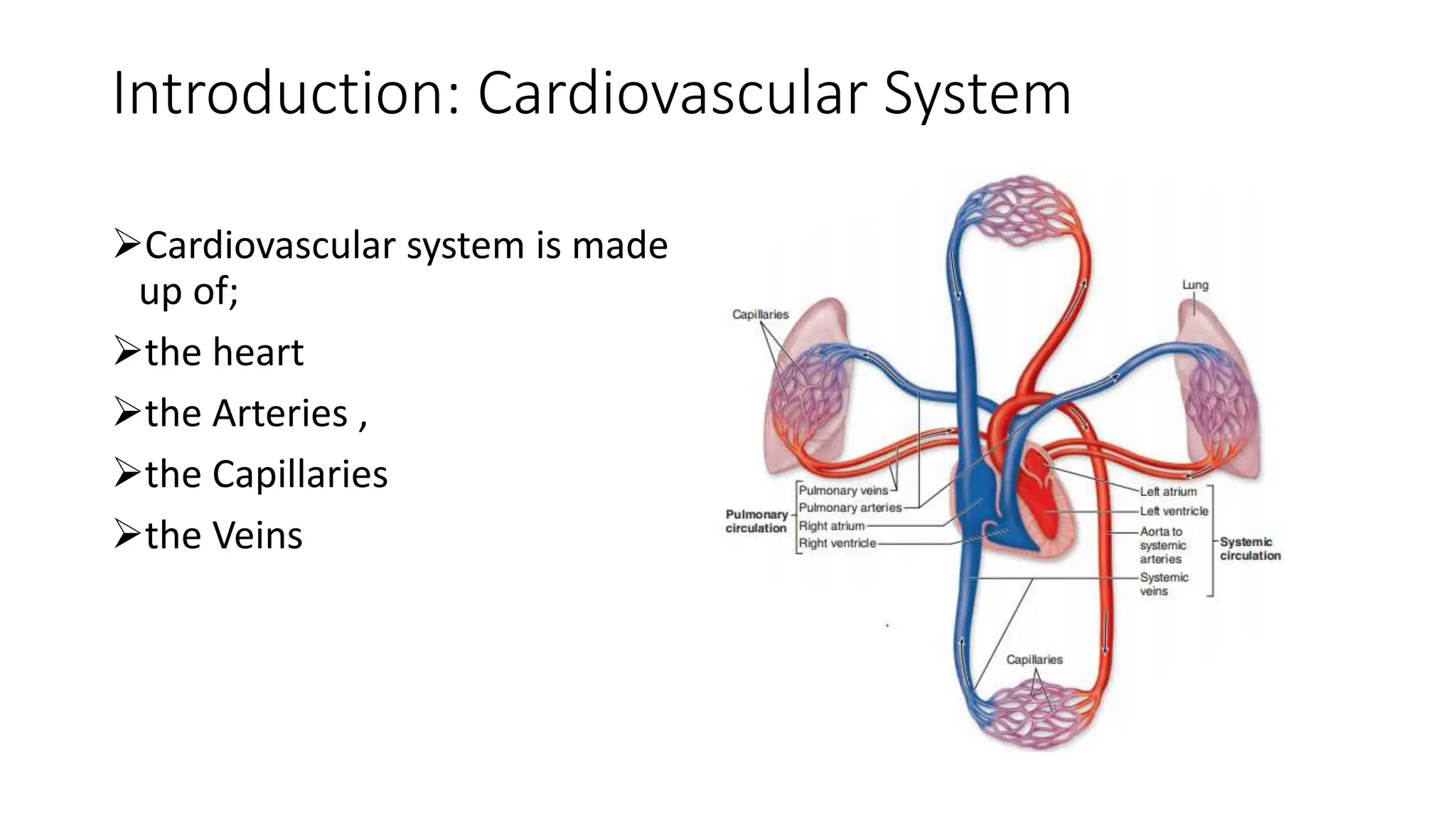
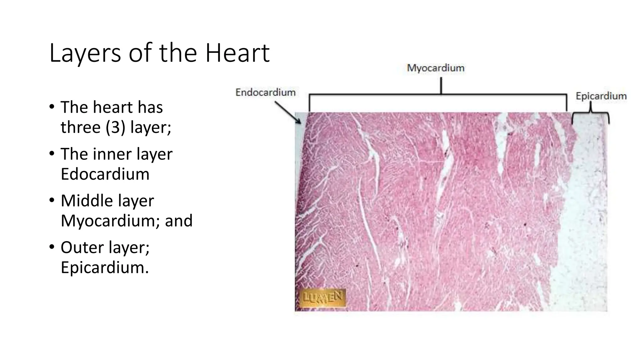
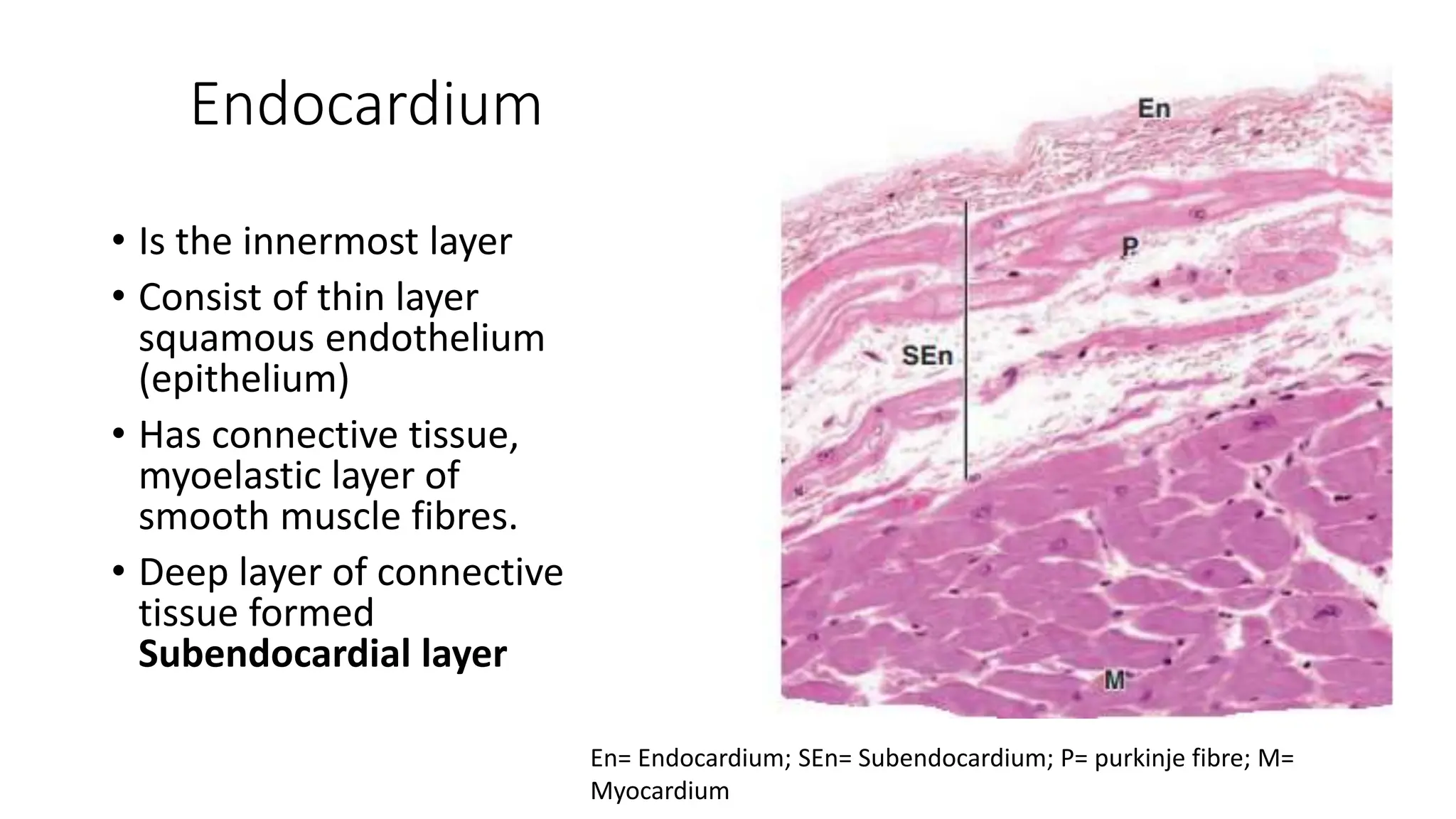
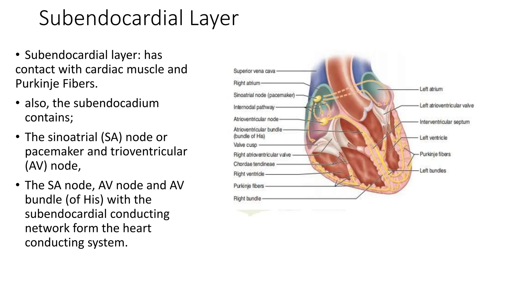
3. The Heart
• The herat has four
chambers namely;
2 Ventricles:
• Right ventricle
• Left ventricle
2 Atrium;
• Right atrium
• Left atrium
4. Layers of the Heart
• The heart has
three (3) layer;
• The inner layer
Edocardium
• Middle layer
Myocardium; and
• Outer layer;
Epicardium.
5. Endocardium
• Is the innermost layer
• Consist of thin layer
squamous endothelium
(epithelium)
• Has connective tissue,
myoelastic layer of
smooth muscle fibres.
• Deep layer of connective
tissue formed
Subendocardial layer
En= Endocardium; SEn= Subendocardium; P= purkinje fibre; M=
Myocardium
6. Subendocardial Layer
• Subendocardial layer: has
contact with cardiac muscle and
Purkinje Fibers.
• also, the subendocadium
contains;
• The sinoatrial (SA) node or
pacemaker and trioventricular
(AV) node,
• The SA node, AV node and AV
bundle (of His) with the
subendocardial conducting
network form the heart
conducting system.
7. Purkinje Fibers.
• These fibers are modified cardiac
muscle cells with 1-2 nuclei
• It is specialized for impulse
conduction rather than contraction.
• Its cytoplasm is filled with glycogen
and myofibrils are found at the
periphery,
• Purkinje fibers typically are more
pale staining than contractile cardiac
muscle fibers due to low myofibril
En= Endocardium; P= purkinje fibre; M= Myocardium
8. Myocardium
• Thick middle layer of the
heart
• Consist majorly cardiac
muscle
• The myocardium is
thicker in the ventricular
wall than the atrial wall
due to high pressure
needed to pump out
blood
En= Endocardium; P= purkinje fibre; M= Myocardium
9. Myocardium
• Consist of cardiac muscle cells =
myocytes
Cardic muscle consisit of nuclei,
cross striations, and intercalated
disks
Intercalated disks
Junctional complexes that
contain fascia adherens,
desmosomes, and gap junction
to provide connection and
communication.
Bind myocytes and allow ion
exchange to facilitate electrical
impulses to pass
N=Nuclei; I= intercalated discs; S= striation; D=
Desmosomes; F=fascia adherentes; M= Mitochondria
10. Epicardium
• Forms the outermost layer of the
heart.
• Covers and protects the heart
• Consist of simple squamous
mesothelium.
• It contains connective tissue with
nerves, vessels, adipocytes.
• The Mesothelium secretes
pericardial fluid
• The pericardial fluid acts as lubricant
that prevents friction. EP= epicardium; CT= connective tissue; N= autonomic
nerves; F= fat; Mes= mesothelium. M= Myocardium.
11. Cardiac Skeleton
• Is made up of irregular dense connective
tissue located in the endocardium
• Forms part of the interventricular and
interatrial septa,
• Anchors the valves and surrounds the
two atrioventricular canals,
• Extends into the valve cusps and the
chordae tendineae
• It maintains the proper shape of
atrioventricular canals Arrow= atrioventricular valve; CT=
chordae tendinae; C= connective tissue,
V= Ventricle
12. Cardiac Valves
• The heart has four (4) valves;
– 2 AV (mitral and tricuspid) in the
chambers
– 2 semilunar (aortic /pulmonary)
• the valve possess connective tissue layers
covered by endothelium on each side;
• they are arranged into 3 layers
Spongiosa: loose collagen
Fibrosa: dense core of connective tissue
Ventricularis: dense connective tissue with
many elastic and collagen fibers
13. Vascular system
• The components of the vascular system
include
• Arteries: transport blood away from the
heart by branching into vessels of smaller
and smaller diameters.
• Capillaries: Thin-walled vessels with the
smallest diameter, forming capillary beds,
where gases and substances are exchange.
• Veins: Vessels that drain capillary beds and
form larger and larger vessels returning
blood to the heart.
14. Tunics or Layers of the Blood vessels
• Wall of the blood vessels has 3
layers or tunics
The innermost layer, the tunica
intima,
The intermediate layer, the
tunica media,
The outermost layer, the tunica
adventitia,
15. Tunica Intima
• Forms the innermost layer of the
blood vessel
• Composed of;
Simple squamous endothelium
(epithelium) whic rest on a
basment membrane
Subendothelial connective
tissue; and
internal elastic lamina
16. Tunica Intima
• The internal elastic lamina
separates the tunica intima
from the tunica media
• It contain elastin that
allows for easy diffusion of
Substances into the deeper
aspect of the vesssel wall
17. Tunica Media
• It the middle and thickest layer
• It maily has concentric layers of helically
arranged smooth muscle cells
• The smooth muscel cells produce elastic
fibers, elastic lamellae, reticular fibers, and
proteoglycans which Interspersed within the
smooth muscle cells.
• The tunica media has a thin external elastic
lamina separating it from the tunica adventitia
• Pericytes replace the tunica media in the
capillaries and postcapillary venules
18. Tunica Adventitia
It forms the outermost layer of
the vessel wall
It consist majorly of fibroblasts,
types I and III collagen fibers, and
elastic fibers.
It is continous with the connective
tissue of the organ via which the
blood flow
19. Vasa Vasorum (vessels of the vessel)
• Supply blood to the wall of the of the
large vessels
• It is formed by the arterioles, capillaries,
and venules in the adventitia and outer
part of the media.
• Because the walls of the large vessels are
thick, the diffusion is not enough to
supply their nutrient needs.
• Vasa vasorum is more in the vein
because they carry deoxygenated blood.
A= arterioles; V= venules; N=
sympathetic nerves; E= elastic lamellae
20. Arteries: Classification
• Arteries are classified; Based on their relative size, morphological
characteristics, or both as follows:
• Elastic (conducting) arteries
• Muscular (distributing) arteries
• Arterioles
21. Elastic Arteries
• Are called conducting arteries
• carrying blood from the heart to smaller arteries
• Elastic arteries include; aorta, the common carotid artery, subclavian
artery, common iliac arteries, and the pulmonary trunk.
22. features of the Elastic Arteries
• In the elastic arteries, the tunica media has
elastic laminae aternate with smooth muscle
fibres
• Well developed tunic intimal with more
smooth muscle cells in the subendothelial
connective tissue
• The adventitia is much thinner than the
media.
• The walls of these vessels may be yellow in the
fresh state because of the abundance of
elastin.
23. Muscular (Distributing) Arteries
• Convey blood to body organs and regulate blood
pressure
• The tunica intima has a very thin subendothelial layer
and a prominent internal elastic lamina
• Relatively thick tunica media is relatively thick and
composed mostly of layers of smooth muscle cells
with variable layers of elastic lamillae
• External elastic lamina is present only in the larger
muscular arteries
• The tunica adventitia consists of connective tissue,
Lymphatic capillaries, vasa vasorum, and nerves.
24. Arterioles
• Arteries with a diameter of less than 0.1
mm
• forms terminal arterial vessels that regulate
blood flow into capillary beds
• have only one or two smooth muscle layers
with thin subendothelial layer’
• No elastic laminae are absent, and the
media consists of the circularly arranged
smooth muscle cells.
• The tunica adventitia of arterioles is scant
• Arterioles branch to form anastomosing
networks or beds of capillaries
25. Capillaries
• Arise from the terminal ends of
arterioles
• Formed capillary beds branching
and anastomosing
• Capillaries permit and regulate
metabolic exchange between
blood and surrounding tissues
26. General Structure of Capillaries
• Capillaries are the smallest blood vessels.
cnsisting of a single layer of endothelial cells,
• The average diameter of capillaries varies from 4
to 10 μm and 50 mm in length,
• pinocytotic vesicles associated with the
endothelial cell plasmalemma of the capillaries.
• Pericytes are cells located along the outside of
the capillaries and small venules.
27. Classification of Capillaries
1. Continuous Capillaries; Also called Somatic
capillaries.
have no pores or fenestrae in their walls.
Na+-K+ ATPase is located in the adluminal cell
membrane only.
Substance such glucose move across the
membrane via carrier-mediated transport
capillaries.
this type of capillaries are found in muscle,
nervous, and connective tissues as well as in the
lungs and exocrine glands.
28. 2. Fenestrated Capillaries; also knwn as
Visceral Capillaries.
They possess pores (fenestrae) in their
walls overed by thin diaphragms of
proteoglycans
The basal lamina is continuous and covers
the fenestrations.
Found in organs such as the lung,
pancrease, kidneys, intestine, choroid
plexus, and endocrine glands.
29. 3. Discontinuous capillaries; also called
Sinusoidal Capillaries
• It allows passage of molecules and cells
between blood and tissue.
• Discontinuous endothelial cells and basal
lamina.
• Endothelial cells here have large
perforations without covering.
• Are found in the liver, spleen, some
endocrine organs, and bone marrow
30. Venules
• The postcapillary venules are similar to the
capillaries
• They possess pericytes and have diameter of
about 15 - 20 µm
• They have endothelial cell layers
• They are surrounded by tunic media with 2 to 3
layers of smoth muscle cells called muscular
venules
• Postcapillary venules form larger collecting vein
with a contractile cells.
• The postcapillary venules form larger collecting
venules with high contractile cells.
31. Veins
• Veins are thin walled vessels
• Return blood from the capillaries to the heart
• Large veins are formed by the union of smaller vein
• They possess valve which prevent reflux of blood
• Veins can be classified into:
• Small or medium veins; and
• Large vein
32. Small or Medium Vein
• They have diameter of about 10 mm or less
• They are located close and parallel to the
corresponding muscular arteries
• Their tunic intima has thin subendothelial layer.
• Tunic media has small bundle of smooth muscle cells
with reticular and elastic fibres in between
• Their tunic adventitial is well debveloped with loose
fibrous connective tissue, vasa vasorum and nerve
fibres
5/10/2024 32
33. Large Vein
• They form big vrnous trunk
• Pair with elastic arteries close to the heart
• They have well developed tunic intima with endothelial
and subendothelial connective tissue
• They have thin tunic media layer with alternate smooth
muscle and connective tissue layers
• Their tunic adventitial is thick with longitudinal layer of
smooth muscles
• They lack both internal and external elastic laminae
35. Lymphatic System
They are thin walled vascular system
The lymphatic system drains excess interstitial fluid (lymph)
Contain single layer of endothelial cells
Openings between the cells are held by filament containing elastic fibres
The fibres keep the vessel open and bind them to connective tissue
The lymphatic capillaries joined to form large lymphatic vessel.
36. Large Lymphatic Vessels
• Have more valve compared to vein
• Have definitive distinction between the tunics
• The tunic media contains smooth muscles
arranged longitudinally and circularly.
• The tunic adventitialcontains vasa vasorum and
nerve
• The lymphatic circulation begins at blind-ended
capillaries and ends at the thoracic duct and the
right lymphatic ducts
37. Clinical Correlate
• Atheroma: due to Formation of fibrofatty plaques by the deposition of fat and collagen
in T Intima
• Atheroma leads to narrowing of the arterial lumen and consequently reduced blood
flow.
• Calcification of T Media (Arteriosclerosis)
• Atherosclerosis (atheroma+arteriosclerosis)
• Damage to the endothelium can cause coagulation of blood forming a thrombus which
can completely obstruct the artery leading to death of the tissue it supplies
• If this happens with Myocardium: Coronary thrombosis leading to Myocardial Infarction
(may manifest as heart attack)
• In brain: Cerebral thrombosis leading to stroke and paralysis