Download to read offline


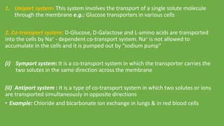


Secondary active transport uses the energy from the electrochemical gradient of ions like Na+ and H+ to move other molecules against their concentration gradient. It involves transporter proteins that symport solutes like glucose into cells along with Na+ ions. This movement is secondary to the primary active transport of Na+ ions out of cells by the Na+/K+ ATPase pump, giving this method of transport its name.