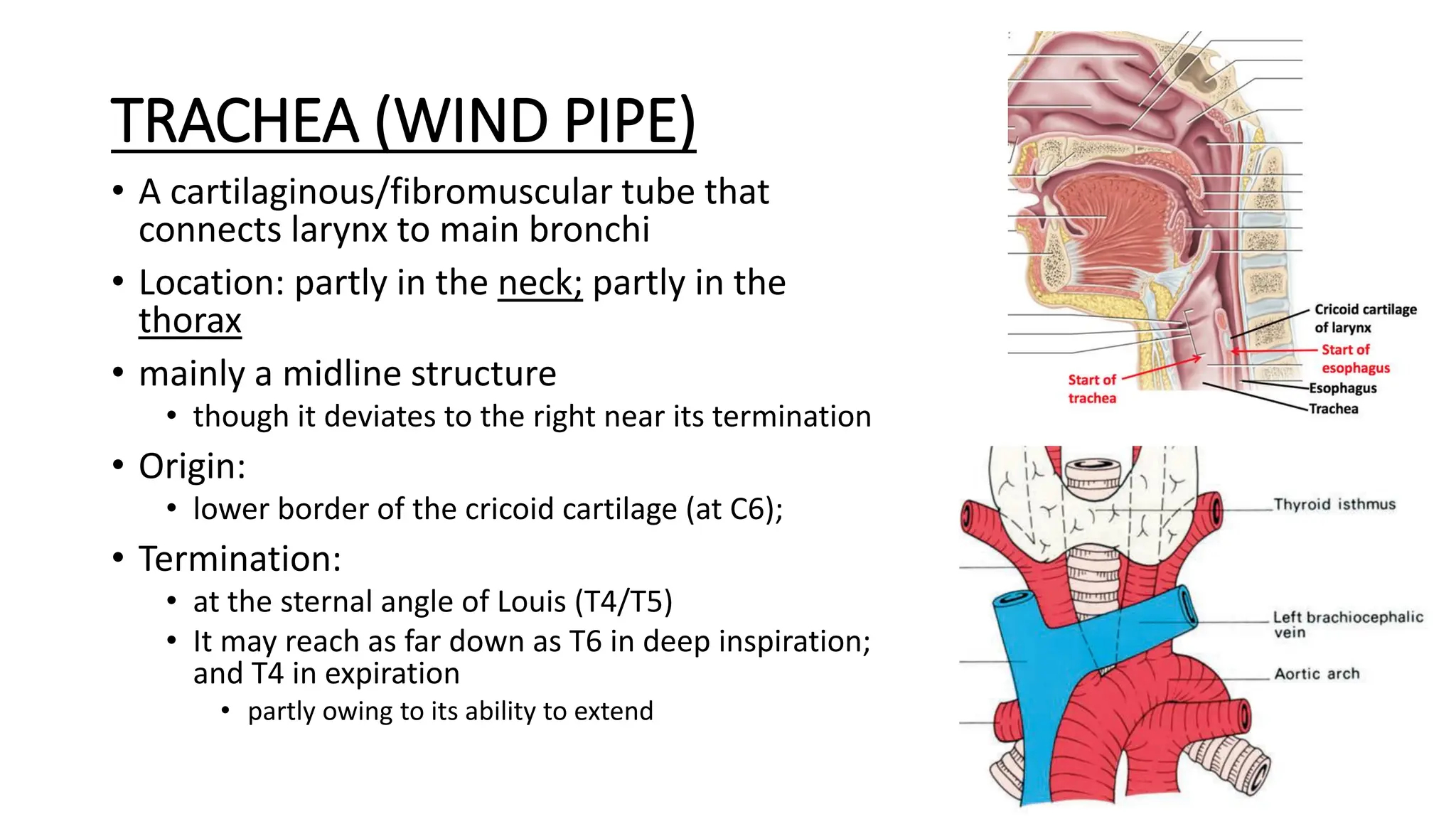
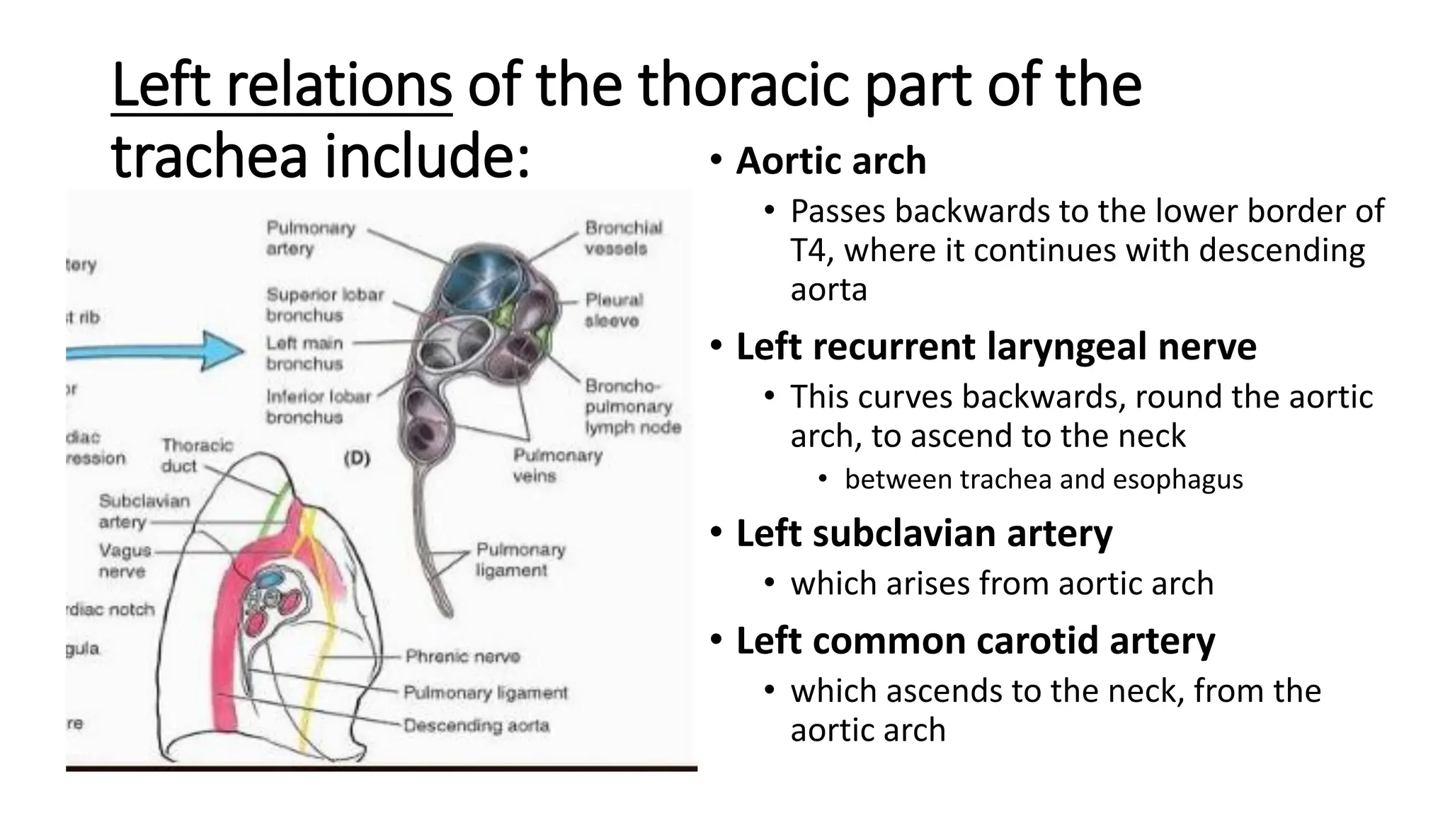
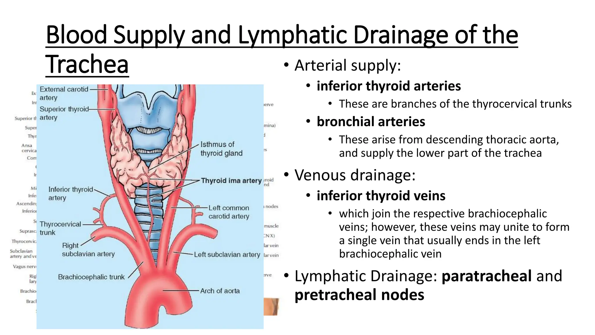
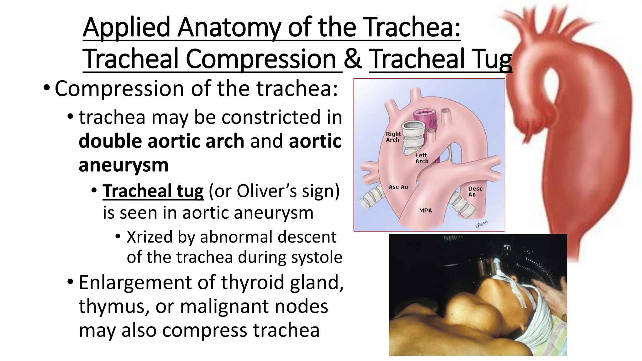
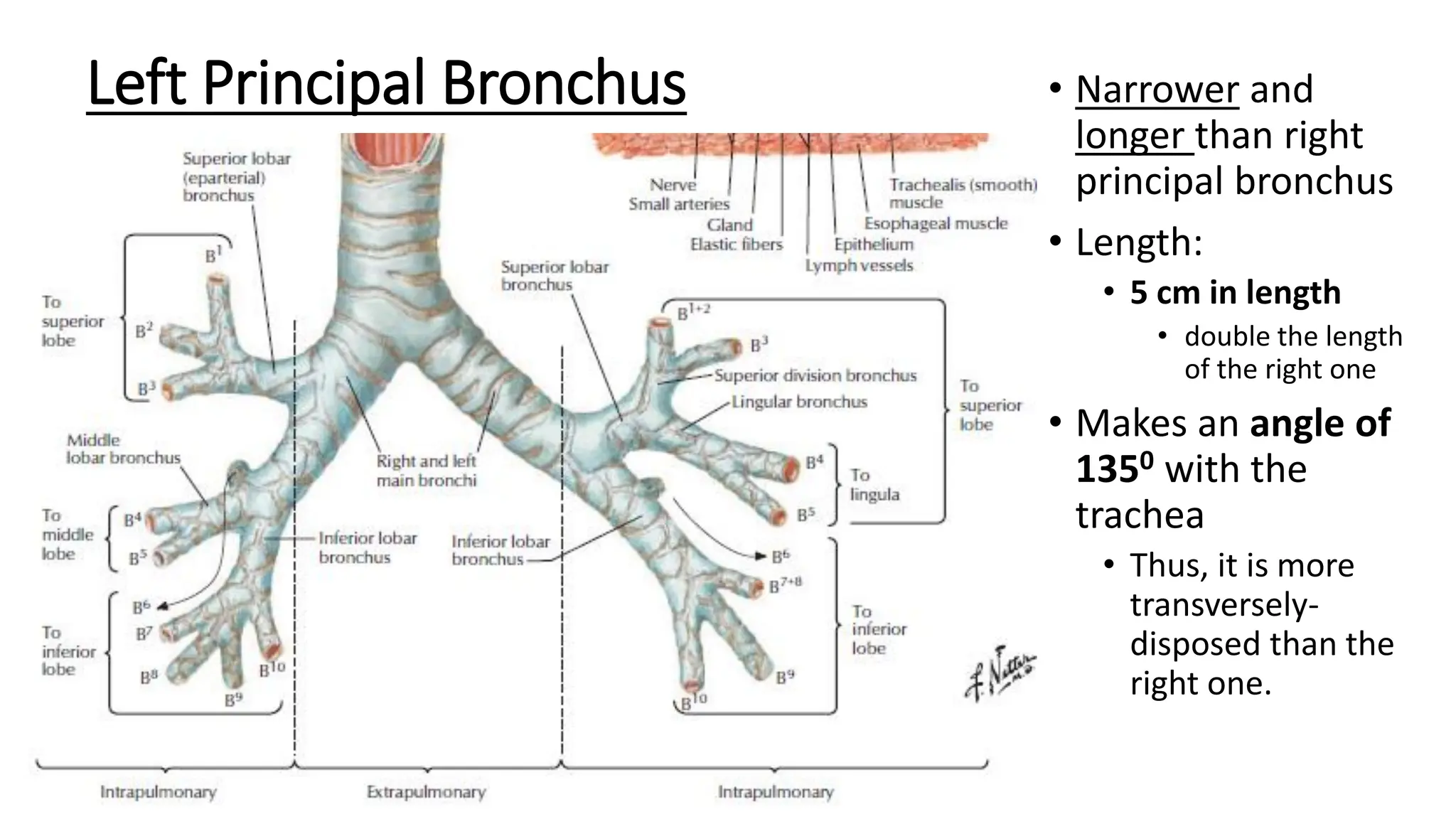
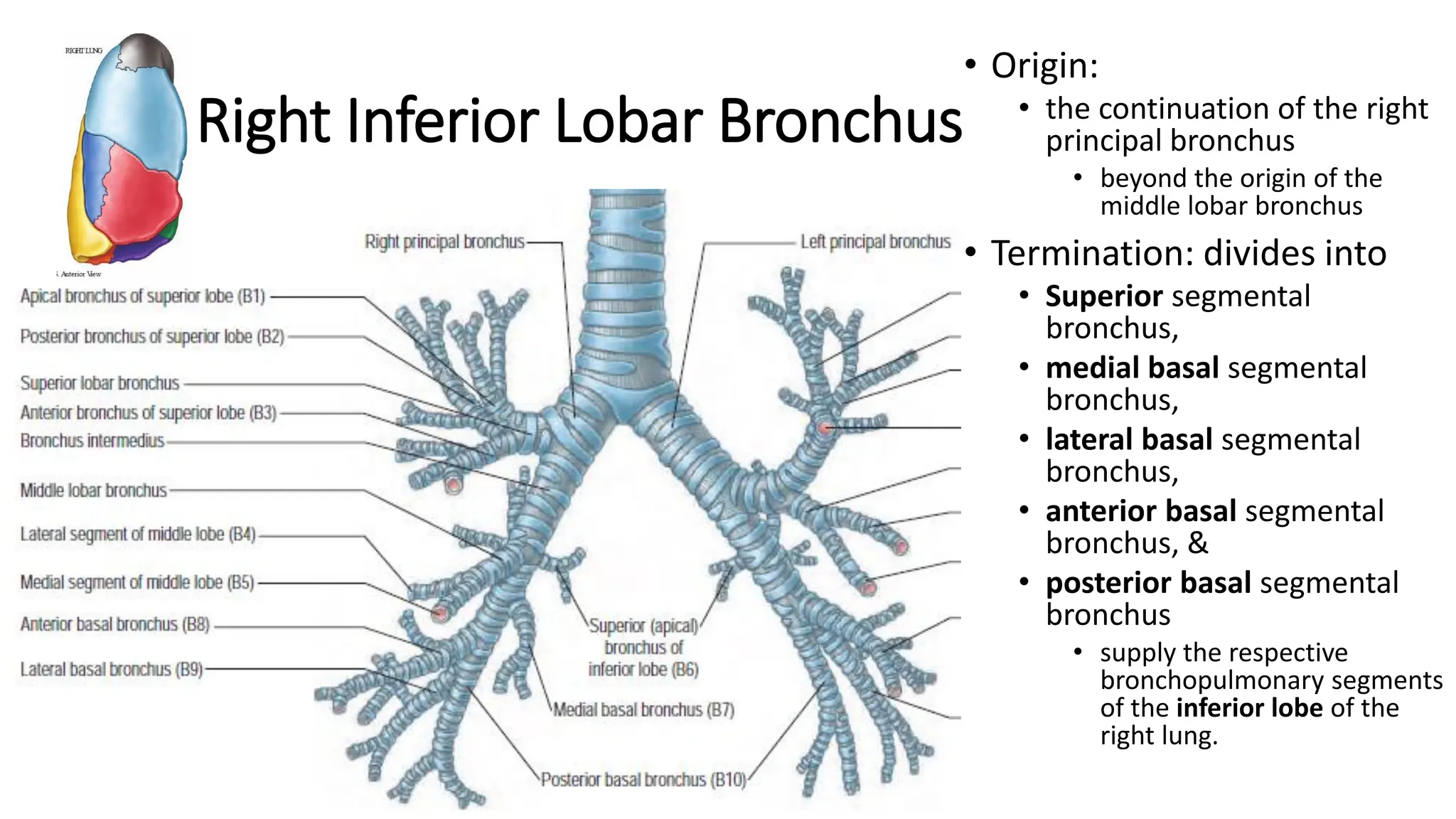
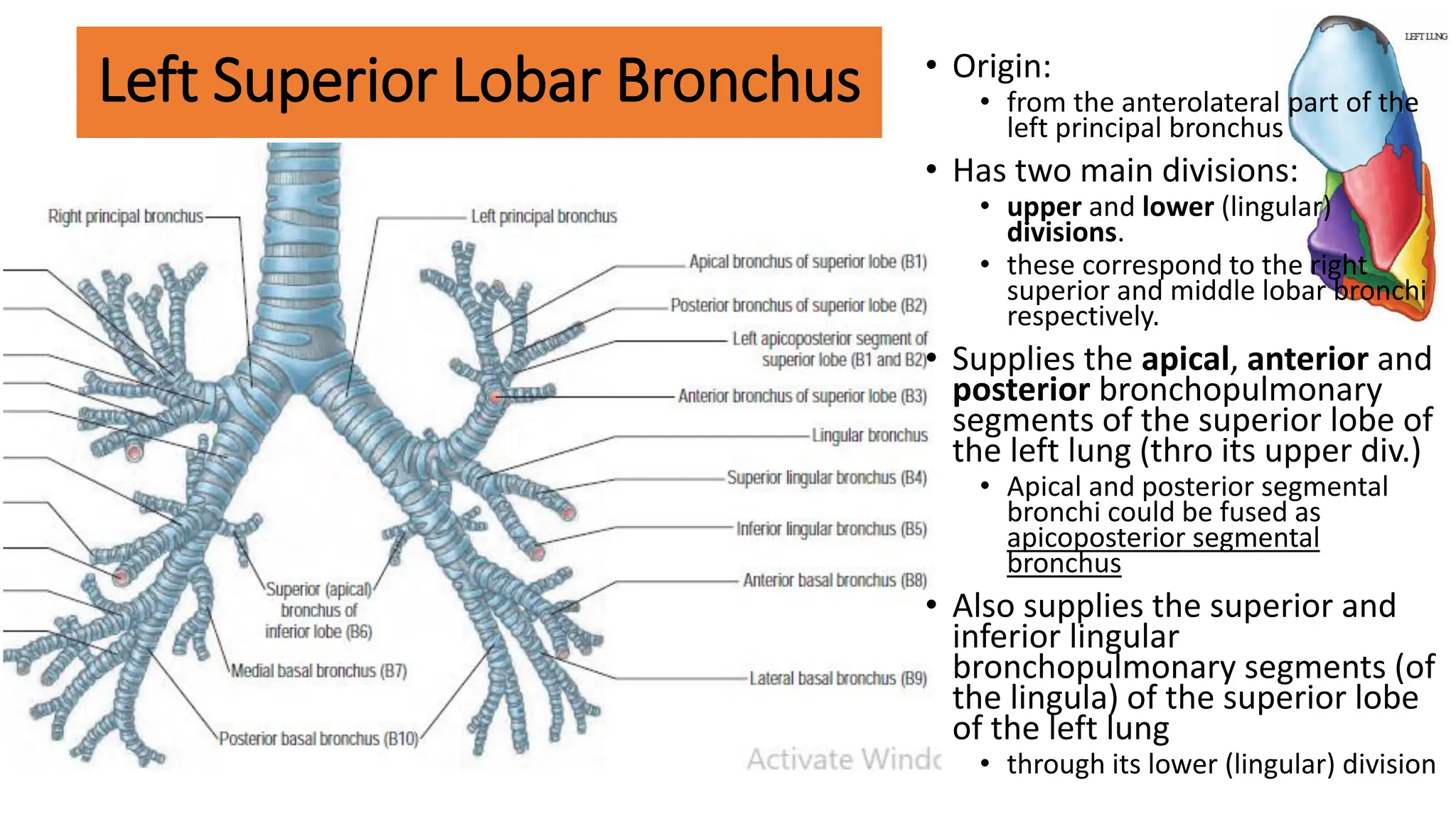
SlideShare a Scribd company logo
1 of 33
Download to read offline
Gross Anatomy of the
TRACHEA AND BRONCHI
PROF. OLUWOLE AKINOLA
Learning Objectives
At the end of the lecture, you should be able to:
• Describe the basic anatomy of the trachea
• Describe the basic anatomy of the different divisions of bronchi
• Describe the clinical anatomy of the trachea and bronchi
Resource Materials
• Textbooks
• Clinically-Oriented Anatomy, Keith L Moore
• Clinical Anatomy, Harrold Ellis
• Grant’s Atlas of Anatomy, by Anne Agur & Arthur Dalley
• Atlas of Human Anatomy, by Frank Netter
• Highlights of Human Anatomy, Akinola & Dosumu. Available online at
https://www.researchgate.net/publication/325870666_highlights_of_human
_anatomy
• Cadaveric dissection
• Museum specimens
TRACHEA (WIND PIPE)
• A cartilaginous/fibromuscular tube that
connects larynx to main bronchi
• Location: partly in the neck; partly in the
thorax
• mainly a midline structure
• though it deviates to the right near its termination
• Origin:
• lower border of the cricoid cartilage (at C6);
• Termination:
• at the sternal angle of Louis (T4/T5)
• It may reach as far down as T6 in deep inspiration;
and T4 in expiration
• partly owing to its ability to extend
Tracheal cartilages • 16–20 C-shaped rings of hyaline
cartilage
• Keep trachea patent
• Concavity of cartilages face
posteriorly
• Possesses no cartilages in its
posterior part
• Here, it adjoins esophagus
• This allows esophagus to expand
during deglutition
• The last cartilage is keel-like, and
is referred to as carina
• Its deviation from midline suggests
metastases in the
tracheobronchial nodes
Dimensions of the Trachea
• Length:
• at birth: 4 cm
• In adult: 9-15 cm
• External diameter:
• 2 cm in adult males
• 1.5 cm in adult females
• Internal diameter:
• 0.3 cm in infants
• 1.2 cm in adults
• Internal diameter (in mm)
corresponds to age (in years)
during childhood
• Wider in cadavers than in the
living
Relations of the Trachea
Anterior Relations of Cervical Part:
• Isthmus of thyroid gland
• lies transversely across 2nd-4th tracheal rings
• Remains of thymus, jugular arch,
• Muscles: sternothyroids and
sternohyoids,
• Fascia:
• Pretracheal fascia
• below the isthmus of thyroid gland
• superficial and investing layers of cervical
fasciae, and the skin
• Inferior thyroid vein and (occasionally)
thyroidea ima artery
• below the level of isthmus of thyroid gland
• Arterial anastomoses formed by the
superior thyroid arteries,
• just above the isthmus of thyroid gland.
• Tracheal lymph nodes
• that drain the trachea and adjoining
structures
Posterior relations of cervical part of the trachea include:
• Cervical part of the esophagus,
• which lies directly behind it
• Recurrent laryngeal nerves
• in the groove between esophagus
and trachea
• Some branches of inferior
thyroid artery
• Vertebral column
Lateral Relations of cervical part of the trachea
• Right and left lobes of thyroid gland
• Each lobe reaches as far down as level
of the 5th or 6th tracheal ring
• Carotid sheath
• located posterolateral to the lobe of
thyroid gland, and
• contains common carotid artery,
internal jugular vein & vagus nerve
• Inferior thyroid artery
• ascends from the thyrocervical trunk
Relations of the Thoracic
Part of the Trachea
Anterior relations of the thoracic part:
• Manubrium sterni,
• bounds superior mediastinum
anteriorly
• Lower part of inferior thyroid vein
• which drains the upper part of the
trachea
• Aortic arch
• Brachiocephalic trunk and left
common carotid artery.
• These arise from the aortic arch
• Left brachiocephalic vein
• This which crosses to the right of the
midline,
• where it joins the right brachiocephalic
vein to form SVC (behind the right 1st
costal cartilage)
Relations of the
Thoracic Part of the Trachea
Anterior relations of trachea
(thoracic part):
• Remains of the thymus
• Deep cardiac plexus of nerves
• This lies anterior to tracheal
bifurcation
• Some lymph nodes
• into which the trachea and
adjacent structures drain
• Origins of sternothyroids &
sternohyoids
Posterior relations of thoracic
part of the trachea include:
• Esophagus,
• which lies directly behind it
• Vertebral column and its
anterior longitudinal ligament
Related to the right of the thoracic part of the trachea are:
• Right pleural cavity
• Right lung
• Superior vena cava (SVC)
• This enters the right atrium at the level of the
right 3rd costal cartilage
• Right brachiocephalic vein
• which joins the left vein at the level of the right 1st
costal cartilage, to form the SVC
• Azygos vein
• ascends over the vertebral column, on the right of
midline
• It ends by arching forwards over the right root of
the lung, to join the SVC at the level of the right
2nd costal cartilage
• Right vagus nerve, which descends behind
the right root of the lung
Left relations of the thoracic part of the
trachea include: • Aortic arch
• Passes backwards to the lower border of
T4, where it continues with descending
aorta
• Left recurrent laryngeal nerve
• This curves backwards, round the aortic
arch, to ascend to the neck
• between trachea and esophagus
• Left subclavian artery
• which arises from aortic arch
• Left common carotid artery
• which ascends to the neck, from the
aortic arch
Blood Supply and Lymphatic Drainage of the
Trachea • Arterial supply:
• inferior thyroid arteries
• These are branches of the thyrocervical trunks
• bronchial arteries
• These arise from descending thoracic aorta,
and supply the lower part of the trachea
• Venous drainage:
• inferior thyroid veins
• which join the respective brachiocephalic
veins; however, these veins may unite to form
a single vein that usually ends in the left
brachiocephalic vein
• Lymphatic Drainage: paratracheal and
pretracheal nodes
Innervation of the Trachea
• Sensory fibres: recurrent
laryngeal nerves
• These supply its mucosa
• Parasympathetic fibres: vagus
nerves
• these are secretomotor to
tracheal glands
• Sympathetic fibres: from
sympathetic chain
• produce vasoconstriction of
tracheal vessels, thereby
reducing the secretory activity
of tracheal glands
•Compression of the trachea:
• trachea may be constricted in
double aortic arch and aortic
aneurysm
• Tracheal tug (or Oliver’s sign)
is seen in aortic aneurysm
• Xrized by abnormal descent
of the trachea during systole
• Enlargement of thyroid gland,
thymus, or malignant nodes
may also compress trachea
Applied Anatomy of the Trachea:
Tracheal Compression & Tracheal Tug
Applied Anatomy of the Trachea:
Tracheotomy & Tracheostomy
• When larynx is obstructed, tracheotomy
may be performed
• By midline incision of the trachea midway btw
thyroid cartilage and suprasternal notch
• A tube could then be introduced to enhance
ventilation (tracheostomy)
• Greater risk is associated with a low
tracheotomy
• owing to the possibility of bleeding from
inferior thyroid vein, thyroidea ima artery
(when present); left brachiocephalic vein or
brachiocephalic trunk (in children)
• Common carotid artery and recurrent
laryngeal nerves are at risk during
tracheotomy
Applied Anatomy of the Trachea: Bronchoscopy
• Biopsies may be taken
and the interior of the
trachea examined
during bronchoscopy
• Carina can be seen
• Foreign bodies and fluid
may also be removed
from the trachea using
a bronchoscope
SURFACE PROJECTION OF THE TRACHEA
•
Bronchi
Bronchi exist as:
• Principal bronchi
• arise from the trachea
• at the level of the
sternal angle
• Secondary or lobar
bronchi
• arise from the principal
bronchi and
• supply the lobes of the
lungs
• Tertiary or segmental
bronchi
• arise from the
secondary bronchi and
• supply
bronchopulmonary
segments
• Series of smaller tubes
that branch repeatedly
to supply the lungs
Right Principal Bronchus
• Shorter, wider & more
vertically-disposed than
left principal bronchus
• Forms an angle of 1550
with the trachea
• Length: 2.5 cm
• half the length of the left
principal bronchus
• Gives off a right superior
lobar (eparterial)
bronchus before it
reaches the hilum of
right lung
• Termination; by diving
into:
• middle lobar bronchus,
and
• inferior lobar bronchus
• at the hilum of the right
lung
Right Principal Bronchus
• Anterior Relations:
• SVC, ascending aorta,
right pulmonary artery
and veins
• Superior Relation:
• Arch of the Azygos vein
• Clinical Anatomy:
• Is the more common site
for the lodgment of
objects that enter the
trachea
• Is easier to approach
surgically than the left,
which is closer to the
aorta
Left Principal Bronchus • Narrower and
longer than right
principal bronchus
• Length:
• 5 cm in length
• double the length
of the right one
• Makes an angle of
1350 with the
trachea
• Thus, it is more
transversely-
disposed than the
right one.
Left Principal Bronchus • Enters the root of the left
lung opposite T6
vertebra
• Posterior Relations:
• esophagus (at T5),
• thoracic duct, and
• descending thoracic
aorta.
• Superior Relations:
• aortic arch
• Left pulmonary artery
• Lies initially behind and
then below the left
pulmonary artery
• Is situated behind and
just above the plane of
the transverse sinus of
the heart
Left Principal Bronchus
•Termination:
• Divides into:
• left superior
lobar bronchus,
&
• left inferior lobar
bronchus
Secondary (Lobar) Bronchi • Arise from the primary
bronchi
• Usually 3 on the right and
2 on the left
• Enter the lungs through
the hila
• Here, they all lie below the
pulmonary arteries, except
the eparterial bronchus,
• that enters hilum above
the right pulmonary artery
• Each supplies a lobe of the
lungs
• Here, each ends by dividing
into series of smaller tubes
called segmental (or
tertiary) bronchi
Right superior lobar bronchus
(Eparterial bronchus)
• Origin:
• Lateral aspect of the right
principal bronchus
• it passes superolaterally,
above the right pulmonary
artery
• Is the highest-placed
structure in the hilum of
the right lung
• Ends about 1 cm from its
origin by dividing into 3
segmental bronchi:
• Apical segmental bronchus
• Anterior segmental
bronchus, and
• posterior segmental bronchi
• These supply the respective
bronchopulmonary
segments of the superior
lobe of the right lung
Middle Lobar Bronchus • Origin:
• Anterior aspect of the right
principal bronchus,
• 2 cm distal to the origin of
eparterial bronchus
• Passes anterolaterally to
the middle lobe of the
right lung
• Termination:
• Divides into medial and
lateral segmental bronchi,
• that supply the respective
bronchopulmonary
segments of the middle
lobe of the right lung
Right Inferior Lobar Bronchus
• Origin:
• the continuation of the right
principal bronchus
• beyond the origin of the
middle lobar bronchus
• Termination: divides into
• Superior segmental
bronchus,
• medial basal segmental
bronchus,
• lateral basal segmental
bronchus,
• anterior basal segmental
bronchus, &
• posterior basal segmental
bronchus
• supply the respective
bronchopulmonary segments
of the inferior lobe of the
right lung.
Left Superior Lobar Bronchus • Origin:
• from the anterolateral part of the
left principal bronchus
• Has two main divisions:
• upper and lower (lingular)
divisions.
• these correspond to the right
superior and middle lobar bronchi
respectively.
• Supplies the apical, anterior and
posterior bronchopulmonary
segments of the superior lobe of
the left lung (thro its upper div.)
• Apical and posterior segmental
bronchi could be fused as
apicoposterior segmental
bronchus
• Also supplies the superior and
inferior lingular
bronchopulmonary segments (of
the lingula) of the superior lobe
of the left lung
• through its lower (lingular) division
Left Inferior
Lobar Bronchus
• Is the continuation of the left
principal bronchus
• Divides in the same manner as does
the right inferior lobar bronchus
• Gives rise to the left superior
segmental bronchus from its
posterior aspect
• This supplies superior
bronchopulmonary segment of the left
inferior lobe
• Also gives rise to the left anterior
basal and medial basal segmental
bronchi (via a common stem)
• They may be fused as anteromedial basal
bronchus
• These supply bronchopulmonary
segments of the same name
• Is also the source of the left posterior
basal and lateral basal segmental
bronchi
• They supply bronchopulmonary
segments of the same name in the
inferior lobe of the left lung
• Each segmental (tertiary) bronchus divides repeatedly and
dichotomously;
• producing several (>20) generations of intrasegmental bronchi; and eventually
smaller tubes devoid of cartilage termed bronchioles
• Bronchioles include terminal and respiratory bronchi.
• Respiratory bronchioles supply alveolar sacs

More Related Content

Similar to respiraaaaatory5b_Trachea and Bronchi.pdf

18-Mediastinum.ppt middle posterior anterior
18-Mediastinum.ppt middle posterior anterior18-Mediastinum.ppt middle posterior anterior
18-Mediastinum.ppt middle posterior anteriorMitchelloJalika
 
Anatomy of trachea, bronchus and lung
Anatomy of trachea, bronchus and lungAnatomy of trachea, bronchus and lung
Anatomy of trachea, bronchus and lungabrahammezgebu1
 
theaorta-180612135504.pdf
theaorta-180612135504.pdftheaorta-180612135504.pdf
theaorta-180612135504.pdfssuserf59e1a
 
Anatomy of lungs and development of lungs
Anatomy of lungs and development of lungsAnatomy of lungs and development of lungs
Anatomy of lungs and development of lungsDrhappyachu
 
Gross anatomy of lungs
Gross anatomy of lungsGross anatomy of lungs
Gross anatomy of lungsSumayyaFarooq1
 
respiratoryyyyy11_Thoracic Diaphragm.pdf
respiratoryyyyy11_Thoracic Diaphragm.pdfrespiratoryyyyy11_Thoracic Diaphragm.pdf
respiratoryyyyy11_Thoracic Diaphragm.pdfAlabiDavid4
 
anatomy_lecture-19-ul.pdf
anatomy_lecture-19-ul.pdfanatomy_lecture-19-ul.pdf
anatomy_lecture-19-ul.pdfshamie8
 
Anatomi Respirasi_RDA_Update.pptx
Anatomi Respirasi_RDA_Update.pptxAnatomi Respirasi_RDA_Update.pptx
Anatomi Respirasi_RDA_Update.pptxpeter269806
 
Cardiovascular Anatomy
Cardiovascular AnatomyCardiovascular Anatomy
Cardiovascular AnatomyJess Little
 
pleura 2022.pptx
pleura 2022.pptxpleura 2022.pptx
pleura 2022.pptxanatomysrs
 

Similar to respiraaaaatory5b_Trachea and Bronchi.pdf (20)

Lungs.pptx
Lungs.pptxLungs.pptx
Lungs.pptx
 
18-Mediastinum.ppt middle posterior anterior
18-Mediastinum.ppt middle posterior anterior18-Mediastinum.ppt middle posterior anterior
18-Mediastinum.ppt middle posterior anterior
 
Mediastinum.pptx
Mediastinum.pptxMediastinum.pptx
Mediastinum.pptx
 
Mediastinum
Mediastinum Mediastinum
Mediastinum
 
Anatomy of trachea, bronchus and lung
Anatomy of trachea, bronchus and lungAnatomy of trachea, bronchus and lung
Anatomy of trachea, bronchus and lung
 
theaorta-180612135504.pdf
theaorta-180612135504.pdftheaorta-180612135504.pdf
theaorta-180612135504.pdf
 
The aorta
The aortaThe aorta
The aorta
 
Anatomy of lungs and development of lungs
Anatomy of lungs and development of lungsAnatomy of lungs and development of lungs
Anatomy of lungs and development of lungs
 
Gross anatomy of lungs
Gross anatomy of lungsGross anatomy of lungs
Gross anatomy of lungs
 
respiratoryyyyy11_Thoracic Diaphragm.pdf
respiratoryyyyy11_Thoracic Diaphragm.pdfrespiratoryyyyy11_Thoracic Diaphragm.pdf
respiratoryyyyy11_Thoracic Diaphragm.pdf
 
anatomy_lecture-19-ul.pdf
anatomy_lecture-19-ul.pdfanatomy_lecture-19-ul.pdf
anatomy_lecture-19-ul.pdf
 
Anatomi Respirasi_RDA_Update.pptx
Anatomi Respirasi_RDA_Update.pptxAnatomi Respirasi_RDA_Update.pptx
Anatomi Respirasi_RDA_Update.pptx
 
Lecture 3 lungs & pleura
Lecture 3 lungs & pleuraLecture 3 lungs & pleura
Lecture 3 lungs & pleura
 
Thoracic ducts
Thoracic ductsThoracic ducts
Thoracic ducts
 
Anatomy of trachea & lungs
Anatomy of  trachea & lungsAnatomy of  trachea & lungs
Anatomy of trachea & lungs
 
Git 01042010 Pdf
Git 01042010 PdfGit 01042010 Pdf
Git 01042010 Pdf
 
Axilla
AxillaAxilla
Axilla
 
Cardiovascular Anatomy
Cardiovascular AnatomyCardiovascular Anatomy
Cardiovascular Anatomy
 
6. Pleura and lung
6. Pleura and lung6. Pleura and lung
6. Pleura and lung
 
pleura 2022.pptx
pleura 2022.pptxpleura 2022.pptx
pleura 2022.pptx
 

More from AlabiDavid4

2cvs.The Heart and. Its Covering 2-1.pdf
2cvs.The Heart and. Its Covering 2-1.pdf2cvs.The Heart and. Its Covering 2-1.pdf
2cvs.The Heart and. Its Covering 2-1.pdfAlabiDavid4
 
respirMUSCLE FATIGUE AND OXYGEN DEBT.pdf
respirMUSCLE  FATIGUE AND OXYGEN DEBT.pdfrespirMUSCLE  FATIGUE AND OXYGEN DEBT.pdf
respirMUSCLE FATIGUE AND OXYGEN DEBT.pdfAlabiDavid4
 
respiratorysy11_Dvt of the Diaphragm.pdf
respiratorysy11_Dvt of the Diaphragm.pdfrespiratorysy11_Dvt of the Diaphragm.pdf
respiratorysy11_Dvt of the Diaphragm.pdfAlabiDavid4
 
IONIC BASIS OF RESTING MEMBRANE AND ACTION POTENTIALS.pptx
IONIC BASIS OF RESTING MEMBRANE AND ACTION POTENTIALS.pptxIONIC BASIS OF RESTING MEMBRANE AND ACTION POTENTIALS.pptx
IONIC BASIS OF RESTING MEMBRANE AND ACTION POTENTIALS.pptxAlabiDavid4
 
8_Nembryologydeveneurulation_Somites.pptx
8_Nembryologydeveneurulation_Somites.pptx8_Nembryologydeveneurulation_Somites.pptx
8_Nembryologydeveneurulation_Somites.pptxAlabiDavid4
 
Cell body defense CBD DIGESTION-GLYCOLYSIS.pptx
Cell body defense CBD DIGESTION-GLYCOLYSIS.pptxCell body defense CBD DIGESTION-GLYCOLYSIS.pptx
Cell body defense CBD DIGESTION-GLYCOLYSIS.pptxAlabiDavid4
 
12_dEvents at Sternal Angle of Louis.pdf
12_dEvents at Sternal Angle of Louis.pdf12_dEvents at Sternal Angle of Louis.pdf
12_dEvents at Sternal Angle of Louis.pdfAlabiDavid4
 
ANATOMY OF THE UPPER LIMBSSAxilla 2.pdf
ANATOMY OF THE UPPER  LIMBSSAxilla 2.pdfANATOMY OF THE UPPER  LIMBSSAxilla 2.pdf
ANATOMY OF THE UPPER LIMBSSAxilla 2.pdfAlabiDavid4
 
respiratory,,......8_Histology of RS.pdf
respiratory,,......8_Histology of RS.pdfrespiratory,,......8_Histology of RS.pdf
respiratory,,......8_Histology of RS.pdfAlabiDavid4
 
respiration.....The blood MBBS 200L.pptx
respiration.....The blood MBBS 200L.pptxrespiration.....The blood MBBS 200L.pptx
respiration.....The blood MBBS 200L.pptxAlabiDavid4
 
THE GROSS ANATOMY OF THE HAND PPPPTT.pptx
THE GROSS ANATOMY OF THE HAND PPPPTT.pptxTHE GROSS ANATOMY OF THE HAND PPPPTT.pptx
THE GROSS ANATOMY OF THE HAND PPPPTT.pptxAlabiDavid4
 
4_Oogenesis gametogenesis lectureeeee.pdf
4_Oogenesis gametogenesis lectureeeee.pdf4_Oogenesis gametogenesis lectureeeee.pdf
4_Oogenesis gametogenesis lectureeeee.pdfAlabiDavid4
 
BUCCAL SMEAR _LYONIZATION hhhhhhhhh.pptx
BUCCAL SMEAR _LYONIZATION hhhhhhhhh.pptxBUCCAL SMEAR _LYONIZATION hhhhhhhhh.pptx
BUCCAL SMEAR _LYONIZATION hhhhhhhhh.pptxAlabiDavid4
 
acid and base with acid and base disorders
acid and base with acid and base disordersacid and base with acid and base disorders
acid and base with acid and base disordersAlabiDavid4
 
Impulse conduction of Nervous tissue hhh
Impulse conduction of Nervous tissue hhhImpulse conduction of Nervous tissue hhh
Impulse conduction of Nervous tissue hhhAlabiDavid4
 
Functional classification of proteins- -pry, sec, tert, and quartenary- struc...
Functional classification of proteins- -pry, sec, tert, and quartenary- struc...Functional classification of proteins- -pry, sec, tert, and quartenary- struc...
Functional classification of proteins- -pry, sec, tert, and quartenary- struc...AlabiDavid4
 

More from AlabiDavid4 (16)

2cvs.The Heart and. Its Covering 2-1.pdf
2cvs.The Heart and. Its Covering 2-1.pdf2cvs.The Heart and. Its Covering 2-1.pdf
2cvs.The Heart and. Its Covering 2-1.pdf
 
respirMUSCLE FATIGUE AND OXYGEN DEBT.pdf
respirMUSCLE  FATIGUE AND OXYGEN DEBT.pdfrespirMUSCLE  FATIGUE AND OXYGEN DEBT.pdf
respirMUSCLE FATIGUE AND OXYGEN DEBT.pdf
 
respiratorysy11_Dvt of the Diaphragm.pdf
respiratorysy11_Dvt of the Diaphragm.pdfrespiratorysy11_Dvt of the Diaphragm.pdf
respiratorysy11_Dvt of the Diaphragm.pdf
 
IONIC BASIS OF RESTING MEMBRANE AND ACTION POTENTIALS.pptx
IONIC BASIS OF RESTING MEMBRANE AND ACTION POTENTIALS.pptxIONIC BASIS OF RESTING MEMBRANE AND ACTION POTENTIALS.pptx
IONIC BASIS OF RESTING MEMBRANE AND ACTION POTENTIALS.pptx
 
8_Nembryologydeveneurulation_Somites.pptx
8_Nembryologydeveneurulation_Somites.pptx8_Nembryologydeveneurulation_Somites.pptx
8_Nembryologydeveneurulation_Somites.pptx
 
Cell body defense CBD DIGESTION-GLYCOLYSIS.pptx
Cell body defense CBD DIGESTION-GLYCOLYSIS.pptxCell body defense CBD DIGESTION-GLYCOLYSIS.pptx
Cell body defense CBD DIGESTION-GLYCOLYSIS.pptx
 
12_dEvents at Sternal Angle of Louis.pdf
12_dEvents at Sternal Angle of Louis.pdf12_dEvents at Sternal Angle of Louis.pdf
12_dEvents at Sternal Angle of Louis.pdf
 
ANATOMY OF THE UPPER LIMBSSAxilla 2.pdf
ANATOMY OF THE UPPER  LIMBSSAxilla 2.pdfANATOMY OF THE UPPER  LIMBSSAxilla 2.pdf
ANATOMY OF THE UPPER LIMBSSAxilla 2.pdf
 
respiratory,,......8_Histology of RS.pdf
respiratory,,......8_Histology of RS.pdfrespiratory,,......8_Histology of RS.pdf
respiratory,,......8_Histology of RS.pdf
 
respiration.....The blood MBBS 200L.pptx
respiration.....The blood MBBS 200L.pptxrespiration.....The blood MBBS 200L.pptx
respiration.....The blood MBBS 200L.pptx
 
THE GROSS ANATOMY OF THE HAND PPPPTT.pptx
THE GROSS ANATOMY OF THE HAND PPPPTT.pptxTHE GROSS ANATOMY OF THE HAND PPPPTT.pptx
THE GROSS ANATOMY OF THE HAND PPPPTT.pptx
 
4_Oogenesis gametogenesis lectureeeee.pdf
4_Oogenesis gametogenesis lectureeeee.pdf4_Oogenesis gametogenesis lectureeeee.pdf
4_Oogenesis gametogenesis lectureeeee.pdf
 
BUCCAL SMEAR _LYONIZATION hhhhhhhhh.pptx
BUCCAL SMEAR _LYONIZATION hhhhhhhhh.pptxBUCCAL SMEAR _LYONIZATION hhhhhhhhh.pptx
BUCCAL SMEAR _LYONIZATION hhhhhhhhh.pptx
 
acid and base with acid and base disorders
acid and base with acid and base disordersacid and base with acid and base disorders
acid and base with acid and base disorders
 
Impulse conduction of Nervous tissue hhh
Impulse conduction of Nervous tissue hhhImpulse conduction of Nervous tissue hhh
Impulse conduction of Nervous tissue hhh
 
Functional classification of proteins- -pry, sec, tert, and quartenary- struc...
Functional classification of proteins- -pry, sec, tert, and quartenary- struc...Functional classification of proteins- -pry, sec, tert, and quartenary- struc...
Functional classification of proteins- -pry, sec, tert, and quartenary- struc...
 

Recently uploaded

Separation of Lanthanides/ Lanthanides and Actinides
Separation of Lanthanides/ Lanthanides and ActinidesSeparation of Lanthanides/ Lanthanides and Actinides
Separation of Lanthanides/ Lanthanides and ActinidesFatimaKhan178732
 
1029-Danh muc Sach Giao Khoa khoi 6.pdf
1029-Danh muc Sach Giao Khoa khoi  6.pdf1029-Danh muc Sach Giao Khoa khoi  6.pdf
1029-Danh muc Sach Giao Khoa khoi 6.pdfQucHHunhnh
 
Activity 01 - Artificial Culture (1).pdf
Activity 01 - Artificial Culture (1).pdfActivity 01 - Artificial Culture (1).pdf
Activity 01 - Artificial Culture (1).pdfciinovamais
 
Z Score,T Score, Percential Rank and Box Plot Graph
Z Score,T Score, Percential Rank and Box Plot GraphZ Score,T Score, Percential Rank and Box Plot Graph
Z Score,T Score, Percential Rank and Box Plot GraphThiyagu K
 
Beyond the EU: DORA and NIS 2 Directive's Global Impact
Beyond the EU: DORA and NIS 2 Directive's Global ImpactBeyond the EU: DORA and NIS 2 Directive's Global Impact
Beyond the EU: DORA and NIS 2 Directive's Global ImpactPECB
 
Mastering the Unannounced Regulatory Inspection
Mastering the Unannounced Regulatory InspectionMastering the Unannounced Regulatory Inspection
Mastering the Unannounced Regulatory InspectionSafetyChain Software
 
CARE OF CHILD IN INCUBATOR..........pptx
CARE OF CHILD IN INCUBATOR..........pptxCARE OF CHILD IN INCUBATOR..........pptx
CARE OF CHILD IN INCUBATOR..........pptxGaneshChakor2
 
Hybridoma Technology ( Production , Purification , and Application )
Hybridoma Technology  ( Production , Purification , and Application  ) Hybridoma Technology  ( Production , Purification , and Application  )
Hybridoma Technology ( Production , Purification , and Application ) Sakshi Ghasle
 
Software Engineering Methodologies (overview)
Software Engineering Methodologies (overview)Software Engineering Methodologies (overview)
Software Engineering Methodologies (overview)eniolaolutunde
 
How to Make a Pirate ship Primary Education.pptx
How to Make a Pirate ship Primary Education.pptxHow to Make a Pirate ship Primary Education.pptx
How to Make a Pirate ship Primary Education.pptxmanuelaromero2013
 
“Oh GOSH! Reflecting on Hackteria's Collaborative Practices in a Global Do-It...
“Oh GOSH! Reflecting on Hackteria's Collaborative Practices in a Global Do-It...“Oh GOSH! Reflecting on Hackteria's Collaborative Practices in a Global Do-It...
“Oh GOSH! Reflecting on Hackteria's Collaborative Practices in a Global Do-It...Marc Dusseiller Dusjagr
 
Measures of Central Tendency: Mean, Median and Mode
Measures of Central Tendency: Mean, Median and ModeMeasures of Central Tendency: Mean, Median and Mode
Measures of Central Tendency: Mean, Median and ModeThiyagu K
 
Q4-W6-Restating Informational Text Grade 3
Q4-W6-Restating Informational Text Grade 3Q4-W6-Restating Informational Text Grade 3
Q4-W6-Restating Informational Text Grade 3JemimahLaneBuaron
 
Arihant handbook biology for class 11 .pdf
Arihant handbook biology for class 11 .pdfArihant handbook biology for class 11 .pdf
Arihant handbook biology for class 11 .pdfchloefrazer622
 
Industrial Policy - 1948, 1956, 1973, 1977, 1980, 1991
Industrial Policy - 1948, 1956, 1973, 1977, 1980, 1991Industrial Policy - 1948, 1956, 1973, 1977, 1980, 1991
Industrial Policy - 1948, 1956, 1973, 1977, 1980, 1991RKavithamani
 
Privatization and Disinvestment - Meaning, Objectives, Advantages and Disadva...
Privatization and Disinvestment - Meaning, Objectives, Advantages and Disadva...Privatization and Disinvestment - Meaning, Objectives, Advantages and Disadva...
Privatization and Disinvestment - Meaning, Objectives, Advantages and Disadva...RKavithamani
 
Contemporary philippine arts from the regions_PPT_Module_12 [Autosaved] (1).pptx
Contemporary philippine arts from the regions_PPT_Module_12 [Autosaved] (1).pptxContemporary philippine arts from the regions_PPT_Module_12 [Autosaved] (1).pptx
Contemporary philippine arts from the regions_PPT_Module_12 [Autosaved] (1).pptxRoyAbrique
 
POINT- BIOCHEMISTRY SEM 2 ENZYMES UNIT 5.pptx
POINT- BIOCHEMISTRY SEM 2 ENZYMES UNIT 5.pptxPOINT- BIOCHEMISTRY SEM 2 ENZYMES UNIT 5.pptx
POINT- BIOCHEMISTRY SEM 2 ENZYMES UNIT 5.pptxSayali Powar
 
Student login on Anyboli platform.helpin
Student login on Anyboli platform.helpinStudent login on Anyboli platform.helpin
Student login on Anyboli platform.helpinRaunakKeshri1
 

Recently uploaded (20)

Separation of Lanthanides/ Lanthanides and Actinides
Separation of Lanthanides/ Lanthanides and ActinidesSeparation of Lanthanides/ Lanthanides and Actinides
Separation of Lanthanides/ Lanthanides and Actinides
 
1029-Danh muc Sach Giao Khoa khoi 6.pdf
1029-Danh muc Sach Giao Khoa khoi  6.pdf1029-Danh muc Sach Giao Khoa khoi  6.pdf
1029-Danh muc Sach Giao Khoa khoi 6.pdf
 
Activity 01 - Artificial Culture (1).pdf
Activity 01 - Artificial Culture (1).pdfActivity 01 - Artificial Culture (1).pdf
Activity 01 - Artificial Culture (1).pdf
 
Z Score,T Score, Percential Rank and Box Plot Graph
Z Score,T Score, Percential Rank and Box Plot GraphZ Score,T Score, Percential Rank and Box Plot Graph
Z Score,T Score, Percential Rank and Box Plot Graph
 
Beyond the EU: DORA and NIS 2 Directive's Global Impact
Beyond the EU: DORA and NIS 2 Directive's Global ImpactBeyond the EU: DORA and NIS 2 Directive's Global Impact
Beyond the EU: DORA and NIS 2 Directive's Global Impact
 
Mastering the Unannounced Regulatory Inspection
Mastering the Unannounced Regulatory InspectionMastering the Unannounced Regulatory Inspection
Mastering the Unannounced Regulatory Inspection
 
Código Creativo y Arte de Software | Unidad 1
Código Creativo y Arte de Software | Unidad 1Código Creativo y Arte de Software | Unidad 1
Código Creativo y Arte de Software | Unidad 1
 
CARE OF CHILD IN INCUBATOR..........pptx
CARE OF CHILD IN INCUBATOR..........pptxCARE OF CHILD IN INCUBATOR..........pptx
CARE OF CHILD IN INCUBATOR..........pptx
 
Hybridoma Technology ( Production , Purification , and Application )
Hybridoma Technology  ( Production , Purification , and Application  ) Hybridoma Technology  ( Production , Purification , and Application  )
Hybridoma Technology ( Production , Purification , and Application )
 
Software Engineering Methodologies (overview)
Software Engineering Methodologies (overview)Software Engineering Methodologies (overview)
Software Engineering Methodologies (overview)
 
How to Make a Pirate ship Primary Education.pptx
How to Make a Pirate ship Primary Education.pptxHow to Make a Pirate ship Primary Education.pptx
How to Make a Pirate ship Primary Education.pptx
 
“Oh GOSH! Reflecting on Hackteria's Collaborative Practices in a Global Do-It...
“Oh GOSH! Reflecting on Hackteria's Collaborative Practices in a Global Do-It...“Oh GOSH! Reflecting on Hackteria's Collaborative Practices in a Global Do-It...
“Oh GOSH! Reflecting on Hackteria's Collaborative Practices in a Global Do-It...
 
Measures of Central Tendency: Mean, Median and Mode
Measures of Central Tendency: Mean, Median and ModeMeasures of Central Tendency: Mean, Median and Mode
Measures of Central Tendency: Mean, Median and Mode
 
Q4-W6-Restating Informational Text Grade 3
Q4-W6-Restating Informational Text Grade 3Q4-W6-Restating Informational Text Grade 3
Q4-W6-Restating Informational Text Grade 3
 
Arihant handbook biology for class 11 .pdf
Arihant handbook biology for class 11 .pdfArihant handbook biology for class 11 .pdf
Arihant handbook biology for class 11 .pdf
 
Industrial Policy - 1948, 1956, 1973, 1977, 1980, 1991
Industrial Policy - 1948, 1956, 1973, 1977, 1980, 1991Industrial Policy - 1948, 1956, 1973, 1977, 1980, 1991
Industrial Policy - 1948, 1956, 1973, 1977, 1980, 1991
 
Privatization and Disinvestment - Meaning, Objectives, Advantages and Disadva...
Privatization and Disinvestment - Meaning, Objectives, Advantages and Disadva...Privatization and Disinvestment - Meaning, Objectives, Advantages and Disadva...
Privatization and Disinvestment - Meaning, Objectives, Advantages and Disadva...
 
Contemporary philippine arts from the regions_PPT_Module_12 [Autosaved] (1).pptx
Contemporary philippine arts from the regions_PPT_Module_12 [Autosaved] (1).pptxContemporary philippine arts from the regions_PPT_Module_12 [Autosaved] (1).pptx
Contemporary philippine arts from the regions_PPT_Module_12 [Autosaved] (1).pptx
 
POINT- BIOCHEMISTRY SEM 2 ENZYMES UNIT 5.pptx
POINT- BIOCHEMISTRY SEM 2 ENZYMES UNIT 5.pptxPOINT- BIOCHEMISTRY SEM 2 ENZYMES UNIT 5.pptx
POINT- BIOCHEMISTRY SEM 2 ENZYMES UNIT 5.pptx
 
Student login on Anyboli platform.helpin
Student login on Anyboli platform.helpinStudent login on Anyboli platform.helpin
Student login on Anyboli platform.helpin
 

respiraaaaatory5b_Trachea and Bronchi.pdf

  • 1. Gross Anatomy of the TRACHEA AND BRONCHI PROF. OLUWOLE AKINOLA
  • 2. Learning Objectives At the end of the lecture, you should be able to: • Describe the basic anatomy of the trachea • Describe the basic anatomy of the different divisions of bronchi • Describe the clinical anatomy of the trachea and bronchi
  • 3. Resource Materials • Textbooks • Clinically-Oriented Anatomy, Keith L Moore • Clinical Anatomy, Harrold Ellis • Grant’s Atlas of Anatomy, by Anne Agur & Arthur Dalley • Atlas of Human Anatomy, by Frank Netter • Highlights of Human Anatomy, Akinola & Dosumu. Available online at https://www.researchgate.net/publication/325870666_highlights_of_human _anatomy • Cadaveric dissection • Museum specimens
  • 4. TRACHEA (WIND PIPE) • A cartilaginous/fibromuscular tube that connects larynx to main bronchi • Location: partly in the neck; partly in the thorax • mainly a midline structure • though it deviates to the right near its termination • Origin: • lower border of the cricoid cartilage (at C6); • Termination: • at the sternal angle of Louis (T4/T5) • It may reach as far down as T6 in deep inspiration; and T4 in expiration • partly owing to its ability to extend
  • 5. Tracheal cartilages • 16–20 C-shaped rings of hyaline cartilage • Keep trachea patent • Concavity of cartilages face posteriorly • Possesses no cartilages in its posterior part • Here, it adjoins esophagus • This allows esophagus to expand during deglutition • The last cartilage is keel-like, and is referred to as carina • Its deviation from midline suggests metastases in the tracheobronchial nodes
  • 6. Dimensions of the Trachea • Length: • at birth: 4 cm • In adult: 9-15 cm • External diameter: • 2 cm in adult males • 1.5 cm in adult females • Internal diameter: • 0.3 cm in infants • 1.2 cm in adults • Internal diameter (in mm) corresponds to age (in years) during childhood • Wider in cadavers than in the living
  • 7. Relations of the Trachea Anterior Relations of Cervical Part: • Isthmus of thyroid gland • lies transversely across 2nd-4th tracheal rings • Remains of thymus, jugular arch, • Muscles: sternothyroids and sternohyoids, • Fascia: • Pretracheal fascia • below the isthmus of thyroid gland • superficial and investing layers of cervical fasciae, and the skin • Inferior thyroid vein and (occasionally) thyroidea ima artery • below the level of isthmus of thyroid gland • Arterial anastomoses formed by the superior thyroid arteries, • just above the isthmus of thyroid gland. • Tracheal lymph nodes • that drain the trachea and adjoining structures
  • 8. Posterior relations of cervical part of the trachea include: • Cervical part of the esophagus, • which lies directly behind it • Recurrent laryngeal nerves • in the groove between esophagus and trachea • Some branches of inferior thyroid artery • Vertebral column
  • 9. Lateral Relations of cervical part of the trachea • Right and left lobes of thyroid gland • Each lobe reaches as far down as level of the 5th or 6th tracheal ring • Carotid sheath • located posterolateral to the lobe of thyroid gland, and • contains common carotid artery, internal jugular vein & vagus nerve • Inferior thyroid artery • ascends from the thyrocervical trunk
  • 10. Relations of the Thoracic Part of the Trachea Anterior relations of the thoracic part: • Manubrium sterni, • bounds superior mediastinum anteriorly • Lower part of inferior thyroid vein • which drains the upper part of the trachea • Aortic arch • Brachiocephalic trunk and left common carotid artery. • These arise from the aortic arch • Left brachiocephalic vein • This which crosses to the right of the midline, • where it joins the right brachiocephalic vein to form SVC (behind the right 1st costal cartilage)
  • 11. Relations of the Thoracic Part of the Trachea Anterior relations of trachea (thoracic part): • Remains of the thymus • Deep cardiac plexus of nerves • This lies anterior to tracheal bifurcation • Some lymph nodes • into which the trachea and adjacent structures drain • Origins of sternothyroids & sternohyoids
  • 12. Posterior relations of thoracic part of the trachea include: • Esophagus, • which lies directly behind it • Vertebral column and its anterior longitudinal ligament
  • 13. Related to the right of the thoracic part of the trachea are: • Right pleural cavity • Right lung • Superior vena cava (SVC) • This enters the right atrium at the level of the right 3rd costal cartilage • Right brachiocephalic vein • which joins the left vein at the level of the right 1st costal cartilage, to form the SVC • Azygos vein • ascends over the vertebral column, on the right of midline • It ends by arching forwards over the right root of the lung, to join the SVC at the level of the right 2nd costal cartilage • Right vagus nerve, which descends behind the right root of the lung
  • 14. Left relations of the thoracic part of the trachea include: • Aortic arch • Passes backwards to the lower border of T4, where it continues with descending aorta • Left recurrent laryngeal nerve • This curves backwards, round the aortic arch, to ascend to the neck • between trachea and esophagus • Left subclavian artery • which arises from aortic arch • Left common carotid artery • which ascends to the neck, from the aortic arch
  • 15. Blood Supply and Lymphatic Drainage of the Trachea • Arterial supply: • inferior thyroid arteries • These are branches of the thyrocervical trunks • bronchial arteries • These arise from descending thoracic aorta, and supply the lower part of the trachea • Venous drainage: • inferior thyroid veins • which join the respective brachiocephalic veins; however, these veins may unite to form a single vein that usually ends in the left brachiocephalic vein • Lymphatic Drainage: paratracheal and pretracheal nodes
  • 16. Innervation of the Trachea • Sensory fibres: recurrent laryngeal nerves • These supply its mucosa • Parasympathetic fibres: vagus nerves • these are secretomotor to tracheal glands • Sympathetic fibres: from sympathetic chain • produce vasoconstriction of tracheal vessels, thereby reducing the secretory activity of tracheal glands
  • 17. •Compression of the trachea: • trachea may be constricted in double aortic arch and aortic aneurysm • Tracheal tug (or Oliver’s sign) is seen in aortic aneurysm • Xrized by abnormal descent of the trachea during systole • Enlargement of thyroid gland, thymus, or malignant nodes may also compress trachea Applied Anatomy of the Trachea: Tracheal Compression & Tracheal Tug
  • 18. Applied Anatomy of the Trachea: Tracheotomy & Tracheostomy • When larynx is obstructed, tracheotomy may be performed • By midline incision of the trachea midway btw thyroid cartilage and suprasternal notch • A tube could then be introduced to enhance ventilation (tracheostomy) • Greater risk is associated with a low tracheotomy • owing to the possibility of bleeding from inferior thyroid vein, thyroidea ima artery (when present); left brachiocephalic vein or brachiocephalic trunk (in children) • Common carotid artery and recurrent laryngeal nerves are at risk during tracheotomy
  • 19. Applied Anatomy of the Trachea: Bronchoscopy • Biopsies may be taken and the interior of the trachea examined during bronchoscopy • Carina can be seen • Foreign bodies and fluid may also be removed from the trachea using a bronchoscope
  • 20. SURFACE PROJECTION OF THE TRACHEA •
  • 21. Bronchi Bronchi exist as: • Principal bronchi • arise from the trachea • at the level of the sternal angle • Secondary or lobar bronchi • arise from the principal bronchi and • supply the lobes of the lungs • Tertiary or segmental bronchi • arise from the secondary bronchi and • supply bronchopulmonary segments • Series of smaller tubes that branch repeatedly to supply the lungs
  • 22. Right Principal Bronchus • Shorter, wider & more vertically-disposed than left principal bronchus • Forms an angle of 1550 with the trachea • Length: 2.5 cm • half the length of the left principal bronchus • Gives off a right superior lobar (eparterial) bronchus before it reaches the hilum of right lung • Termination; by diving into: • middle lobar bronchus, and • inferior lobar bronchus • at the hilum of the right lung
  • 23. Right Principal Bronchus • Anterior Relations: • SVC, ascending aorta, right pulmonary artery and veins • Superior Relation: • Arch of the Azygos vein • Clinical Anatomy: • Is the more common site for the lodgment of objects that enter the trachea • Is easier to approach surgically than the left, which is closer to the aorta
  • 24. Left Principal Bronchus • Narrower and longer than right principal bronchus • Length: • 5 cm in length • double the length of the right one • Makes an angle of 1350 with the trachea • Thus, it is more transversely- disposed than the right one.
  • 25. Left Principal Bronchus • Enters the root of the left lung opposite T6 vertebra • Posterior Relations: • esophagus (at T5), • thoracic duct, and • descending thoracic aorta. • Superior Relations: • aortic arch • Left pulmonary artery • Lies initially behind and then below the left pulmonary artery • Is situated behind and just above the plane of the transverse sinus of the heart
  • 26. Left Principal Bronchus •Termination: • Divides into: • left superior lobar bronchus, & • left inferior lobar bronchus
  • 27. Secondary (Lobar) Bronchi • Arise from the primary bronchi • Usually 3 on the right and 2 on the left • Enter the lungs through the hila • Here, they all lie below the pulmonary arteries, except the eparterial bronchus, • that enters hilum above the right pulmonary artery • Each supplies a lobe of the lungs • Here, each ends by dividing into series of smaller tubes called segmental (or tertiary) bronchi
  • 28. Right superior lobar bronchus (Eparterial bronchus) • Origin: • Lateral aspect of the right principal bronchus • it passes superolaterally, above the right pulmonary artery • Is the highest-placed structure in the hilum of the right lung • Ends about 1 cm from its origin by dividing into 3 segmental bronchi: • Apical segmental bronchus • Anterior segmental bronchus, and • posterior segmental bronchi • These supply the respective bronchopulmonary segments of the superior lobe of the right lung
  • 29. Middle Lobar Bronchus • Origin: • Anterior aspect of the right principal bronchus, • 2 cm distal to the origin of eparterial bronchus • Passes anterolaterally to the middle lobe of the right lung • Termination: • Divides into medial and lateral segmental bronchi, • that supply the respective bronchopulmonary segments of the middle lobe of the right lung
  • 30. Right Inferior Lobar Bronchus • Origin: • the continuation of the right principal bronchus • beyond the origin of the middle lobar bronchus • Termination: divides into • Superior segmental bronchus, • medial basal segmental bronchus, • lateral basal segmental bronchus, • anterior basal segmental bronchus, & • posterior basal segmental bronchus • supply the respective bronchopulmonary segments of the inferior lobe of the right lung.
  • 31. Left Superior Lobar Bronchus • Origin: • from the anterolateral part of the left principal bronchus • Has two main divisions: • upper and lower (lingular) divisions. • these correspond to the right superior and middle lobar bronchi respectively. • Supplies the apical, anterior and posterior bronchopulmonary segments of the superior lobe of the left lung (thro its upper div.) • Apical and posterior segmental bronchi could be fused as apicoposterior segmental bronchus • Also supplies the superior and inferior lingular bronchopulmonary segments (of the lingula) of the superior lobe of the left lung • through its lower (lingular) division
  • 32. Left Inferior Lobar Bronchus • Is the continuation of the left principal bronchus • Divides in the same manner as does the right inferior lobar bronchus • Gives rise to the left superior segmental bronchus from its posterior aspect • This supplies superior bronchopulmonary segment of the left inferior lobe • Also gives rise to the left anterior basal and medial basal segmental bronchi (via a common stem) • They may be fused as anteromedial basal bronchus • These supply bronchopulmonary segments of the same name • Is also the source of the left posterior basal and lateral basal segmental bronchi • They supply bronchopulmonary segments of the same name in the inferior lobe of the left lung
  • 33. • Each segmental (tertiary) bronchus divides repeatedly and dichotomously; • producing several (>20) generations of intrasegmental bronchi; and eventually smaller tubes devoid of cartilage termed bronchioles • Bronchioles include terminal and respiratory bronchi. • Respiratory bronchioles supply alveolar sacs