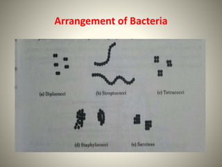
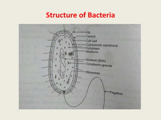
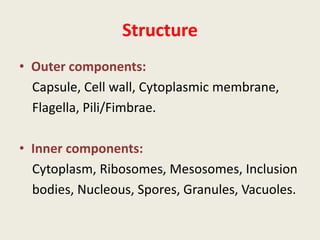
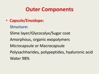
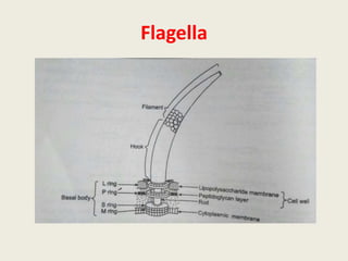
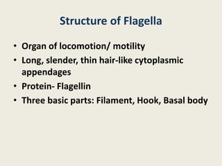
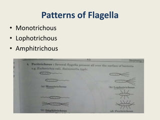
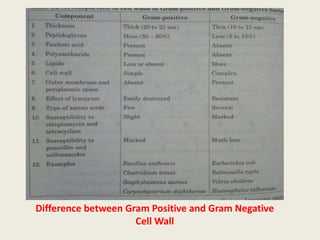
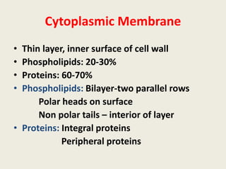
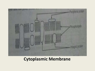
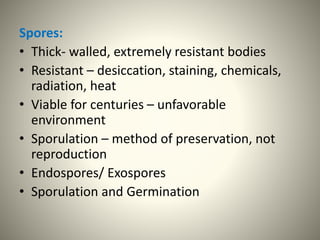
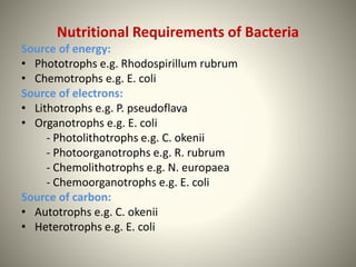
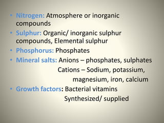
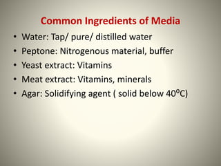
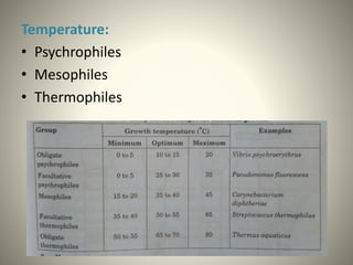
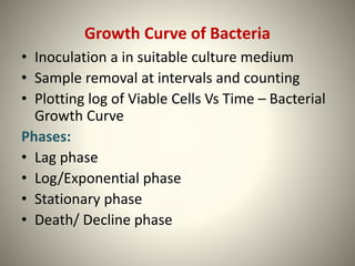
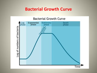
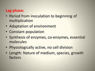
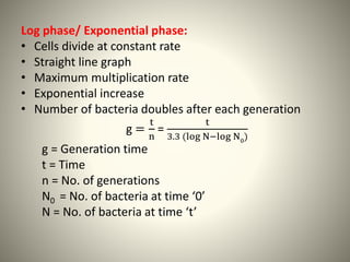
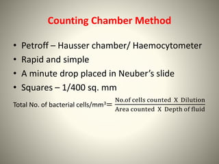
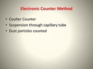
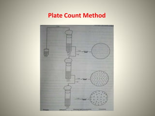
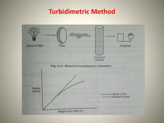
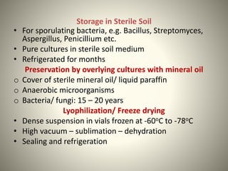
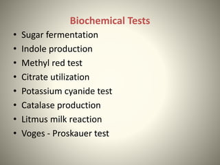
Study of Bacteria
Mr. A.T. Sharma
Assistant Professor
Nanded Pharmacy College, Nanded
Size & Shape of Bacteria
Arrangement of Bacteria
Structure of Bacteria
Structure
• Outer components:
Capsule, Cell wall, Cytoplasmic membrane,
Flagella, Pili/Fimbrae.
• Inner components:
Cytoplasm, Ribosomes, Mesosomes, Inclusion
bodies, Nucleous, Spores, Granules, Vacuoles.
Outer Components
• Capsule/Envolope:
Structure:
Slime layer/Glycocalyx/Sugar coat
Amorphous, organic exopolymers
Microcapsule or Macrocapsule
Polysachharides, polypeptides, hyaluronic acid
Water 98%
Functions of Capsule:
• Protection from Lysozymes
• Protection from phagocytosis
• Prevents temporary drying
• Block attachments of bacteriophages
• Attachment
Flagella
Flagella
Structure of Flagella
• Organ of locomotion/ motility
• Long, slender, thin hair-like cytoplasmic
appendages
• Protein- Flagellin
• Three basic parts: Filament, Hook, Basal body
Patterns of Flagella
• Monotrichous
• Lophotrichous
• Amphitrichous
Pili/ Fimbrae
• Hair-like microfibrils
• Thiner, shorter, more in number
• Protein- Pillin
• Fimbrae: Entire surface, or at poles
100-200
• Pili: Longer than fimbrae
1or 2/cell
DNA transefer
Functions of Pili
• Attachment/ adhession
• Sex pili
• Antigenic
• Agglutination of RBCs
Cell wall
• Rigid structure- shape
• 20-30% of dry weight
• Diaminopimelic acid (DAP) + Muramic acid +
Teichoic acid
• Peptidoglycan/ Murein/Mucopeptide
Three Parts of Peptidoglycan:
A back bone: Alternate N-acetyl glucosamine
and N-acetyl muramic acid
A set of tetrapeptide side chains attached to
N-acetyl muramic acid
A set of pentapeptide cross bridges
Parts of Peptidoglycan
Gram Positive and Gram Negative Cell Wall
• Gram Positive: Teichoic acid, polysaccharides
• Gram Negative: More complex,
lipopolysaccharides
Lipopolysaccharide-
• Region I: O-Antigenicity
• Region II: Core polysaccharides
• Region III: Lipid-A
Outer membrane: OMP-Porins-Diffusion Channels
Components of Cell Wall of Gram Positive and
Gram Negative Bacteria
Difference between Gram Positive and Gram Negative
Cell Wall
Functions of Cell Wall
• Cell growth, cell division
• Shape
• Protection
• Attachment with complement
• Receptors for phages
• Resistance
Cytoplasmic Membrane
• Thin layer, inner surface of cell wall
• Phospholipids: 20-30%
• Proteins: 60-70%
• Phospholipids: Bilayer-two parallel rows
Polar heads on surface
Non polar tails – interior of layer
• Proteins: Integral proteins
Peripheral proteins
Cytoplasmic Membrane
Functions of Cytoplasmic Membrane
• Semipermeable membrane
• Mechanical strength
• DNA replication
• Synthesis of enzymes
Inner Components
Cytoplasm:
• Suspension of Organic and Inorganic solutes
• No ER, Golgi Apparatus, Mitochondria,
Lysosomes
• Nucleus, ribosomes, proteins
• Water soluble components, reserve
materials, plasmid DNA
Cytoplasm:
• Suspension Of organic and inorganic matter
• No ER, Mitochondria, Lysosomes
• Nucleus, ribosomes, proteins, water soluble
components, reserve materials, plasmid DNA
Ribosomes:
• Sites of protein synthesis
• 15000/ cell
• Diameter- 200 A⁰
• 70S subunits
• Polysomes
Mesosomes:
• Infoldings of cytoplasmic membrane
• Two types: Central and Peripheral
• Chondroids
• Photosynthesis
• Sporulation
Cytoplasmic inclusions:
• Round granules – Volutin, Lipid granules,
polysaccharide
• Virus infected bacteria
Nucleus:
• Oval, elongated body
• Double stranded DNA
• Haploid
Plasmid: Extra-chromosomal DNA
• Drug resistance, conjugation ability,
pathogenesis, nitrogen fixation ability
Spores:
• Thick- walled, extremely resistant bodies
• Resistant – desiccation, staining, chemicals,
radiation, heat
• Viable for centuries – unfavorable
environment
• Sporulation – method of preservation, not
reproduction
• Endospores/ Exospores
• Sporulation and Germination
Nutritional Requirements of Bacteria
Source of energy:
• Phototrophs e.g. Rhodospirillum rubrum
• Chemotrophs e.g. E. coli
Source of electrons:
• Lithotrophs e.g. P. pseudoflava
• Organotrophs e.g. E. coli
- Photolithotrophs e.g. C. okenii
- Photoorganotrophs e.g. R. rubrum
- Chemolithotrophs e.g. N. europaea
- Chemoorganotrophs e.g. E. coli
Source of carbon:
• Autotrophs e.g. C. okenii
• Heterotrophs e.g. E. coli
• Nitrogen: Atmosphere or inorganic
compounds
• Sulphur: Organic/ inorganic sulphur
compounds, Elemental sulphur
• Phosphorus: Phosphates
• Mineral salts: Anions – phosphates, sulphates
Cations – Sodium, potassium,
magnesium, iron, calcium
• Growth factors: Bacterial vitamins
Synthesized/ supplied
Common Ingredients of Media
• Water: Tap/ pure/ distilled water
• Peptone: Nitrogenous material, buffer
• Yeast extract: Vitamins
• Meat extract: Vitamins, minerals
• Agar: Solidifying agent ( solid below 40⁰C)
Physical Factors Required for Growth of
Bacteria
• Temperature
• pH
• Gaseous requirement (Oxygen)
• Osmotic pressure
• Light
Temperature:
• Psychrophiles
• Mesophiles
• Thermophiles
pH:
• Acidophiles: 1 - 6.5, Lactobacillus acidophilus
• Neutrophiles: 6.5 - 7.5, E. coli
• Alkalophiles: 7.5 - 14, Vibrio cholerae
Gaseous Requirements:
• Aerobic
• Anaerobic
Tolerant/Non-stringent
Strict/Stringent
• Facultatively anaerobic
• Microaerophilic
Osmotic pressure
• Hypertonic – Plasmolysis
• Hypotonic – Cytolysis
Light
• Darkness favourable
• Sensitive to UV radiations, direct light
Growth Curve of Bacteria
• Inoculation a in suitable culture medium
• Sample removal at intervals and counting
• Plotting log of Viable Cells Vs Time – Bacterial
Growth Curve
Phases:
• Lag phase
• Log/Exponential phase
• Stationary phase
• Death/ Decline phase
Bacterial Growth Curve
Lag phase:
• Period from inoculation to beginning of
multiplication
• Adaptation of environment
• Constant population
• Synthesis of enzymes, co-enzymes, essential
molecules
• Physiologically active, no cell division
• Length: Nature of medium, species, growth
factors
Log phase/ Exponential phase:
• Cells divide at constant rate
• Straight line graph
• Maximum multiplication rate
• Exponential increase
• Number of bacteria doubles after each generation
g =
t
n
=
t
3.3 (log N−log N0)
g = Generation time
t = Time
n = No. of generations
N0 = No. of bacteria at time ‘0’
N = No. of bacteria at time ‘t’
Stationary phase:
• A constant high population
• Balance between cell division and cell death
• Rate of multiplication reduced
Deletion of nutrients
Accumulation of toxic waste products
High cell concentration
Low oxygen tension
• Consumption of reserved food materials,
degradation of ribosomes.
Death/ Decline phase:
• No. of viable cells decreases
• Depletion of nutrients
• Accumulation of toxic waste products
Measurement of Bacterial Growth
1. Determination of Cell Number
a) Total count/ Direct methods
i. Direct microscopic count/ Breed method
ii. Counting chamber method/ Haemocytometer
method
iii. Proportional count method
iv. Electronic counter method
b) Viable count/ Indirect method
i. Plate count technique
ii. Membrane filter count
2. Determination of Cell Mass
a) Direct method
i. Dry weight measurement
ii. Measurement of cell nitrogen
b) Indirect method
i. Turbidimetric method
3. Determination of Cell Activity
Measurement biochemical activity
Breed Method
• A known volume of suspension (0.01ml)
• Area - 1 sq. cm
• Fixed, stained, examined under oil immersion lens
• Counts in microscopic areas – Average
• Total cells/sq. cm counted
Counting Chamber Method
• Petroff – Hausser chamber/ Haemocytometer
• Rapid and simple
• A minute drop placed in Neuber’s slide
• Squares – 1/400 sq. mm
Total No. of bacterial cells/mm3=
No.of cells counted X Dilution
Area counted X Depth of fluid
Neubaur’s Chamber
Proportional Count Method
• Standard suspension of plastic beads (25000/
cell)
• Equal volume of cell suspension
• Spread, fixed, stained
• Particles and cells counted, Average taken
• Example: 10 – Particles, 50 - Cells
50 X25000 = 1,25,000 cells/ml
10
Electronic Counter Method
• Coulter Counter
• Suspension through capillary tube
• Dust particles counted
Viable count/ Indirect method
Plate Count Technique
• Principle
a) Plate count method
b) Membrane filter count
Plate Count Method
Determination of Cell Mass
• Direct Method
- Dry weight measurement
- Measurement of cell nitrogen
• Indirect Method
Turbidimetric method
o Colorimeter/Spectrophotometer
o Beer and Lambert law
o Absorbance in terms of Optical Density
o Calibration curve
Turbidimetric Method
Determination of Cell Activity
• Biochemical activity
• Chemical change by metabolic activities
• Lactic acid, H2S, CO2, Enzymes etc.
Isolation and Preservation Methods for
Pure Cultures
Preservation of Cultures:
• Maintaining an isolated pure culture for extended
periods in a viable condition, without any genetic
change or contamination
• Culture collection centres – Stock-culture collection
Methods:
o Periodic transfer to fresh media
o Storage at low temperature
o Storage in sterile soil
o Preservation by overlying cultures with mineral oil
o Lyophilization/ Freeze drying
Periodic Transfer to Fresh Media
• Agar slants incubated for 24hrs and stored in
refrigerator
• Periodic transfer
• Risk of contamination – Genetic/ biochemical
changes
Storage at Low Temperature
• Short time preservation: Refrigerator/ Cold
rooms at 4oC
• Long term preservation:
o Liquid nitrogen
o Dense suspension (Glycerol/ Dimethyl sulfoxide)
o Sealed ampoules/ vials
o Frozen to -150oC
o Stored in a liquid nitrogen refrigerator at -196oC
o 10 to 30 years
Ampoules Vials
Storage in Sterile Soil
• For sporulating bacteria, e.g. Bacillus, Streptomyces,
Aspergillus, Penicillium etc.
• Pure cultures in sterile soil medium
• Refrigerated for months
Preservation by overlying cultures with mineral oil
o Cover of sterile mineral oil/ liquid paraffin
o Anaerobic microorganisms
o Bacteria/ fungi: 15 – 20 years
Lyophilization/ Freeze drying
• Dense suspension in vials frozen at -60oC to -78oC
• High vacuum – sublimation – dehydration
• Sealing and refrigeration
Biochemical Tests
• Sugar fermentation
• Indole production
• Methyl red test
• Citrate utilization
• Potassium cyanide test
• Catalase production
• Litmus milk reaction
• Voges - Proskauer test
Thank You…!!!

Study of bacteria

  • 1.
    Study of Bacteria Mr.A.T. Sharma Assistant Professor Nanded Pharmacy College, Nanded
  • 2.
    Size & Shapeof Bacteria
  • 3.
  • 4.
  • 5.
    Structure • Outer components: Capsule,Cell wall, Cytoplasmic membrane, Flagella, Pili/Fimbrae. • Inner components: Cytoplasm, Ribosomes, Mesosomes, Inclusion bodies, Nucleous, Spores, Granules, Vacuoles.
  • 6.
    Outer Components • Capsule/Envolope: Structure: Slimelayer/Glycocalyx/Sugar coat Amorphous, organic exopolymers Microcapsule or Macrocapsule Polysachharides, polypeptides, hyaluronic acid Water 98%
  • 7.
    Functions of Capsule: •Protection from Lysozymes • Protection from phagocytosis • Prevents temporary drying • Block attachments of bacteriophages • Attachment
  • 8.
  • 9.
  • 10.
    Structure of Flagella •Organ of locomotion/ motility • Long, slender, thin hair-like cytoplasmic appendages • Protein- Flagellin • Three basic parts: Filament, Hook, Basal body
  • 11.
    Patterns of Flagella •Monotrichous • Lophotrichous • Amphitrichous
  • 12.
    Pili/ Fimbrae • Hair-likemicrofibrils • Thiner, shorter, more in number • Protein- Pillin • Fimbrae: Entire surface, or at poles 100-200 • Pili: Longer than fimbrae 1or 2/cell DNA transefer
  • 13.
    Functions of Pili •Attachment/ adhession • Sex pili • Antigenic • Agglutination of RBCs
  • 14.
    Cell wall • Rigidstructure- shape • 20-30% of dry weight • Diaminopimelic acid (DAP) + Muramic acid + Teichoic acid • Peptidoglycan/ Murein/Mucopeptide
  • 15.
    Three Parts ofPeptidoglycan: A back bone: Alternate N-acetyl glucosamine and N-acetyl muramic acid A set of tetrapeptide side chains attached to N-acetyl muramic acid A set of pentapeptide cross bridges
  • 16.
  • 17.
    Gram Positive andGram Negative Cell Wall • Gram Positive: Teichoic acid, polysaccharides • Gram Negative: More complex, lipopolysaccharides Lipopolysaccharide- • Region I: O-Antigenicity • Region II: Core polysaccharides • Region III: Lipid-A Outer membrane: OMP-Porins-Diffusion Channels
  • 18.
    Components of CellWall of Gram Positive and Gram Negative Bacteria
  • 19.
    Difference between GramPositive and Gram Negative Cell Wall
  • 20.
    Functions of CellWall • Cell growth, cell division • Shape • Protection • Attachment with complement • Receptors for phages • Resistance
  • 21.
    Cytoplasmic Membrane • Thinlayer, inner surface of cell wall • Phospholipids: 20-30% • Proteins: 60-70% • Phospholipids: Bilayer-two parallel rows Polar heads on surface Non polar tails – interior of layer • Proteins: Integral proteins Peripheral proteins
  • 22.
  • 23.
    Functions of CytoplasmicMembrane • Semipermeable membrane • Mechanical strength • DNA replication • Synthesis of enzymes
  • 24.
    Inner Components Cytoplasm: • Suspensionof Organic and Inorganic solutes • No ER, Golgi Apparatus, Mitochondria, Lysosomes • Nucleus, ribosomes, proteins • Water soluble components, reserve materials, plasmid DNA
  • 25.
    Cytoplasm: • Suspension Oforganic and inorganic matter • No ER, Mitochondria, Lysosomes • Nucleus, ribosomes, proteins, water soluble components, reserve materials, plasmid DNA Ribosomes: • Sites of protein synthesis • 15000/ cell • Diameter- 200 A⁰ • 70S subunits • Polysomes
  • 26.
    Mesosomes: • Infoldings ofcytoplasmic membrane • Two types: Central and Peripheral • Chondroids • Photosynthesis • Sporulation
  • 27.
    Cytoplasmic inclusions: • Roundgranules – Volutin, Lipid granules, polysaccharide • Virus infected bacteria Nucleus: • Oval, elongated body • Double stranded DNA • Haploid Plasmid: Extra-chromosomal DNA • Drug resistance, conjugation ability, pathogenesis, nitrogen fixation ability
  • 28.
    Spores: • Thick- walled,extremely resistant bodies • Resistant – desiccation, staining, chemicals, radiation, heat • Viable for centuries – unfavorable environment • Sporulation – method of preservation, not reproduction • Endospores/ Exospores • Sporulation and Germination
  • 29.
    Nutritional Requirements ofBacteria Source of energy: • Phototrophs e.g. Rhodospirillum rubrum • Chemotrophs e.g. E. coli Source of electrons: • Lithotrophs e.g. P. pseudoflava • Organotrophs e.g. E. coli - Photolithotrophs e.g. C. okenii - Photoorganotrophs e.g. R. rubrum - Chemolithotrophs e.g. N. europaea - Chemoorganotrophs e.g. E. coli Source of carbon: • Autotrophs e.g. C. okenii • Heterotrophs e.g. E. coli
  • 30.
    • Nitrogen: Atmosphereor inorganic compounds • Sulphur: Organic/ inorganic sulphur compounds, Elemental sulphur • Phosphorus: Phosphates • Mineral salts: Anions – phosphates, sulphates Cations – Sodium, potassium, magnesium, iron, calcium • Growth factors: Bacterial vitamins Synthesized/ supplied
  • 31.
    Common Ingredients ofMedia • Water: Tap/ pure/ distilled water • Peptone: Nitrogenous material, buffer • Yeast extract: Vitamins • Meat extract: Vitamins, minerals • Agar: Solidifying agent ( solid below 40⁰C)
  • 32.
    Physical Factors Requiredfor Growth of Bacteria • Temperature • pH • Gaseous requirement (Oxygen) • Osmotic pressure • Light
  • 33.
  • 34.
    pH: • Acidophiles: 1- 6.5, Lactobacillus acidophilus • Neutrophiles: 6.5 - 7.5, E. coli • Alkalophiles: 7.5 - 14, Vibrio cholerae Gaseous Requirements: • Aerobic • Anaerobic Tolerant/Non-stringent Strict/Stringent • Facultatively anaerobic • Microaerophilic
  • 35.
    Osmotic pressure • Hypertonic– Plasmolysis • Hypotonic – Cytolysis Light • Darkness favourable • Sensitive to UV radiations, direct light
  • 36.
    Growth Curve ofBacteria • Inoculation a in suitable culture medium • Sample removal at intervals and counting • Plotting log of Viable Cells Vs Time – Bacterial Growth Curve Phases: • Lag phase • Log/Exponential phase • Stationary phase • Death/ Decline phase
  • 37.
  • 38.
    Lag phase: • Periodfrom inoculation to beginning of multiplication • Adaptation of environment • Constant population • Synthesis of enzymes, co-enzymes, essential molecules • Physiologically active, no cell division • Length: Nature of medium, species, growth factors
  • 39.
    Log phase/ Exponentialphase: • Cells divide at constant rate • Straight line graph • Maximum multiplication rate • Exponential increase • Number of bacteria doubles after each generation g = t n = t 3.3 (log N−log N0) g = Generation time t = Time n = No. of generations N0 = No. of bacteria at time ‘0’ N = No. of bacteria at time ‘t’
  • 40.
    Stationary phase: • Aconstant high population • Balance between cell division and cell death • Rate of multiplication reduced Deletion of nutrients Accumulation of toxic waste products High cell concentration Low oxygen tension • Consumption of reserved food materials, degradation of ribosomes.
  • 41.
    Death/ Decline phase: •No. of viable cells decreases • Depletion of nutrients • Accumulation of toxic waste products
  • 42.
    Measurement of BacterialGrowth 1. Determination of Cell Number a) Total count/ Direct methods i. Direct microscopic count/ Breed method ii. Counting chamber method/ Haemocytometer method iii. Proportional count method iv. Electronic counter method b) Viable count/ Indirect method i. Plate count technique ii. Membrane filter count 2. Determination of Cell Mass a) Direct method i. Dry weight measurement ii. Measurement of cell nitrogen b) Indirect method i. Turbidimetric method 3. Determination of Cell Activity Measurement biochemical activity
  • 43.
    Breed Method • Aknown volume of suspension (0.01ml) • Area - 1 sq. cm • Fixed, stained, examined under oil immersion lens • Counts in microscopic areas – Average • Total cells/sq. cm counted
  • 44.
    Counting Chamber Method •Petroff – Hausser chamber/ Haemocytometer • Rapid and simple • A minute drop placed in Neuber’s slide • Squares – 1/400 sq. mm Total No. of bacterial cells/mm3= No.of cells counted X Dilution Area counted X Depth of fluid
  • 45.
  • 46.
    Proportional Count Method •Standard suspension of plastic beads (25000/ cell) • Equal volume of cell suspension • Spread, fixed, stained • Particles and cells counted, Average taken • Example: 10 – Particles, 50 - Cells 50 X25000 = 1,25,000 cells/ml 10
  • 47.
    Electronic Counter Method •Coulter Counter • Suspension through capillary tube • Dust particles counted
  • 48.
    Viable count/ Indirectmethod Plate Count Technique • Principle a) Plate count method b) Membrane filter count
  • 49.
  • 50.
    Determination of CellMass • Direct Method - Dry weight measurement - Measurement of cell nitrogen • Indirect Method Turbidimetric method o Colorimeter/Spectrophotometer o Beer and Lambert law o Absorbance in terms of Optical Density o Calibration curve
  • 51.
  • 52.
    Determination of CellActivity • Biochemical activity • Chemical change by metabolic activities • Lactic acid, H2S, CO2, Enzymes etc.
  • 53.
    Isolation and PreservationMethods for Pure Cultures Preservation of Cultures: • Maintaining an isolated pure culture for extended periods in a viable condition, without any genetic change or contamination • Culture collection centres – Stock-culture collection Methods: o Periodic transfer to fresh media o Storage at low temperature o Storage in sterile soil o Preservation by overlying cultures with mineral oil o Lyophilization/ Freeze drying
  • 54.
    Periodic Transfer toFresh Media • Agar slants incubated for 24hrs and stored in refrigerator • Periodic transfer • Risk of contamination – Genetic/ biochemical changes
  • 55.
    Storage at LowTemperature • Short time preservation: Refrigerator/ Cold rooms at 4oC • Long term preservation: o Liquid nitrogen o Dense suspension (Glycerol/ Dimethyl sulfoxide) o Sealed ampoules/ vials o Frozen to -150oC o Stored in a liquid nitrogen refrigerator at -196oC o 10 to 30 years
  • 56.
  • 57.
    Storage in SterileSoil • For sporulating bacteria, e.g. Bacillus, Streptomyces, Aspergillus, Penicillium etc. • Pure cultures in sterile soil medium • Refrigerated for months Preservation by overlying cultures with mineral oil o Cover of sterile mineral oil/ liquid paraffin o Anaerobic microorganisms o Bacteria/ fungi: 15 – 20 years Lyophilization/ Freeze drying • Dense suspension in vials frozen at -60oC to -78oC • High vacuum – sublimation – dehydration • Sealing and refrigeration
  • 58.
    Biochemical Tests • Sugarfermentation • Indole production • Methyl red test • Citrate utilization • Potassium cyanide test • Catalase production • Litmus milk reaction • Voges - Proskauer test
  • 59.