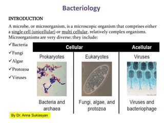
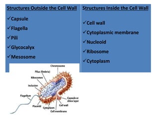
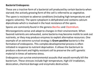
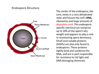
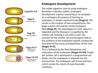
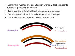
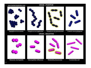
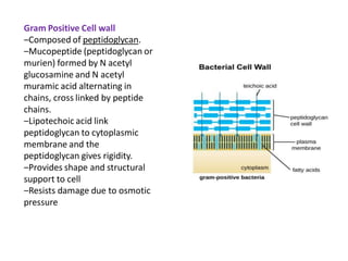
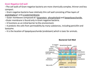
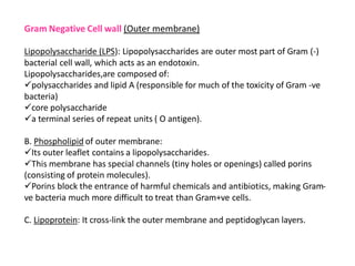
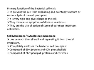
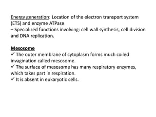
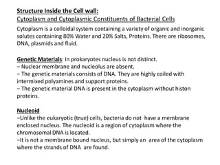
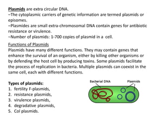
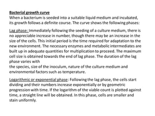
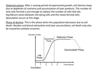
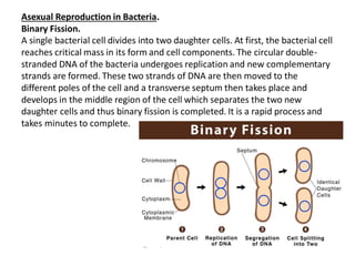
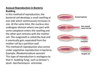
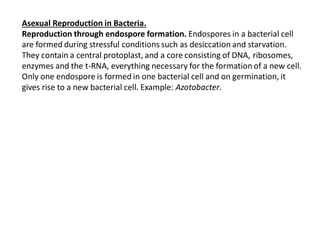
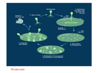
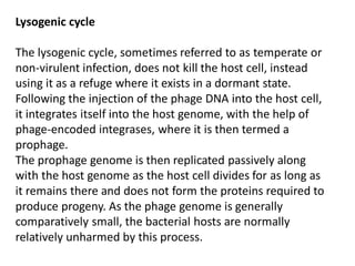
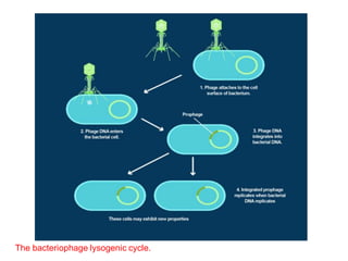
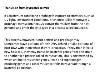
This document provides an overview of microorganisms and bacteria. It discusses that microorganisms are unicellular or multicellular organisms that include bacteria, fungi, algae, protozoa, and viruses. Bacteria are specifically unicellular prokaryotic organisms that lack membrane-bound organelles. The document describes bacterial cell structure both inside and outside the cell wall, including shapes, flagella, pili, capsules, cell membrane, cytoplasm, nucleoid, plasmids, and ribosomes. It also discusses endospore formation in certain bacteria.
EubacteriaDefinitionBacteria are prokaryotic single-celled or BetseyCalderon89
Eubacteria
Definition
Bacteria are prokaryotic single-celled or colonial microorganisms
Characteristics of Bacteria
Lack Green Pigment Chlorophyll
Reproduce by Transverse Fission
Morphology
Bacteria display a wide diversity of shapes and sizes.
Size
0.5 µm diameter
Length 0.5 µm - 80 µm
Bacterial cells are about one-tenth the size of eukaryotic cells and are typically 0.5–5.0 micrometers in length. However, a few species are visible to the unaided eye—for example, Thiomargarita namibiensis is up to half a millimeter long] and Epulopiscium fishelsoni reaches 0.7 mm.] Among the smallest bacteria are members of the genus Mycoplasma, which measure only 0.3 micrometers, as small as the largest viruses.] Some bacteria may be even smaller, but these ultramicrobacteria are not well-studied.
Shape
Spherical – coccus
Rod-shaped - bacillus
Vibrio - Comma shaped
Spiral-shaped -spirillum
Spherical bacteria are known as cocci (singular coccus, Rod-shaped bacteria are called bacilli. Some bacteria, called vibrio, are shaped like slightly curved rods or comma-shaped; others can be spiral-shaped, called spirilla, or tightly coiled, called spirochaetes. A small number of other unusual shapes have been described, such as star-shaped bacteria.
Arrangements of Bacterial Cells
Bacteria are unicellular or colonial
Colonial – cells remain together after division
Colony type – depends on plane of cleavage and planes of successive cleavage.
Bacillus – can only divide in one plane, at right angles to the long axis of the cell.
Streptobacillus
Diplobacillus – remain in pairs following division. after 4 chain fragments
Spirillum- (spiral) divides in one plane
2 types:
Strepto spirillum
Diplo spirillum
Spherical (coccus) can initially divide in any plane. Great variation in colony types.
Streptococcus
Cells divide simultaneously
Diplococcus
If after 4 unit, chain fragments into chains of 2 organisms each – diplococcus
Tetrad Gaffkya
Cells divide at right angles to the preceding division
Sarcina – 3 planes of division. Successive planes are at right angles
Sarcina colonies are cuboidal. All dimensions are the same.
Staphylococcus – irregular cluster of spherical cells. Cells divide in any plane. No pattern
Coccus organism –the type of colony is a species characteristic. It can be used to identify an organism. The colony type is often indicated by the generic name. This is not true of bacillus or spirillum. The colony type can be varied by environment or temperature.
Many bacterial species exist simply as single cells; others associate in characteristic patterns: Neisseria forms diploids (pairs), streptococci form chains, and staphylococci group together in "bunch of grapes" clusters. Bacteria can also group to form larger multicellular structures, such as the elongated filaments of Actinobacteria species, the aggregates of Myxobacteria species, and the complex hyphae of Streptomyces species. These multicellular structures are often only seen in cert ...
Ultrastructure and characterstic features of bacteria.Archana Shaw
Ultrastructure and characterstic features of bacteria: BACTERIA AS A MODEL ORGANISM
THIS WAS MY PRESENTATION TOPIC IN CLASS. THOUGHT OF SHARING IT AND HOPE IT HELPS.
This presentation includes all the general characteristics of fungi, types, structure of a fungi, classifications, and reproduction. Different types of fungi and its classification, its reproduction are all included.
Cell biology is the study of cell structure and function, and it revolves around the concept that the cell is the fundamental unit of life. Focusing on the cell permits a detailed understanding of the tissues and organisms that cells compose.
Embark on a captivating journey into the world of microorganisms with "Exploring the Microscopic Marvels: An Introduction to General Microbiology." This engaging Slideshare presentation provides a comprehensive overview of the fascinating field of microbiology, unraveling the mysteries of the unseen microbial universe that surrounds us.
Delve into the diverse realm of microorganisms and gain a deeper understanding of their structure, function, and significance. From bacteria and viruses to fungi and protozoa, each class of microbe holds its own captivating story, shaping the intricate tapestry of life on Earth. Witness the astonishing complexity of microbial communities and explore their profound impact on human health, ecosystems, and industry.
Uncover the fundamental principles underlying microbiology through visually stunning slides and concise, easy-to-follow explanations. Learn about the techniques and tools used to study microorganisms, including microscopy, culturing methods, and molecular biology approaches. Discover the key characteristics of different microbial groups, such as their morphological diversity, metabolism, and genetic traits.
Dive into the world of microbial interactions and explore the intricate relationships between microorganisms and their environments. Gain insights into microbial pathogenesis, antibiotic resistance, and the vital roles of microbes in ecological processes, biotechnology, and food production. Understand the concepts of microbial control and the strategies employed to combat infectious diseases.
Whether you are a student, a professional in the field, or simply curious about the invisible world of microorganisms, "Exploring the Microscopic Marvels: An Introduction to General Microbiology" will captivate and enlighten you. Expand your knowledge, ignite your curiosity, and embrace the profound impact that microorganisms have on our lives.
Join us on this enthralling journey through the microscopic wonders of general microbiology. Unveil the hidden secrets of the microbial world and gain a new appreciation for the profound influence of these tiny yet mighty organisms. Get ready to witness the remarkable diversity, complexity, and significance of microorganisms through this visually engaging and informative Slideshare presentation.
Food Microorganisms Food microbiology encompasses the stu.docxAKHIL969626
Food Microorganisms
Food microbiology encompasses the study of microorganisms that have either a
beneficial or deleterious effect on the quality and safety of foods.
Food microbiology focuses on the microorganisms that are found in and around foods.
It includes studies of microbial growth characteristics, identification, and prevention.
Specific areas of interest are: foodborne illness, food spoilage, food preservation,
sanitation, and food legislation. These interest areas comprise this course.
The microbiological world consists of bacteria, yeasts, molds, viruses, parasites,
algae and prions. All except algae are considered food microorganisms. Let’s look at
each group briefly.
Bacteria
Bacteria are prokaryotes because they do not possess nuclei. They have a normal
double stranded DNA genome. Sometimes there may be extracellular DNA fragments
known as plasmids. Plasmids can often encode proteins that make the cell enhanced
such as antibiotic resistance.
Plasmids can be shared with other
cells usually of a similar genus or
species. The DNA clusters in the
center of the cell forming a nucleoid.
(If it had a membrane around it, it
would be a nucleus). Closely
associated with the nucleoid are the
ribosomes. Ribosomes translate
DNA into amino acids which are the
building blocks of proteins.
Discuss plasmids and food
microbiology
The main interior is called the
cytoplasm. This is where water,
enzymes, and nutrients all work to
power the cell. There are three main
functions, energy production,
metabolism and catabolism. Energy
naturally is in the form of ATP
generated from metabolic reactions.
Metabolism also consists of enzymes
that build up the components of the
cell. Catabolism consists of enzymes
that break down nutrients. All three
work congruently.
Figure 1. Diagram of bacterial structures.
Discuss catabolic enzymes and food
microbiology
The cytoplasm is surrounded by a
cytoplasmic membrane which
determines what goes in and out of
the organism. Water, dissolved gases
(e.g. CO2 and O2) and lipid-soluble
molecules simply diffuse across the
phospholipid bilayer. Water-soluble
ions generally pass through small
pores in the membrane. All other
molecules require carrier molecules to
transport them through the
membrane.
Discuss membrane function and food
microbiology
Immediately outside of the
cytoplasmic membrane is the cell
wall. This is the component that
stains differently in the Gram stain.
Red is gram negative a thinner peptidoglycan cell wall. Blue or purple is gram positive
usually a much thicker peptidoglycan cell wall. Its main function is structural. The cell
wall provides the bacteria shape: cocci, rods, spirals, etc.
External to the cell wall in SOME bacteria is a capsule layer.
This is a polysaccharide/protein layer that can protect the cell
from desiccation. There are other benefits. Similar to a
capsule, s ...
Living material is organized in unit and microorganism were living form of microscopical size and usually unicellular in structure originally classification is unsatisfied.
Microorganisms, those minuscule entities that elude the naked eye, take centre stage in Class 8 Science Chapter 2, titled "Microorganisms: Friend and Foe." This chapter delves into the intricate world of these tiny beings, exploring their dual nature as both friends and foes, with profound implications for our environment, health, and daily life.
Abhishek Yadav
Seiko college of Pharmacy,
Lucknow
.
.
.
Ultra structure and marphological classification of bacteria, microbiology
B.Pharm 3rd semester
.
.
.
.
Prepared by Abhishek Yadav
Attend 2 international conference in two year of pharmacy.
Read| The latest issue of The Challenger is here! We are thrilled to announce that our school paper has qualified for the NATIONAL SCHOOLS PRESS CONFERENCE (NSPC) 2024. Thank you for your unwavering support and trust. Dive into the stories that made us stand out!
How to Make a Field invisible in Odoo 17Celine George
It is possible to hide or invisible some fields in odoo. Commonly using “invisible” attribute in the field definition to invisible the fields. This slide will show how to make a field invisible in odoo 17.
Instructions for Submissions thorugh G- Classroom.pptxJheel Barad
This presentation provides a briefing on how to upload submissions and documents in Google Classroom. It was prepared as part of an orientation for new Sainik School in-service teacher trainees. As a training officer, my goal is to ensure that you are comfortable and proficient with this essential tool for managing assignments and fostering student engagement.
The Art Pastor's Guide to Sabbath | Steve ThomasonSteve Thomason
What is the purpose of the Sabbath Law in the Torah. It is interesting to compare how the context of the law shifts from Exodus to Deuteronomy. Who gets to rest, and why?
The Indian economy is classified into different sectors to simplify the analysis and understanding of economic activities. For Class 10, it's essential to grasp the sectors of the Indian economy, understand their characteristics, and recognize their importance. This guide will provide detailed notes on the Sectors of the Indian Economy Class 10, using specific long-tail keywords to enhance comprehension.
For more information, visit-www.vavaclasses.com
Welcome to TechSoup New Member Orientation and Q&A (May 2024).pdfTechSoup
In this webinar you will learn how your organization can access TechSoup's wide variety of product discount and donation programs. From hardware to software, we'll give you a tour of the tools available to help your nonprofit with productivity, collaboration, financial management, donor tracking, security, and more.
How to Create Map Views in the Odoo 17 ERPCeline George
The map views are useful for providing a geographical representation of data. They allow users to visualize and analyze the data in a more intuitive manner.
Students, digital devices and success - Andreas Schleicher - 27 May 2024..pptxEduSkills OECD
Andreas Schleicher presents at the OECD webinar ‘Digital devices in schools: detrimental distraction or secret to success?’ on 27 May 2024. The presentation was based on findings from PISA 2022 results and the webinar helped launch the PISA in Focus ‘Managing screen time: How to protect and equip students against distraction’ https://www.oecd-ilibrary.org/education/managing-screen-time_7c225af4-en and the OECD Education Policy Perspective ‘Students, digital devices and success’ can be found here - https://oe.cd/il/5yV
Palestine last event orientationfvgnh .pptxRaedMohamed3
An EFL lesson about the current events in Palestine. It is intended to be for intermediate students who wish to increase their listening skills through a short lesson in power point.
Synthetic Fiber Construction in lab .pptxPavel ( NSTU)
Synthetic fiber production is a fascinating and complex field that blends chemistry, engineering, and environmental science. By understanding these aspects, students can gain a comprehensive view of synthetic fiber production, its impact on society and the environment, and the potential for future innovations. Synthetic fibers play a crucial role in modern society, impacting various aspects of daily life, industry, and the environment. ynthetic fibers are integral to modern life, offering a range of benefits from cost-effectiveness and versatility to innovative applications and performance characteristics. While they pose environmental challenges, ongoing research and development aim to create more sustainable and eco-friendly alternatives. Understanding the importance of synthetic fibers helps in appreciating their role in the economy, industry, and daily life, while also emphasizing the need for sustainable practices and innovation.
1. Bacteriology
INTRODUCTION
A microbe, or microorganism, is a microscopic organism that comprises either
a single cell (unicellular) or multi cellular, relatively complex organisms.
Microorganisms are very diverse; they include:
Bacteria
Fungi
Algae
Protozoa
Viruses
By Dr. Anna Sukiasyan
2. These organisms are vital to humans and the environment, as they participate in
the Earth’s element cycles, such as the carbon cycle and the nitrogen cycle.
Microorganisms live in all parts of the biosphere:
water
soil
hot springs
on the ocean floor
in the atmosphere
deep inside the rocks, within the Earth’s crust
‒Microbes can multiply in all habitats except in the atmosphere.
‒Together their numbers far exceed all other living cells on this planet.
‒The influence of microorganism in human life is both beneficial as well as
detrimental also.
‒For example microorganisms are required for the production of bread, cheese,
yogurt, alcohol, wine, beer, antibiotics (e.g. penicillin, streptomycin,
chloromycetin), vaccines, vitamins, enzymes and many more important
products.
‒Microorganisms are indispensable components of our ecosystem.
‒Many microbes spoil food and deteriorate materials like iron pipes, glass lenses,
computer chips, jet fuel, paints, metal, plastic and etc.
3. Bacteria are unicellular prokaryotic organism where the organisms lack a few
organelles and a true nucleus.
Bacterial cell have simpler internal structure, which lacks all membrane
bound cell organelles such as mitochondria, lysosome, golgi complex,
endoplasmic reticulum, chloroplast, vacuole etc.
All the action takes place in the cytosol or cytoplasmic membrane
Bacteria also lacks true membrane bound nucleus and nucleolus. The
bacterial nucleus is known as nucleoid.
Most bacteria possess peptidoglycan, a unique polymer that makes its
synthesis
‒peptidoglycan is a good target for antibiotics.
Protein synthesis takes place in the cytosol with structurally different
ribosome’s.
Shape
Coccus (cocci)
Rod (bacillus, bacilli)
Spiral shapes (spirochetes; spirillum)
Filamentous and various odd shapes.
5. Structures Outside the Cell Wall
Capsule
These are structures surrounding the outside of the cell envelope. When more
defined, they are referred to as a capsule when less defined as a slime layer or
glycocalyx. They usually consist of polysaccharide.
They are not essential to cell viability and some strains within a species will
produce a capsule, whilst others do not.
Capsule is 98% water and 2% polysaccharide or glycoprotein/ polypeptide or
both.
There are two types of capsule.
‒Macro-capsule: thickness of 0.2µm or more, visible under light microscope
‒Microcapsule: thickness less than 0.2µm, visible under Electron microscope
Capsule is very delicate structure, It can be removed by vigorous washing.
Capsule is most important virulence factor of bacteria.
Function of capsule:
It helps in attachments as well as it prevent the cell from desiccation and
drying.
Capsule resist phagocytosis by WBCs
6. Some bacterial species are mobile and have (possess) locomotory organelles
– Flagella
They are able to taste their environment and respond to specific chemical
foodstuffs or toxic materials and move towards or away from them
(chemotaxis). Flagella have the cell membrane. Flagella consist of a number
of proteins including flagellin known as H antigen.
Binding proteins in the periplasmic space or cell membrane bind food
sources (such as sugars and amino acids) causing methylation of other cell
membrane proteins which in turn affect the movement of the cell by
flagella.
Function of Flagella
‒Flagella are fully responsible for the bacterial motility.
‒Deflagellation by mechanical means renders the motile cells immotile.
‒The apparent movement of the bacterial cell usually takes place by the
rotation of the flagella either in the clockwise or anticlockwise direction
along its long axis.
7. Pili / Fimbriae
Hair-like proteinaceous structures that extend from the cell
membrane to external environment are pili which are otherwise
known as fimbriae.
They are thinner, shorter and more numerous than flagella and they
do not function in motility.
There are two types of pili namely:
1) Non-sex pili (Common pili) eg.
fimbriae. The fimbriae are
antigenic and mediate their
adhesion which inhibits
phagocytosis.
2) The sex pili help in conjugation.
8. Wall-less forms of Bacteria
When bacteria are treated with
1) enzymes that are lytic for the cell wall e.g. lysozyme or
2) 2) antibiotics that interfere with biosynthesis of peptidoglycan, wall-
less bacteria are often produced. (Usually these treatments generate
non-viable organisms).
Wall-less bacteria that can not replicate are called (referred to) as
spheroplasts (when an outer membrane is present) or protoplasts (if an
outer membrane is not present).
Occasionally wall-less bacteria that can replicate are generated by these
treatments (L forms).
L-form bacteria
9. Bacterial Endospores
These are a inactive form of a bacterial cell produced by certain bacteria when
starved; the actively growing form of the cell is referred to as vegetative.
The spore is resistant to adverse conditions (including high temperatures and
organic solvents). The spore cytoplasm is dehydrated and contains calcium
dipicolinate which is involved in the heat resistance of the spore.
Spores are commonly found in the genera Bacillus and Clostridium.
Microorganisms sense and adapt to changes in their environment. When
favored nutrients are exhausted, some bacteria may become motile to seek out
nutrients, or they may produce enzymes to exploit alternative resources. One
example of an extreme survival strategy is Gram-positive bacteria is the
formation of endospores. This complex developmental process is often
initiated in response to nutrient deprivation. It allows the bacterium to
produce a dormant and highly resistant cell to preserve the cell's genetic
material in times of extreme stress.
Endospores can survive environmental assaults that would normally kill the
bacterium. These stresses include high temperature, high UV irradiation,
desiccation, chemical damage and enzymatic destruction.
10. Endospore Structure
The center of the endospore, the
core, exists in a very dehydrated
state and houses the cell's DNA,
ribosomes and large amounts of
dipicolinic acid. This endospore-
specific chemical can comprise
up to 10% of the spore's dry
weight and appears to play a role
in maintaining spore dormancy.
Small acid-soluble proteins
(SASPs) are also only found in
endospores. These proteins
tightly bind and condense the
DNA, and are in part responsible
for resistance to UV light and
DNA-damaging chemicals.
11. Endospore Development
The model organism used to study endospore
formation is Bacillus subtilis. Endospore
development requires several hours to complete.
As a cell begins the process of forming an
endospore, it divides asymmetrically (Stage II). This
results in the creation of two compartments, the
larger mother cell and the smaller forespore.
Next (Stage III), the peptidoglycan in the septum is
degraded and the forespore is engulfed by the
mother cell, forming a cell within a cell. The
activities of the mother cell and forespore lead to
the synthesis of the endospore-specific compounds,
formation of the cortex and deposition of the coat
(Stages IV+V).
This is followed by the final dehydration and
maturation of the endospore (Stages VI+VII). Finally,
the mother cell is destroyed in a programmed cell
death, and the endospore is released into the
environment. The endospore will remain dormant
until it senses the return of more favorable
conditions.
12. Cell Wall
Beneath the external structures is the cell wall.
Animal cells do not have a cell wall outside the cell membrane.
Plant cells and fungal cells do have a cell wall.
Most bacteria have a cell wall, they are essential structures in
bacteria.
A bacterium is referred as a protoplast when it is without cell wall.
Cell wall may be lost due to the action of lysozyme enzyme, which
destroys peptidoglycan.
This cell is easily lysed and it is metabolically active but unable to
reproduce.
A bacterium with a damaged cell wall is referred as spheroplasts.
It is caused by the action of toxic chemical or an antibiotic, they show
a variety of forms and they are able to change into their normal form
when the toxic agent is removed, i.e. when grown on a culture media.
They are made of chemical components, which are found nowhere
else in nature.
13. Gram stain invented by Hans Christian Gram divides bacteria into
two main groups based on stain.
Gram-positive cell wall is thick homogeneous monolayer
Gram-negative cell wall is thin heterogeneous multilayer
Correlates with two types of cell wall architecture.
Gram positive
Gram negative
14.
15. Gram Positive Cell wall
‒Composed of peptidoglycan.
‒Mucopeptide (peptidoglycan or
murien) formed by N acetyl
glucosamine and N acetyl
muramic acid alternating in
chains, cross linked by peptide
chains.
‒Lipotechoic acid link
peptidoglycan to cytoplasmic
membrane and the
peptidoglycan gives rigidity.
‒Provides shape and structural
support to cell
‒Resists damage due to osmotic
pressure
16. Gram Negative Cell wall
‒The cell walls of Gram-negative bacteria are more chemically complex, thinner and less
compact.
‒ Gram-negative bacteria have relatively thin cell wall consisting of few layers of
peptidoglycan and a outermembrane.
‒ Outer membrane composed of: lipoprotein, phospholipid and lipopolysaccharide.
‒Outer membrane is found only in Gram negative bacteria.
‒ It functions as an initial barrier to the environment.
‒ It protects the cells from permeability by many substances, including penicillin and
lysozyme.
‒ It is the location of lipopolysaccharide (endotoxin) which is toxic for animals.
17. Gram Negative Cell wall (Outer membrane)
Lipopolysaccharide (LPS): Lipopolysaccharides are outer most part of Gram (-)
bacterial cell wall, which acts as an endotoxin.
Lipopolysaccharides,are composed of:
polysaccharides and lipid A (responsible for much of the toxicity of Gram -ve
bacteria)
core polysaccharide
a terminal series of repeat units ( O antigen).
B. Phospholipid of outer membrane:
Its outer leaflet contains a lipopolysaccharides.
This membrane has special channels (tiny holes or openings) called porins
(consisting of protein molecules).
Porins block the entrance of harmful chemicals and antibiotics, making Gram-
ve bacteria much more difficult to treat than Gram+ve cells.
C. Lipoprotein: It cross-link the outer membrane and peptidoglycan layers.
18. Primary function of the bacterial cell wall:
To prevent the cell from expanding and eventually rupture or
osmotic lysis of the cell protoplast.
It is very rigid and gives shape to the cell.
They may cause symptoms of disease in animals.
They are the site of action of some of our most important
antibiotics.
Cell Membrane/ Cytoplasmic membrane
Lies beneath the cell wall and separating it from the cell
cytoplasm.
Completely encloses the bacterial cell protoplast
Composed of 60% protein and 40% phospholipid
Composed of Phospholipid, proteins and enzymes
19. Energy generation: Location of the electron transport system
(ETS) and enzyme ATPase
‒ Specialized functions involving: cell wall synthesis, cell division
and DNA replication.
Mesosome
The outer membrane of cytoplasm forms much coiled
invagination called mesosome.
The surface of mesosome has many respiratory enzymes,
which takes part in respiration.
It is absent in eukaryotic cells.
20. Structure Inside the Cell wall:
Cytoplasm and Cytoplasmic Constituents of Bacterial Cells
Cytoplasm is a colloidal system containing a variety of organic and inorganic
solutes containing 80% Water and 20% Salts, Proteins. There are ribosomes,
DNA, plasmids and fluid.
Genetic Materials: In prokaryotes nucleus is not distinct.
‒ Nuclear membrane and nucleolus are absent.
‒ The genetic materials consists of DNA. They are highly coiled with
intermixed polyamines and support proteins.
‒ The genetic material DNA is present in the cytoplasm without histon
proteins.
Nucleoid
‒Unlike the eukaryotic (true) cells, bacteria do not have a membrane
enclosed nucleus. The nucleoid is a region of cytoplasm where the
chromosomal DNA is located.
‒It is not a membrane bound nucleus, but simply an area of the cytoplasm
where the strands of DNA are found.
21. Plasmids are extra circular DNA.
‒The cytoplasmic carriers of genetic information are termed plasmids or
episomes.
‒Plasmides are small extra-chromosomal DNA contain genes for antibiotic
resistance or virulence.
‒Number of plasmids: 1-700 copies of plasmid in a cell.
Functions of Plasmids
Plasmids have many different functions. They may contain genes that
enhance the survival of an organism, either by killing other organisms or
by defending the host cell by producing toxins. Some plasmids facilitate
the process of replication in bacteria. Multiple plasmids can coexist in the
same cell, each with different functions.
Types of plasmids:
1. fertility F-plasmids,
2. resistance plasmids,
3. virulence plasmids,
4. degradative plasmids,
5. Col plasmids.
22. Fertility F-plasmids
Fertility plasmids, also known as F-plasmids, contain transfer genes that allow
genes to be transferred from one bacteria to another through conjugation.
Resistance Plasmids
Resistance or R plasmids contain genes that help a bacterial cell defend
against environmental factors such as poisons or antibiotics. Some resistance
plasmids can transfer themselves through conjugation. When this happens, a
strain of bacteria can become resistant to antibiotics.
Virulence Plasmids
When a virulence plasmid is inside a bacterium, it turns that bacterium into a
pathogen, which is an agent of disease. Bacteria that cause disease can be
easily spread and replicated among affected individuals. The bacterium
Escherichia coli (E. coli) has several virulence plasmids. E. coli is found
naturally in the human gut and in other animals, but certain strains of E. coli
can cause severe diarrhea and vomiting. Salmonella enterica is another
bacterium that contains virulence plasmids.
23. Degradative Plasmids
Degradative plasmids help the host bacterium to digest
compounds that are not commonly found in nature, such as
camphor, xylene, toluene, and salicylic acid. These plasmids
contain genes for special enzymes that break down specific
compounds.
Col Plasmids
Col plasmids contain genes that make bacteriocins (also
known as colicins), which are proteins that kill other bacteria
and thus defend the host bacterium. Bacteriocins are found
in many types of bacteria including E. coli, which gets them
from the plasmid ColE
24. Ribosome
‒ The procaryotic ribosome (L) is 70S in size, being composed of a 50S
(large) subunit and a and 30S (small) subunit. The eucaryotic
ribosome (R) is 80S in size and is composed of a 60S and a 40S
subunit.
‒ Ribosomes are made of two subunits, a large subunit and a small
subunit. Each subunit is made up of RNA and various proteins.
Function of Ribosome:
Ribosomes function in protein synthesis.
‒Amino acids are assembled into proteins according to the genetic
code on the surfaces of ribosomes during the process of translation.
25. CLASSIFICATION OF BACTERIA
Bacteria can be classified into various categories based on their
features and characteristics.
The classification of bacteria is mainly based on the following
characters:
1) Morphological Classification (Based on Shape)
2) Composition of the cell wall
3) Mode of respiration
4) Mode of nutrition
26. Bacteria and Archaea are
classified by direct examination
with the light microscope
according to their morphology
and arrangement.
The basic morphologies are:
1) Spheres (coccus)
2) Round-ended cylinders
(bacillus).
3) Helically twisted cylinders
(spirochetes)
4) Cylinders curved in one plane
(selenomonads)
5) Unusual morphologies (such
as the square, flat box-shaped
cells of the archaean genus).
Morphological Classification (Based on Shape)
27. Coccus (Pleural – Cocci): Spherical bacteria
‒may occur in pairs (diplococci)
‒in groups of four (tetracocci)
‒in grape-like clusters (Staphylococci)
‒in chains (Streptococci)
‒in cubical arrangements of eight or more (Sarcinae).
Example: Staphylococcus aureus, Neisseria gonorrhoeae,
Streptococcus pneumoniae, Streptococcus pyogenes
Bacillus (Pleural–Bacilli): Rod-shaped bacteria
Example s – Bacillus cereus, Clostridium tetani, E.Coli and Salmonella.
‒generally occur singly
‒but may occasionally be found in pairs (diplo-bacilli)
‒chains (streptobacilli).
28. Spirillum (Pleural–Spirilla): Spiral-shaped bacteria.
Spiral bacteria can be sub-classified on the basis of number of
twists per cell, cell thickness, cell flexibility, and motility.
Examples – Spirillum, Vibrio, Spirochete species.
Bacteria have Other Shapes such as:
Coccobacilli – Elongated spherical or ovoid form.
Filamentous – Bacilli that occur in long chains or threads.
29. Classification of bacteria by Composition of the cell wall (G+ and G-)
Gram positive
bacteria - take
up crystal
violet dye and
retain their
blue or violet
color.
Gram negative
bacteria - do
not take up
crystal violet
dye, and thus
appear red or
pink.
30. Nutritional requirements of Bacteria
For growth and nutrition of bacteria, the minimum nutritional requirements
are water, a source of carbon, a source of nitrogen and some inorganic salts.
Bacteria can be classified nutritionally based on their energy requirements and
on their ability to synthesise essential metabolites:
Bacteria which derive energy from sunlight are called phototrophs.
Those that obtain energy from chemical reactions are called chemotrophs.
Bacteria that can synthesise all their organic compounds are called autotrophs.
They are able to use atmospheric carbon dioxide and nitrogen. They are
capable of independent existence in water and soil. They are of no medical
importance.
Some bacteria are unable to synthesise their own metabolites. They depend
on preformed organic compounds. They are called heterotrophs. These
bacteria are unable to grow with carbon dioxide as the sole source of carbon.
Their nutritional requirements vary widely.
Some may require only a single organic substance like glucose.
Others may need a large number of different compounds like amino acids,
nucleotides, lipids, carbohydrates and coenzymes.
31. Microorganism can be classified on the basis of their oxygen requirements:
1)Obligate Aerobes: Require oxygen to live. Example: Pseudomonas, common
nosocomial pathogen.
2)Facultative Anaerobes: Can use oxygen, but can grow in its absence. Have
complex set of enzymes. Examples: E. coli, Staphylococcus, yeasts, and many
intestinal bacteria.
3)Obligate Anaerobes: Cannot use oxygen and are harmed by the presence of
toxic forms of oxygen. Examples: Clostridium bacteria that cause tetanus and
botulism.
4)Aerotolerant Anaerobes: Can’t use oxygen, but tolerate its presence. Can
break down toxic forms of oxygen. Example: Lactobacillus carries out
fermentation regardless of oxygen presence.
5)Microaerophiles: Require oxygen, but at low concentrations. Sensitive to toxic
forms of oxygen. Example: Campylobacter.
32. Temperature Requirements
Bacteria vary in their requirement of temperature for growth.
The temperature at which growth occurs best is known as the “optimum
temperature”.
In the case of most pathogenic bacteria, the optimum temperature is 37oC.
Bacteria which grow best at temperatures of 25-40oC are called mesophilic,
for example Escherichia coli.
Psychrophilic bacteria are those that grow best at temperatures below 20oC.
They are soil and water saprophytes and may cause
spoilage of refrigerated food.
Thermophilic bacteria are those which grow best at high temperatures, 55-
80oC. They may cause spoilage of underprocessed canned food. Some
thermophiles, for example Geobacillus stearothermophilus, form spores that
are highly thermoresistant.
Note: Majority of pathogenic bacteria grow best at neutral or slightly alkaline
pH.
33. Bacterial growth curve
When a bacterium is seeded into a suitable liquid medium and incubated,
its growth follows a definite course. The curve shows the following phases:
Lag phase: Immediately following the seeding of a culture medium, there is
no appreciable increase in number, though there may be an increase in the
size of the cells. This initial period is the time required for adaptation to the
new environment. The necessary enzymes and metabolic intermediates are
built up in adequate quantities for multiplication to proceed. The maximum
cell size is obtained towards the end of lag phase. The duration of the lag
phase varies with
the species, size of the inoculum, nature of the culture medium and
environmental factors such as temperature.
Logarithmic or exponential phase: Following the lag phase, the cells start
dividing and their numbers increase exponentially or by geometric
progression with time. If the logarithm of the viable count is plotted against
time, a straight line will be obtained. In this phase, cells are smaller and
stain uniformly.
34. Stationary phase: After a varying period of exponential growth, cell division stops
due to depletion of nutrients and accumulation of toxic products. The number of
new cells formed is just enough to replace the number of cells that die.
Equilibrium exists between the dying cells and the newly formed cells.
Sporulation occurs at this stage.
Phase of decline: This is the phase when the population decreases due to cell
death. Besides nutritional exhaustion and toxic accumulation, cel death may also
be caused by autolytic enzymes.
35. Bacterial reproduction
Bacterial cell division is studied in many research laboratories
throughout the world. These investigations are uncovering the
genetic mechanisms that regulate and drive bacterial cell division.
Understanding the mechanics of this process is of great interest
because it may allow for the design of new chemicals or novel
antibiotics that specifically target and interfere with cell division in
bacteria.
Bacteria reproduce through both asexual and sexual means.
36. Asexual Reproduction in Bacteria.
Binary Fission.
A single bacterial cell divides into two daughter cells. At first, the bacterial cell
reaches critical mass in its form and cell components. The circular double-
stranded DNA of the bacteria undergoes replication and new complementary
strands are formed. These two strands of DNA are then moved to the
different poles of the cell and a transverse septum then takes place and
develops in the middle region of the cell which separates the two new
daughter cells and thus binary fission is completed. It is a rapid process and
takes minutes to complete.
37. Asexual Reproduction in Bacteria
Budding.
In this method of reproduction, the
bacterial cell develops a small swelling at
one side which continuously increases in
size. At the same time, the nucleus also
undergoes division where one part with
some cytoplasm enters the swelling and
the other part remains with the mother
cell. The outgrowth is called the bud and
it eventually gets separated from the
mother cell by a partition wall.
This method of reproduction also comes
under vegetative reproduction in bacteria.
Example: Rhodomicrobium vannielii
This type of reproduction is analogous to
that in budding fungi, such as brewer’s
yeast -Saccharomyces cerevisiae.
38. Asexual Reproduction in Bacteria.
Reproduction through endospore formation. Endospores in a bacterial cell
are formed during stressful conditions such as desiccation and starvation.
They contain a central protoplast, and a core consisting of DNA, ribosomes,
enzymes and the t-RNA, everything necessary for the formation of a new cell.
Only one endospore is formed in one bacterial cell and on germination, it
gives rise to a new bacterial cell. Example: Azotobacter.
39. Sexual Reproduction in Bacteria (mechanisms of genetic recombination).
Transformation.
In transformation, a bacterium takes up DNA from its environment and
often DNA that’s been shed by another bacteria. The phenomenon was
first discovered by Frederick Griffith in 1928.
In this process bacteria can acquire new genes by taking up DNA
molecules (ex: plasmid) from their surroundings. When bacteria
undergo lysis, they release considerable amounts of DNA into the
environment.
This DNA may be picked up by a competent cell- one capable of taking
up the DNA and undergoing a transformation.
To be competent, bacteria must be in the logarithmic stage of growth,
and a competence factor needed for the transformation must be
present.
40. Sexual Reproduction in Bacteria (mechanismsof genetic recombination).
Transduction.
In this type of sexual reproduction of bacteria, foreign genes are
transferred into a bacterial cell with the help of a virus. These viruses
are called bacteriophage and they are not virulent. The virus acts as a
carrier vehicle and passes over genes from one host to another.
Bacteriophages transfer DNA fragments from one bacterium (the
donor) to another bacterium (the recipient).
The viruses involved contain a strand of DNA enclosed in an outer
coat of protein.
After a bacteriophage enters a bacterium, it may encourage the
bacterium to make copies of the phage. There are two stages/cycles
are lytic and lysogeni.
41. Sexual Reproduction in Bacteria (mechanismsof genetic recombination).
Conjugation.
This process was first discovered in Escherichia coli by Joshua Lederberg and
Edward Tatum in 1946. They found that two different types of nutritional
mutants grown together on minimal medium produced an occasional wild type.
Bacteria that show conjugation are dimorphic, meaning that they have two
types of cells, one male (F+) or donor cell and a female (F-) or recipient cell.
The male or donor cell possesses 1 to 4 sex
pili on the surface and fertility factor
(transfer factor, sex factor) in its plasmid. It
contains genes for producing sex pili and
other characters needed for gene transfer.
The sex pili and fertility factor are absent
from the female or recipient cells.
If these two types of cells happen to come
nearer, a pilus of a male cell establishes a
protoplasmic bridge or conjugation tube
with the female cell. It takes 6-8 minutes
for the process to complete.
42. These were the three types of sexual reproduction in bacteria
and it introduces genetic variation in a bacterial species which is
important for the survival of any species and allows groups to
adapt to environmental changes.
43. Bacteriophages
Bacteria have parasites, the viruses called bacteriophages which are
obligate (only) intracellular parasites that multiply inside bacteria.
These lyze the infected bacterial cell liberating new infection phage
particles.
Bacteriophages are bacterial viruses with a DNA or RNA genome
usually protected by a membrane or protein shell.
These extrachromosomal genetic elements can survive outside of a
host cell and be transmitted from one cell to another.
Bacteriophages infect bacterial cells and either replicate to large
numbers and cause the cell to lyse (lytic infection) or, in some cases,
integrate into the host genome without killing the host (the lysogenic
state), such as the E.coli bacteriophage lambda. Some lysogenic
bacteriophages carry toxin genes (e.g., corynephage beta carries the
gene for the diphtheria toxin).
44. Lytic cycle
In the lytic cycle sometimes referred to as virulent infection,
the infecting phage ultimately kill the host cell to produce
many of their own progeny. Immediately following injection
into the host cell, the phage genome synthesizes early
proteins that break down the host DNA, allowing the phage to
take control of the cellular machinery.
The phage then uses the host cell to synthesize the remaining
proteins required to build new phage particles. During this
process, the host cells gradually become weakened by phage
enzymes and eventually burst, releasing on average 100-200
new phage progeny into the surrounding environment.
46. Lysogenic cycle
The lysogenic cycle, sometimes referred to as temperate or
non-virulent infection, does not kill the host cell, instead
using it as a refuge where it exists in a dormant state.
Following the injection of the phage DNA into the host cell,
it integrates itself into the host genome, with the help of
phage-encoded integrases, where it is then termed a
prophage.
The prophage genome is then replicated passively along
with the host genome as the host cell divides for as long as
it remains there and does not form the proteins required to
produce progeny. As the phage genome is generally
comparatively small, the bacterial hosts are normally
relatively unharmed by this process.
48. Transition from lysogenic to lytic
If a bacterium containing prophage is exposed to stressors, such as
UV light, low nutrient conditions, or chemicals like mitomycin C,
prophage may spontaneously extract themselves from the host
genome and enter the lytic cycle in a process called induction.
This process, however, is not perfect and prophage may
sometimes leave portions of their DNA behind or take portions of
host DNA with them when they re-circularize. If they then infect a
new host cell, they may transport bacterial genes from one strain
to another in a process called transduction. This is one method by
which antibiotic resistance genes, toxin and superantigen-
encoding genes and other virulence traits may spread through a
bacterial population.
49. Latency Period
Viruses that infect plant or animal cells may also undergo infections where they
are not producing virions for long periods. An example is the animal herpes
viruses, including herpes simplex viruses, which cause oral and genital herpes in
humans. In a process called latency, these viruses can exist in nervous tissue for
long periods of time without producing new virions, only to leave latency
periodically and cause lesions in the skin where the virus replicates.
Phage therapy
Prior to the discovery of antibiotics by Alexander Fleming in 1928, phage were
being explored as a method for treating bacterial infections. In the post-
antibiotic era, the convenient broad-spectrum activity of antibiotic treatment
meant that in most organization’s research into phage therapy was abandoned.
Whilst phage are able to infect and destroy bacteria and have been successfully
used to treat life-threatening infection, their species and even strain specificity
and potential for pre-existing immunity of some bacteria mean targeting a
phage treatment is currently not a trivial process and must be tailored to the
individual infection. This makes it costly and lengthy. Consequently, it is
currently a last resort and there is still much work required in this field.
50. Main steps in diagnostic isolation and identification of
bacteria
Step 1. Samples of body fluids (e.g.
blood, urine, cerebrospinal fluid
(marrow)) are streaked (lane) on
culture plates and isolated colonies of
bacteria (which are visible to the naked
eye) appear after incubation for one to
several days .
Each colony consists of millions of
bacterial cells. Observation of these
colonies for size, texture, color, and (if
grown on blood agar) hemolysis
reactions is highly important as a first
step in bacterial identification.
Whether the organism requires oxygen
for growth is another important
differentiating characteristic.
Bicarbonate and blood agar plate cultures
of Bacillus anthracis. Smooth colonies on
bicarbonate (left) and rough colonies on
blood agar (right).
51. Gram positive vs Gram negative stain
Step 2. Colonies are Gram stained and individual bacterial cells
observed under the microscope.
52. Gram positive bacteria have a distinctive purple appearance when observed
under a light microscope following Gram staining.
Gram negative bacteria appear a pale reddish color when observed under a
light microscope following Gram staining.
53. Bacillus brevis. Gram stain.
Streptococcus mutans. Gram stain. Blood agar
plate culture yields coccal-like morphology
without chains. Organism can cause dental
caries
Gram-positive
55. Step 3. The bacteria are characterized using these isolated
colonies. This often requires an additional 24 hours of growth.
Step 4. Another similar colony from the primary isolation plate
is then examined for biochemical properties; for example, will
the bacteria ferment a sugar such as lactose? In some
instances, the bacteria are identified (e.g. by aggregation) with
commercially available antibodies recognizing defined
(specially) surface antigens.
56. Bacterial Infections
Signs and symptoms of a bacterial infection may vary depending
on the location of the infection and the type of bacteria that’s
causing it.
The severity of bacterial infections can vary widely and depends
on the type of bacteria involved.
On one hand, there are relatively minor illnesses like strep throat
and ear infections. But bacterial infections can also cause
potentially life-threatening conditions like meningitis and
encephalitis.
57. Cuts
Your skin is your body’s first defense against infection. Breaks in the
skin, like cuts, scrapes, or surgical incisions, can provide an entryway
into the body for bacteria.
Symptoms:
‒redness in the area of the wound,
‒swelling or warmth in the affected area,
‒pain or tenderness at or around the site of the wound,
‒pus forming around the wound,
‒fever.
Burns
‒People with burns are at risk for developing complications, such as a
bacterial infection. Symptoms that a burn has become infected
include:
‒an increase in pain or discomfort around the affected area
‒redness in the area of the burn,
‒fluid or pus oozing from the burn site,
‒a bad smell around the burn.
58. Strep throat
Strep throat is an infection of the throat caused by a type of bacteria
called group A Streptococcus. Symptoms include:
‒sore throat
‒difficulty swallowing
‒red or white patches on the back of the throat
‒headache
‒loss of appetite
Urinary tract infection occur when bacteria from your rectum or skin
enter your urinary tract, symptoms can include:
‒a burning sensation when urinating
‒having to urinate frequently
‒cloudy urine
‒abdominal cramps
‒fever
59. Pneumonia is an infection that inflames the air sacs in the lungs.
Bacteria such as Streptococcus pneumoniae can cause it. Symptoms of
pneumonia include:
‒cough
‒pain in your chest
‒fever
‒sweating or chills
‒shortness of breath
‒feeling tired or fatigued
Food poisoning can happen when you consume food or water that’s
been contaminated with bacteria. Some types of bacteria that cause
food poisoning include Escherichia coli, Listeria, and Salmonella.
Symptoms can include:
‒nausea or vomiting
‒diarrhea
‒abdominal cramps
‒fever
60. Bacterial meningitis
Meningitis is inflammation of the tissues that surround the brain and spinal cord.
Bacterial meningitis can develop from several types of bacteria, including
Streptococcus pneumoniae and Neisseria meningitidis. Symptoms include:
‒headache
‒stiff neck
‒fever
‒nausea or vomiting
‒confusion
‒sensitivity to light
Sepsis
An untreated bacterial infection can also put you at risk for developing a life-
threatening condition called sepsis. The bacteria most likelyTrusted Source to cause
sepsis include Staphylococcus aureus, E. coli, and some types of Streptococcus.
Symptoms:
‒shortness of breath
‒fast heart rate
‒fever
‒being in severe pain or discomfort
‒chills or sweating
‒confusion