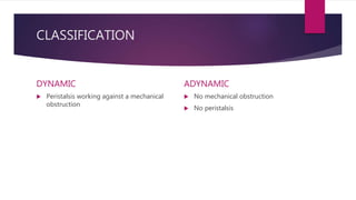
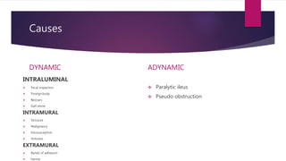
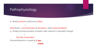
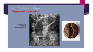
INTESTINAL OBSTRUCTION
CLASSIFICATION
DYNAMIC
 Peristalsis working against a mechanical
obstruction
ADYNAMIC
 No mechanical obstruction
 No peristalsis
Causes
DYNAMIC
INTRALUMINAL
 Fecal impaction
 Foreignbody
 Bezoars
 Gall stone
INTRAMURAL
 Stricture
 Malignancy
 Intususception
 Volvulus
EXTRAMURAL
 Bands of adhesion
 hernia
ADYNAMIC
 Paralytic ileus
 Pseudo obstruction
Pathophysiology
 Bowel proximal to obstruction dilates
↓
Distal bowel - normal peristalsis & absorption→later empty &collapses
 Initially proximal peristalsis increased→later reduction in peristaltic strength
↓
flaccidity and paralysis
Proximal distention is caused by 1. gas
2.fluid
gas
 Significant overgrowth of both aerobic
and anaerobic bacteria
 Reabsorption – oxygen & corbondioxide
 Nitrogen and hydrogen sulphide
fluid
 24 hrs
 Saliva -500ml, bile- 500ml ,pancreatic secretion -
500ml ,gastric secretion-1L
 Accumulates & absorption –retarded
Dehydration & electrolyte loss
Reduced oral intake
Defective absorption
Vomiting
Sequestration in the bowel lumen
Transudation of fluid in to peritoneal cavity
Strangulation – Blood supply is
compromised and the bowel becomes
ischaemic
 CAUSES
DIRECT PRESSURE ON THE BOWEL WALL
 Hernial orifices
 Adhesions / Bands
INTERRUPTED MESENTERIC BLOOD FLOW
 Volvulus
 Intussuseption
INCREASED INTRALUMINAL PRESSURE
 Closed loop obstruction
Closed loop obstruction
 Bowel is obstructed at both the proximal and distal points
 Malignant stricture of the colon with a competent ileocaecal valve
↓
Inability of distended colon to decompress itself in to the small bowel
↓
Increase in luminal pressure
↓
necrosis and perforation
Internal hernia
 Portion of small intestine becomes entrapped in one of the retroperitoneal fossae
or in a congenital mesenteric defect
 SITES OF INTERNAL HERNIA
 the foramen of winslow
 a defect in the mesentery / transverse mesocolon/ broad ligament
 congenital or acquired diaphragmatic hernia
 Duodenal retro peritoneal fossae- left paraduodenal and right duodenojejunal
 Caecal /appendiceal retroperitonel fossae- sup, inf, and retrocaecal
 Intersigmoid fossa
Obstruction from enteric strictures
 Small bowel – secondary to TB / Crohns disease
 Subacute or chronic presentation
 Mx- resection and anastomosis
Bolus obstruction
 Gallstone
 Food
 Stercolith
 Worms
 Trychobezoars and phytobezoars
Gallstone ileus
 60 cm proximal to ileo-caecal valve
Rigler’s triad
 Small bowel obstruction
 Pneumobilia
 Atypical metallic shadow on abdominal x ray
Trichobezoar
 Usually seen in psychiatric patients
 Ingestion of hair
phytobezaors
 High fibre intake
 Inadequate chewing
 Previous gastric Sx
 Hypochlorhydria
 Loss of gastric pump mechanism
Stercoliths
 Jejunal diverticulum and ileal stricture
Worms
 Ascaris lumbricoids
Common causes of intra-abdominal
adhesins
Acute inflammation Sites of anastomoses,
of raw areas, trauma, ischaemia
Foreign material Talc ,starch ,gauze, silk
infection Peritonitis, TB
Chronic inflammatory condditions Crohn’s disease
Radiation enteritis
Prevention of adhesions
 Good surgical technigue
 Washing of the peritonela cavity with saline to remove clots
 Minimising contact with gauze
 Covering anastomosis and raw peritoneal surfaces
Laproscopic technique
 May reduce post op adhesins
 Others
 Hyaluronidase
 Hydrocortisone
 Silicone
 Dextran
 Polyvinylpropylene
 Chondroitin
 Streptomycin
 Anticoagulants
 Antihistamines
 NSAIDS
 Streptokinase
Bands
 Usually only one band is culpable
 Congenital, e.g. obliterated vitellointestinal duct;
 A string band following previous bacterial peritonitis
 A portion of greater omentum, usually adherent to the parietes
Acute intussusception
 One portion of the gut invaginate in to an immediately adherent segment;
 Almost invariably-proximal in to the distal
 MC in children ( 5-10 months ; peak incidence)
 Hyperplasia of Peyer’s patches in the terminal ileum
 Weaning – loss of passively acquired maternal immunity
 Common viral pathogens
Associated pathological lead points in
children
 Meckels diverticulum
 Polyp
 Duplication
 HSP
 Appendix
Lead points in adults
 Polyp eg; Peutz jeghers syndrome
 Submucosal lipoma
 Other tumors
Pathology
3 parts
 The Entering or inner tube- intussusceotum
 The returning or middle tube
 The sheath or outer tube – intussuscipiens
 The part that advances apex, the mass- intussusception and neck is the junction
of entering layer with the mass
Volvulus
 Twisting or axial rotation of a portion of bowel abouts its mesentery
 Causes obstruction ( > 180 degree tortion)
 If tight – vasocular occlusion in the mesentery( 360 degree)
 Primary or secondary
 May involve small intestine, caecum or sihmoid colon
 Neonatal midgut volvulus sec to midgut malrotation is life threatening
 Commonest spontaneous type in adult – SIGMOID
 Sigmoid volvulus – relieved by decompression per anum
 Surgery – to prevent or relieve ischaemia
Clinical features
 Abdominal pain
 Abdominal distention
 Vomiting
 Absolute constipation
classifications
High small bowel
Low small bowel
Large bowel
Simple
strangulated
Complete
Incomplete
Intestinal obstruction with out
constipation
 Richter’s hernia
 Gallstone ileus
 Mesenteric vascular occlusion
 Functional obstruction associated with pelvic abcess
 Partial obstruction
Other manifestations
 Dehydration
 Hypokalaemia
 Pyrexia
 Abdominal tenderness
Pyrexia
 Oncet of ischaemia
 Intestinal perforation
 Infalammation oe abcess associated with obstructive disease
Abdominal tenderness
 Localised tenderness- impending ischaemia
 Diffuse tenderness- perforation peritonitis
c/f; strangulation
 Constant severe pain
 Tenderness with rigidity and peritinism
 Shock
c/f; intussusception
 Redcurrant jelly stool
 Sousage shaped lump
 Empty right iliac fossa- sign of dance
c/f; sigmoid volvulus
 Abdominal distention
 constipation
investigations
 Abdominal x ray – erect- ; multiple air fluid level
 abdominal xray - supine ; jejunanum –conniventose
; ileum- featureless
colon-haustral fold
CECT ABDOMEN
IMAGING IN INTUSSUSCEPTION
 Barium enema – claw sign
 USG-DOUGHNUT APPEARANCE OF CONCENTRIC RINGS IN TRANSVRSE COLON
 CT – target sign
X ray finding – volvulus
 Coffee bean appearance
Treatment
 Supportive management
 Surgical management
Supportive management
 NPO
 RT- aspiration
 IV - Fluids
Surgical management
 INDICATION FOR EARLY SURGICAL INTERVENTION
 Obstructed external hernia
 Clinical features s/o strangulation
 Obstruction in a ‘virgin’ abdomen
 In complete obstruction with out evidence of intestinal ischaemia, surgery may be
deferred until the patient is fully resuscitated
 In adhesive obstruction conservative management may be adviced for up to 72
hrs
Surgical interventions
Laparotomy and assess
 Site of obstruction
 Nature of obstruction
 Viability of the gut
Operative decomression
 Retrograde milking of small bowel content to stomach
 Savage’s decompresser with in a seromuscular purse string suture
 Adhesiolysis
 Resection
 Bypass
 Proximal decompression
Treatment of adhesions
 adheiolysis
Rx of recurrent intestinal obstructiom
caused by adhesions
 Noble’s plication
 Child Philips transmesenteric plication
 Intestinal intubation
Rx of intussusception
 Non operative reduction- air or barium enema
 CONTRA-INDICATIONS
 Signs of perforation/ peritonitis
 Known pathological lead point
 Profound shock
 Surgery - when radiological reduction is failed or contra –indicated
 Reduction- gently compressing the most distal part of the intussusception
towards its origin
 Resection and anastomosis- irreducible or one complicated by infarction or
pathological lead point
Rx of acute large bowel obstruction
 Emergency right hemicolectomy
Removal of lesion in the caecum, ascending colon, hepatic flexure, proximal
transverse colon
If the lesion is irremovable – proximal stoma or ileo- transverse bypass
 For lesion in the left colon or recto sigmoid- resection should be done followed by
a) anastomosis
 b)paul mikulicz procedure
 c)hartmann’s peocedure
Rx of sigmoid colon
Sigmoidoscopy +insertion
of flatus tube
detortion of sigmoid and
fixation to anterior
abdominal wall
sigmoidectomy

Intestinal obstruction

  • 1.
  • 2.
    CLASSIFICATION DYNAMIC  Peristalsis workingagainst a mechanical obstruction ADYNAMIC  No mechanical obstruction  No peristalsis
  • 3.
    Causes DYNAMIC INTRALUMINAL  Fecal impaction Foreignbody  Bezoars  Gall stone INTRAMURAL  Stricture  Malignancy  Intususception  Volvulus EXTRAMURAL  Bands of adhesion  hernia ADYNAMIC  Paralytic ileus  Pseudo obstruction
  • 4.
    Pathophysiology  Bowel proximalto obstruction dilates ↓ Distal bowel - normal peristalsis & absorption→later empty &collapses  Initially proximal peristalsis increased→later reduction in peristaltic strength ↓ flaccidity and paralysis Proximal distention is caused by 1. gas 2.fluid
  • 5.
    gas  Significant overgrowthof both aerobic and anaerobic bacteria  Reabsorption – oxygen & corbondioxide  Nitrogen and hydrogen sulphide fluid  24 hrs  Saliva -500ml, bile- 500ml ,pancreatic secretion - 500ml ,gastric secretion-1L  Accumulates & absorption –retarded Dehydration & electrolyte loss Reduced oral intake Defective absorption Vomiting Sequestration in the bowel lumen Transudation of fluid in to peritoneal cavity
  • 6.
    Strangulation – Bloodsupply is compromised and the bowel becomes ischaemic  CAUSES DIRECT PRESSURE ON THE BOWEL WALL  Hernial orifices  Adhesions / Bands INTERRUPTED MESENTERIC BLOOD FLOW  Volvulus  Intussuseption INCREASED INTRALUMINAL PRESSURE  Closed loop obstruction
  • 7.
    Closed loop obstruction Bowel is obstructed at both the proximal and distal points  Malignant stricture of the colon with a competent ileocaecal valve ↓ Inability of distended colon to decompress itself in to the small bowel ↓ Increase in luminal pressure ↓ necrosis and perforation
  • 8.
    Internal hernia  Portionof small intestine becomes entrapped in one of the retroperitoneal fossae or in a congenital mesenteric defect  SITES OF INTERNAL HERNIA  the foramen of winslow  a defect in the mesentery / transverse mesocolon/ broad ligament  congenital or acquired diaphragmatic hernia  Duodenal retro peritoneal fossae- left paraduodenal and right duodenojejunal  Caecal /appendiceal retroperitonel fossae- sup, inf, and retrocaecal  Intersigmoid fossa
  • 9.
    Obstruction from entericstrictures  Small bowel – secondary to TB / Crohns disease  Subacute or chronic presentation  Mx- resection and anastomosis
  • 10.
    Bolus obstruction  Gallstone Food  Stercolith  Worms  Trychobezoars and phytobezoars
  • 11.
    Gallstone ileus  60cm proximal to ileo-caecal valve Rigler’s triad  Small bowel obstruction  Pneumobilia  Atypical metallic shadow on abdominal x ray
  • 12.
    Trichobezoar  Usually seenin psychiatric patients  Ingestion of hair
  • 13.
    phytobezaors  High fibreintake  Inadequate chewing  Previous gastric Sx  Hypochlorhydria  Loss of gastric pump mechanism
  • 14.
  • 15.
  • 16.
    Common causes ofintra-abdominal adhesins Acute inflammation Sites of anastomoses, of raw areas, trauma, ischaemia Foreign material Talc ,starch ,gauze, silk infection Peritonitis, TB Chronic inflammatory condditions Crohn’s disease Radiation enteritis
  • 17.
    Prevention of adhesions Good surgical technigue  Washing of the peritonela cavity with saline to remove clots  Minimising contact with gauze  Covering anastomosis and raw peritoneal surfaces
  • 18.
    Laproscopic technique  Mayreduce post op adhesins  Others  Hyaluronidase  Hydrocortisone  Silicone  Dextran  Polyvinylpropylene  Chondroitin  Streptomycin  Anticoagulants  Antihistamines  NSAIDS  Streptokinase
  • 19.
    Bands  Usually onlyone band is culpable  Congenital, e.g. obliterated vitellointestinal duct;  A string band following previous bacterial peritonitis  A portion of greater omentum, usually adherent to the parietes
  • 20.
    Acute intussusception  Oneportion of the gut invaginate in to an immediately adherent segment;  Almost invariably-proximal in to the distal  MC in children ( 5-10 months ; peak incidence)  Hyperplasia of Peyer’s patches in the terminal ileum  Weaning – loss of passively acquired maternal immunity  Common viral pathogens
  • 21.
    Associated pathological leadpoints in children  Meckels diverticulum  Polyp  Duplication  HSP  Appendix
  • 22.
    Lead points inadults  Polyp eg; Peutz jeghers syndrome  Submucosal lipoma  Other tumors
  • 23.
  • 24.
    3 parts  TheEntering or inner tube- intussusceotum  The returning or middle tube  The sheath or outer tube – intussuscipiens  The part that advances apex, the mass- intussusception and neck is the junction of entering layer with the mass
  • 25.
    Volvulus  Twisting oraxial rotation of a portion of bowel abouts its mesentery  Causes obstruction ( > 180 degree tortion)  If tight – vasocular occlusion in the mesentery( 360 degree)  Primary or secondary  May involve small intestine, caecum or sihmoid colon  Neonatal midgut volvulus sec to midgut malrotation is life threatening  Commonest spontaneous type in adult – SIGMOID  Sigmoid volvulus – relieved by decompression per anum  Surgery – to prevent or relieve ischaemia
  • 27.
    Clinical features  Abdominalpain  Abdominal distention  Vomiting  Absolute constipation
  • 28.
    classifications High small bowel Lowsmall bowel Large bowel Simple strangulated Complete Incomplete
  • 29.
    Intestinal obstruction without constipation  Richter’s hernia  Gallstone ileus  Mesenteric vascular occlusion  Functional obstruction associated with pelvic abcess  Partial obstruction
  • 30.
    Other manifestations  Dehydration Hypokalaemia  Pyrexia  Abdominal tenderness
  • 31.
    Pyrexia  Oncet ofischaemia  Intestinal perforation  Infalammation oe abcess associated with obstructive disease
  • 32.
    Abdominal tenderness  Localisedtenderness- impending ischaemia  Diffuse tenderness- perforation peritonitis
  • 33.
    c/f; strangulation  Constantsevere pain  Tenderness with rigidity and peritinism  Shock
  • 34.
    c/f; intussusception  Redcurrantjelly stool  Sousage shaped lump  Empty right iliac fossa- sign of dance
  • 35.
    c/f; sigmoid volvulus Abdominal distention  constipation
  • 36.
    investigations  Abdominal xray – erect- ; multiple air fluid level  abdominal xray - supine ; jejunanum –conniventose ; ileum- featureless colon-haustral fold CECT ABDOMEN
  • 40.
    IMAGING IN INTUSSUSCEPTION Barium enema – claw sign  USG-DOUGHNUT APPEARANCE OF CONCENTRIC RINGS IN TRANSVRSE COLON  CT – target sign
  • 41.
    X ray finding– volvulus  Coffee bean appearance
  • 43.
  • 44.
    Supportive management  NPO RT- aspiration  IV - Fluids
  • 45.
    Surgical management  INDICATIONFOR EARLY SURGICAL INTERVENTION  Obstructed external hernia  Clinical features s/o strangulation  Obstruction in a ‘virgin’ abdomen
  • 46.
     In completeobstruction with out evidence of intestinal ischaemia, surgery may be deferred until the patient is fully resuscitated  In adhesive obstruction conservative management may be adviced for up to 72 hrs
  • 47.
    Surgical interventions Laparotomy andassess  Site of obstruction  Nature of obstruction  Viability of the gut
  • 49.
    Operative decomression  Retrogrademilking of small bowel content to stomach  Savage’s decompresser with in a seromuscular purse string suture
  • 52.
     Adhesiolysis  Resection Bypass  Proximal decompression
  • 54.
  • 55.
    Rx of recurrentintestinal obstructiom caused by adhesions  Noble’s plication  Child Philips transmesenteric plication  Intestinal intubation
  • 56.
    Rx of intussusception Non operative reduction- air or barium enema  CONTRA-INDICATIONS  Signs of perforation/ peritonitis  Known pathological lead point  Profound shock
  • 57.
     Surgery -when radiological reduction is failed or contra –indicated  Reduction- gently compressing the most distal part of the intussusception towards its origin  Resection and anastomosis- irreducible or one complicated by infarction or pathological lead point
  • 58.
    Rx of acutelarge bowel obstruction  Emergency right hemicolectomy Removal of lesion in the caecum, ascending colon, hepatic flexure, proximal transverse colon If the lesion is irremovable – proximal stoma or ileo- transverse bypass
  • 59.
     For lesionin the left colon or recto sigmoid- resection should be done followed by a) anastomosis  b)paul mikulicz procedure  c)hartmann’s peocedure
  • 62.
    Rx of sigmoidcolon Sigmoidoscopy +insertion of flatus tube detortion of sigmoid and fixation to anterior abdominal wall sigmoidectomy