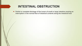
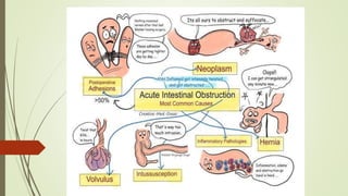
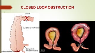
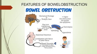
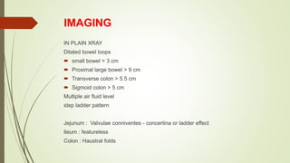
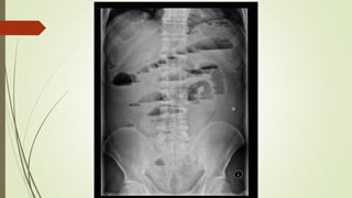
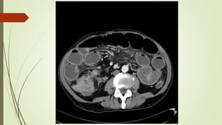
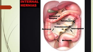
INTESTINAL OBSTRUCTION
PRESENTOR - DR NITU KUMARI
POST GRADUATE, SURGERY
INTESTINAL OBSTRUCTION
 Partial or complete blockage of the lumen of small or large intestine causing an
interruption in the normal flow of intestinal contents along the intestinal tract
 First 12 hours – decreased absorption; after 12 hours – increased secretion
 Distal to intestinal obstruction – normal peristalsis and absorption until it becomes
empty and collapse
STRANGULATION
Blood supply to bowel is compromised and the bowel becomes ischemic
The morbidity and mortality depend on duration of ischemia and its extent
CAUSES
 Direct pressure on bowel wall – Hernial orifices, adhesions/bands
 Interrupted mesenteric blood flow – volvulus, intussusception
 Increased intra-luminal pressure – closed loop obstruction
CLOSED LOOP OBSTRUCTION
 Bowel is obstructed at both proximal and distal points
 Distension is primarily confined to the closed loop
CLOSED LOOP OBSTRUCTION
FEATURES OF BOWELOBSTRUCTION
IMAGING
IN PLAIN XRAY
Dilated bowel loops
 small bowel > 3 cm
 Proximal large bowel > 9 cm
 Transverse colon > 5.5 cm
 Sigmoid colon > 5 cm
Multiple air fluid level
step ladder pattern
Jejunum : Valvulae conniventes - concertina or ladder effect
Ileum : featureless
Colon : Haustral folds
ADHESIONS
 Most common cause of intestinal obstruction
 Lifetime risk of requiring hospital admission following abdominal surgery is 4% and
the risk of requiring a laparotomy is around 2%
 Early fibrinous adhesions - disappear when the cause is removed
 Late fibrous adhesions - mature fibrous tissue; dense
BANDS
 Congenital : eg. obliterated vitellointestinal duct
 A string band following previous bacterial peritonitis
 A portion of greater omentum usually adherent to the parietes
VOLVULUS
 Twisting or axial rotation of a portion of bowel about its mesentery
 >180 degree torsion - obstruction to the lumen
 >360 degree torsion - vascular occlusion in the mesentery
 Bacterial fermentation adds to distension --> increased intraluminal pressure -->
impairs capillary perfusion
 Primary - d/t congenital malrotation of gut, abnormal mesenteric attachments,
congenital bands
 Secondary - d/t rotation of a segment of bowel around an acquired adhesion or
stoma
SIGMOID VOLVULUS
 Rotation nearly always occurs in anti-clockwise direction
 seen most often in elderly patients with chronic constipation
 a/w chronic psychotropic drug use
 Younger patients present earlier and the prognosis is inversely related to duration of
symptoms
 Fulminant presentation - sudden onset, severe pain, early vomitting, rapidly
deteriorating clinical course
 Indolent presentation - insidious onset. slow progressive course, less pain, less
vomiting
INTERNAL HERNIA
A portion of small intestine becomes entrapped in one of the retroperitoneal fossae or in a
congenital mesenteric defect
Potential sites of internal herniation
 The foramen of Winslow
 A defect in the mesentery
 A defect in the transverse mesocolon
 Defects in the broad ligament
 Congenital or acquired diaphragmatic hernia
 Duodenal retroperitoneal fossae
 Cecal/appendiceal retroperitonreal fossa
 Intersigmoid fossa
OBSTRUCTION FROM ENTERIC
STRICTURES
 Tuberculosis or Crohn’s disease
 Subacute or chronic presentation
 Management : Resection and anastomosis. In Crohn’s disease Strictureplasty may
be considered in presence of short multiple strictures without active sepsis
Bolus Obstruction
 Gall stones
 Trichobezoar, phytobezoar
 Stercoliths - a/w jejunal diverticulum or ileal stricture
 Worms – Ascaris lumbricoides
Gall stone ileus
 Elderly
 Fistula formation d/t direct erosion of GB wall by a large gallstone
 MC – Cholecystoduodenal fistula
 2nd MC – Cholecystocolic fistula
 Impaction 60cm proximal to IC valve
 Riggler’s triad : small bowel obstruction, pneumobilia, ectopic gall stones
Management
 Milk the stone proximally
 Intraluminal crushing
 Enterotomy by Longitudinal incision
 Elderly patient with comorbidity : Enterotomy
 Young patient with no comorbidity : Enterotomy, cholecystectomy, CBD
exploration
Bouveret syndrome – gall stones causing duodenal obstruction
 One portion of the gut invaginated into an immediately adjacent sement;
almost invariably the proximal into the distal.
 Peak incidence : between 5 and 10 months of age
CAUSES
 INFANTS : Peyer’s patch enlargement - weaning, common viral pathogens
 OLDER CHILDREN : Meckel’s diverticulum, Polyp, appendix, Henoch-
Schonlein purpura
 ADULTS : Polyp, Tumour, Submucosal lipoma
TYPES : Ileo-ileal, Ileo-colic, Colo-colic
CLINICAL FEATURES
 Intermittent pain
 Red currant jelly stool
 O/E :-
 right iliac fossa empty (Dance sign)
 Sausage or banana shaped lump with concavity towards umbilicus
CLAW SIGN
TARGET SIGN
TREATMENT OF INTUSSUSCEPTION
 Enema/ Air reduction
 Exploratory laparotomy and manual reduction
 Gangrene - Resection and anastamosis
Monitor
 Hematocrit
 Urine output
 RFT
 ABG analysis
 Total count
 Sr electrolytes
 serum D Lactate, CPK-BB, Intestinal fatty acid binding protein
 Nasogastric aspiration/ Tube decompression
 Replacement of IV fluids and electrolytes
 Abdominal girth charting
 Antibiotics
 Inotrope support if the patient develops shock
 Contrast challenge
Surgical Management
 Emergency laparotomy
 Assess
1.The site of obstruction
2.The Nature of Obstruction
3.The viability of the bowel
 Large bore orogastric tube insertion and retrograde milking
 Savage’s decompressor
 Nonviable - Resection and anastamosis
Sigmoid volvulus
 Flexible or Rigid sigmoidoscope - decompression
 Young patients - elective sigmoid colectomy
 Elderly - Resection or sigmoidopexy
 INTUSSUSCEPTION
 ENema/ Air reduction
 Exploratory laparotomy and manual reduction
 Gangrene - Resection and anastamosis
ADHESIONS
 conservative management upto 72 hrs
 At operation, divide only the causative adhesions; limit dissection
 Repair serosal tears; invaginate or resect areas of doubtful viability
 Laparoscopic adhesiolysis
CROHN’S DISEASE - conservative management, strictureplasty if chronic fibrotic
stricture is present
Intra-abdominal abscess
 Percutaneous drainage and abdominal wash
 Laparoscopic wash
Advanced malignant tumours
 Intestinal bypass - Ileostomy/ Colostomy
 Resection and anastamosis
Radiation enteropathy
 corticosteroid and tube decompression
PRINCIPLE OF ILEOSTOMY STOMA
FORMATION
PRINCIPLE OF COLOSTOMY STOMA
FORMATION
PARALYTIC ILEUS
 Failure of transmission of peristatic waves secondary to neuromuscular failure
TYPES
 Post-operative - 24 to 72 hrs
 Infection - intra-abdominal sepsis
 Metabolic - uremia or hypokalemia
 Reflex ileus
 Abdominal Distension - marked
 Colicky pain is not a feature
 Effortless vomiting
TREATMENT OF PARALYTIC ILEUS
 Treat the cause
 Decompression
 Nasogastric suction and delaying oral intake until bowel sounds and passage of
flatus return
 No evidence favouring the use of prokinetic agents
Intestinal Obstruction.pptx

Intestinal Obstruction.pptx

  • 1.
    INTESTINAL OBSTRUCTION PRESENTOR -DR NITU KUMARI POST GRADUATE, SURGERY
  • 3.
    INTESTINAL OBSTRUCTION  Partialor complete blockage of the lumen of small or large intestine causing an interruption in the normal flow of intestinal contents along the intestinal tract
  • 5.
     First 12hours – decreased absorption; after 12 hours – increased secretion  Distal to intestinal obstruction – normal peristalsis and absorption until it becomes empty and collapse
  • 6.
    STRANGULATION Blood supply tobowel is compromised and the bowel becomes ischemic The morbidity and mortality depend on duration of ischemia and its extent CAUSES  Direct pressure on bowel wall – Hernial orifices, adhesions/bands  Interrupted mesenteric blood flow – volvulus, intussusception  Increased intra-luminal pressure – closed loop obstruction
  • 8.
    CLOSED LOOP OBSTRUCTION Bowel is obstructed at both proximal and distal points  Distension is primarily confined to the closed loop
  • 9.
  • 10.
  • 11.
    IMAGING IN PLAIN XRAY Dilatedbowel loops  small bowel > 3 cm  Proximal large bowel > 9 cm  Transverse colon > 5.5 cm  Sigmoid colon > 5 cm Multiple air fluid level step ladder pattern Jejunum : Valvulae conniventes - concertina or ladder effect Ileum : featureless Colon : Haustral folds
  • 14.
    ADHESIONS  Most commoncause of intestinal obstruction  Lifetime risk of requiring hospital admission following abdominal surgery is 4% and the risk of requiring a laparotomy is around 2%  Early fibrinous adhesions - disappear when the cause is removed  Late fibrous adhesions - mature fibrous tissue; dense
  • 15.
    BANDS  Congenital :eg. obliterated vitellointestinal duct  A string band following previous bacterial peritonitis  A portion of greater omentum usually adherent to the parietes
  • 16.
    VOLVULUS  Twisting oraxial rotation of a portion of bowel about its mesentery  >180 degree torsion - obstruction to the lumen  >360 degree torsion - vascular occlusion in the mesentery  Bacterial fermentation adds to distension --> increased intraluminal pressure --> impairs capillary perfusion  Primary - d/t congenital malrotation of gut, abnormal mesenteric attachments, congenital bands  Secondary - d/t rotation of a segment of bowel around an acquired adhesion or stoma
  • 17.
    SIGMOID VOLVULUS  Rotationnearly always occurs in anti-clockwise direction  seen most often in elderly patients with chronic constipation  a/w chronic psychotropic drug use  Younger patients present earlier and the prognosis is inversely related to duration of symptoms  Fulminant presentation - sudden onset, severe pain, early vomitting, rapidly deteriorating clinical course  Indolent presentation - insidious onset. slow progressive course, less pain, less vomiting
  • 20.
    INTERNAL HERNIA A portionof small intestine becomes entrapped in one of the retroperitoneal fossae or in a congenital mesenteric defect Potential sites of internal herniation  The foramen of Winslow  A defect in the mesentery  A defect in the transverse mesocolon  Defects in the broad ligament  Congenital or acquired diaphragmatic hernia  Duodenal retroperitoneal fossae  Cecal/appendiceal retroperitonreal fossa  Intersigmoid fossa
  • 22.
    OBSTRUCTION FROM ENTERIC STRICTURES Tuberculosis or Crohn’s disease  Subacute or chronic presentation  Management : Resection and anastomosis. In Crohn’s disease Strictureplasty may be considered in presence of short multiple strictures without active sepsis
  • 23.
    Bolus Obstruction  Gallstones  Trichobezoar, phytobezoar  Stercoliths - a/w jejunal diverticulum or ileal stricture  Worms – Ascaris lumbricoides
  • 24.
    Gall stone ileus Elderly  Fistula formation d/t direct erosion of GB wall by a large gallstone  MC – Cholecystoduodenal fistula  2nd MC – Cholecystocolic fistula  Impaction 60cm proximal to IC valve  Riggler’s triad : small bowel obstruction, pneumobilia, ectopic gall stones
  • 25.
    Management  Milk thestone proximally  Intraluminal crushing  Enterotomy by Longitudinal incision  Elderly patient with comorbidity : Enterotomy  Young patient with no comorbidity : Enterotomy, cholecystectomy, CBD exploration Bouveret syndrome – gall stones causing duodenal obstruction
  • 26.
     One portionof the gut invaginated into an immediately adjacent sement; almost invariably the proximal into the distal.  Peak incidence : between 5 and 10 months of age CAUSES  INFANTS : Peyer’s patch enlargement - weaning, common viral pathogens  OLDER CHILDREN : Meckel’s diverticulum, Polyp, appendix, Henoch- Schonlein purpura  ADULTS : Polyp, Tumour, Submucosal lipoma TYPES : Ileo-ileal, Ileo-colic, Colo-colic
  • 28.
    CLINICAL FEATURES  Intermittentpain  Red currant jelly stool  O/E :-  right iliac fossa empty (Dance sign)  Sausage or banana shaped lump with concavity towards umbilicus
  • 29.
  • 30.
  • 31.
    TREATMENT OF INTUSSUSCEPTION Enema/ Air reduction  Exploratory laparotomy and manual reduction  Gangrene - Resection and anastamosis
  • 32.
    Monitor  Hematocrit  Urineoutput  RFT  ABG analysis  Total count  Sr electrolytes  serum D Lactate, CPK-BB, Intestinal fatty acid binding protein
  • 33.
     Nasogastric aspiration/Tube decompression  Replacement of IV fluids and electrolytes  Abdominal girth charting  Antibiotics  Inotrope support if the patient develops shock  Contrast challenge
  • 34.
    Surgical Management  Emergencylaparotomy  Assess 1.The site of obstruction 2.The Nature of Obstruction 3.The viability of the bowel  Large bore orogastric tube insertion and retrograde milking  Savage’s decompressor
  • 36.
     Nonviable -Resection and anastamosis Sigmoid volvulus  Flexible or Rigid sigmoidoscope - decompression  Young patients - elective sigmoid colectomy  Elderly - Resection or sigmoidopexy
  • 37.
     INTUSSUSCEPTION  ENema/Air reduction  Exploratory laparotomy and manual reduction  Gangrene - Resection and anastamosis
  • 38.
    ADHESIONS  conservative managementupto 72 hrs  At operation, divide only the causative adhesions; limit dissection  Repair serosal tears; invaginate or resect areas of doubtful viability  Laparoscopic adhesiolysis CROHN’S DISEASE - conservative management, strictureplasty if chronic fibrotic stricture is present
  • 39.
    Intra-abdominal abscess  Percutaneousdrainage and abdominal wash  Laparoscopic wash Advanced malignant tumours  Intestinal bypass - Ileostomy/ Colostomy  Resection and anastamosis Radiation enteropathy  corticosteroid and tube decompression
  • 42.
    PRINCIPLE OF ILEOSTOMYSTOMA FORMATION
  • 43.
    PRINCIPLE OF COLOSTOMYSTOMA FORMATION
  • 44.
    PARALYTIC ILEUS  Failureof transmission of peristatic waves secondary to neuromuscular failure TYPES  Post-operative - 24 to 72 hrs  Infection - intra-abdominal sepsis  Metabolic - uremia or hypokalemia  Reflex ileus
  • 45.
     Abdominal Distension- marked  Colicky pain is not a feature  Effortless vomiting
  • 46.
    TREATMENT OF PARALYTICILEUS  Treat the cause  Decompression  Nasogastric suction and delaying oral intake until bowel sounds and passage of flatus return  No evidence favouring the use of prokinetic agents