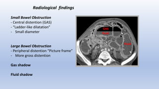
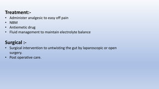
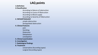
2. Defination :-
Partial or complete blockage the lumen of small or large intestine causing as
interruption in the normal flow of intestinal contents along the intestinal tract is
called as intestinal obstruction.
The block may to complete or incomplete, may be mechanical or paralytic and it
may or may not compromise the vascular supply.
OR
Restriction of the normal flow of intestinal passage is know as intestinal
obstruction
3. Classification
A) According to Nature of obstruction :-
• Dynamic / Mechanical obstruction :-
when something physically block the intestine i.e. adhesions , fibrous
tissue, tumor , volvulus , intussusception
where Peristalsis movement is working against mechanical obstruction.
• Adynamic Obstruction :-
Obstruction due to Neurological cause
Mechanical element is absent
Peristalsis movement
Peristalsis Absent = Paralytic ileus
peristalsis may be present = Pseudo-obstruction
5. ADYNAMIC OBSTRUCTION causes
Small intestine
- Postoperative
- Intra-abdominal abscess or peritonitis
- Mesenteric embolism or thrombosis
Large intestine
- Retroperitoneal hematoma
- Drugs - (those drug effect on muscles &Nerves drug like
antidepressant , opioid analgesic and Antidiarrheal
drug lead to paralytic ileus)
- Hypokalemia (Pseudo Obstruction or Oglivies syndrome)
- Idiopathic (Unknown cause)
6. B) According to Cause of Obstruction :-
1. Inter Mural (In the lumen) -
Due to Food bolus , worms , Foreign bodies , hard stool
Gall stones.
1. Intra Mural (in the intestinal wall ) -
Due to Adhesions , Strictures , Any Growth
2. Extra Mural (Out side the wall) -
Volvulus – Twisting or knotting of the GI tract (intestine)
Intussusception – inversion of one portion of the intestine
with in another
(common in 3rd month to 6 yr age )
Diverticulum – inflamed out pockets of colonic mucosa
through weakness of muscle layer
Obstructed Hernia , Bands , Abscess , Adhesions ,
Compression
Worms Hard Faces
Gall Stone
7. MOST COMMON CAUSES
SMALL INTESTINE
- ADHESIONS
- EXTERNAL HERNIAS (both are more than 75% of cases)
- TB, TUMORS, CONGENITAL………
LARGE INTESTINE
- TUMORS & VOLVULUS (both are 90% of cases )
- DIVERTIDULITIS (rare)
- ADHESIONS (extremely rare if at all)
8. C) According to Blood supply :-
Simple obstruction –
blood supply is not seriously hampered.
Strangulation –
Blood supply is seriously hampered and
it Crete medical emergency condition.
9. D) According to Severity of Obstruction :-
Acute :-
Early symptom is vomiting and colic pain.
mostly affect the small intestine.
sign & symptoms appear very fast .
Chronic:-
Early sign is constipation and pain.
Mostly affect the large intestine.
Sub-Acute :-
It develops due to growth of colon and
suddenly accumulation of food or feacal material.
Abdomen distension is present.
10. PATHOPYSIOLOGY
SIMPLE OBSTRUCTION :
1-ABOVE THE OBSTRUCTION
OBSTRUCTION Peristalsis increases Intestine dilates Reduction in peristaltic strength
Flaccidity and paralysis (protective but late)
2- BELOW THE OBSTRUCTION
NORMAL PERISTALSIS & ABSORBTION Until it becomes empty It contracts & becomes immobile..
11. Strangulated Obstruction :-
Obstruction Distension Venous congestion Edema Progressive arterial
compression Loss of blood supply Gangrene collection of fluid Bacteria
growth Bacteria produce toxins toxins irritate to peritoneal cavity Peritonitis
Septic shock
12. Clinical Features :-
Pain in abdomen :-
Small intestinal obstruction – Central abdomen colic pain
Large intestinal obstruction - Peripheral dull pain
Strangulated obstruction – Continues and severe pain
Paralytic ileus – Pain is absent
13. Vomiting :-
Time of onset :
Early: High small bowel obstruction
Late: Low small bowel obstruction
Delayed or absent: Large bowel obstruction
Nature of vomitus :-
Clear gastric: Pyloric obstruction
Bilious: High small bowel obstruction
Feculent: Low small bowel obstruction or late colonic
14. Abdominal Distension :-
Central distension – Small intestine obstruction
Peripheral Distension – Large intestinal obstruction
Localized Distension – Volvulus Or intussusception
15. Distention of the intestine is caused by accumulation of:
1- GAS
2- FLUIDS GAS
fluids
Distention
16. Gas in the intestine is due to :-
1. Swallowed air
2. Bacterial overgrowth
3. Diffusion from blood
Fluids come from :
1. Saliva
2. Gastric and intestinal juice
3. Bile & Pancreatic secretions
17. Dehydration :-
Dehydration caused by
• Reduce the intake of water
• Doesn’t absorb the fluid from GI system
• Increased loss of water due to vomiting , sweating
• Dry skin , dry tongue , low urine output
• Patient goes hypovolemic shock
• Tachycardia , fever , low BP , mental confusion ,Renal failure, drowsiness
18. General signs
• Rebound tenderness / Blumberg sign – Pain and tenderness that
occurs upon sudden release pressure on the abdomen.
• Absence of bowel sound or sluggish
• Guarding and rigidity of abdominal muscles.
• Tachycardia and fever.
19. Clinical examination
General examination-
Vital signs
Signs of dehydration –tachycardia, hypotension
dry mucus membrane, decreased skin turgor, decreased urine output
Inspection
distension, scars, peristalsis, masses, hernial orifices
Palpation
tenderness, masses, rigidity
Auscultation
high pitched bowel sound or silent abdomen
*Examine rectum for mass, blood, feces or it may be empty in case of complete obstruction
21. Radiological findings
Small Bowel Obstruction
- Central distention (GAS)
- “Ladder-like dilatation”
- Small diameter
Large Bowel Obstruction
- Peripheral distention “Picture frame”
- More gross distention
Gas shadow
Fluid shadow
GAS
Fluid
Fluid
22. • Three main measures-
- GI drainage
-Fluid &Electrolyte replacement
- Relief of obstruction, usually surgical
24. • Some cases will settle by using this conservative regimen, other need surgical
intervention.
• Surgery should be delayed till resuscitation is complete unless signs of
strangulation and evidence of closed-loop obstruction.
• Cases that show reasons for delay should be monitored continuously for 72 hours
in hope of spontaneous resolution e.g. adhesions with radiological findings but no
pain or tenderness
Indication for surgery:
failure of conservative management
tender, irreducible hernia
strangulation
25. • The type of surgical procedure depend upon the cause of obstruction viz
division of bands , adhesiolysis , excision ,or bypass
• Once obstruction relieved, the bowel is inspected for viability, and if non-
viable, resection is required.
Indication of non-viability
1. Absent peristalsis
2. Loss of normal shine
3. Loss of pulsation in mesentery
4. Green or black color of bowel
26. • If in doubt of viability, bowel is wrapped in hot packs for 10 minutes
with increased oxygen and reassessed for viability.
• Resection of non viable gut should be done followed by stoma.
• Sometimes a second look laparotomy is required in 24-48 hours e.g.
multiple ischemic areas.
27. • Most common cause of intestinal obstruction.
• Peritoneal irritation results in local fibrin production, which produce
adhesions.
Causes of adhesions :
• Abdominal operation : anastomosis, raw peritoneal surfaces
• Foreign material: talc, starch, gauze, silk
• Infection: peritonitis, T.B.
• Inflammatory conditions
• Radiation
• enteritis.
28. Prevention:-
• Good surgical technique.
• Washing the peritoneal cavity with saline to remove the clots.
• Minimizing contact with gauze.
• Covering the anastomosis & raw peritoneal surfaces.
Treatment
• Usually conservative treatment is curative. (i.v. rehydration &
nasogastric decompression)
• It should not be prolonged beyond 72 hrs.
Surgery
Division of band.
Minimal adhisiolysis .
29. • It tends to occur in elderly.
• Erosion of large gallstone into duodenum.
• Present with recurrent obstruction.
• X-ray: small bowel obstruction with air in billiary tree.
-may show a radio opaque gall stone.
• Treatment : laparotomy & removal /crushing of stone.
30. • After partial /total gastrectomy.
• Unchewed food can cause obstruction.
• Treatment similar to gall stone.
WORMS
• Ascaris lumbricoides
• Frequently follows initiation of antihelminthic therapy.
• Laparotomy.
31. • One portion of gut becomes invaginated with in adjacent segment.
• Most common in children(3-9 months.)
• Ideopathic-70%
• Associated gastroenteritis/UTI- 30%
Clinical Feauturs :-
• Severe colic pain.
• vomiting as time progress
• blood & mucus (the ‘redcurrant’ jelly stool).
• Abdominal lump (sausage shaped)
• Emptiness in RIF (the sign of Dance).
• Death may occur from bowel obstruction or peritonitis secondary to
gangrene.
32. Treatment :-
• Nasogastric aspiration by Ryle's tube
• IV fluids- volume varies depending on dehydration
• NBM
• Urinary catheter
• Enema if flatus pass
• Flatus not pass & Pain not reduced then post for surgery.
Surgical treatment :-
• Laparotomy for resolve intussusception .
• Irreducible/ gangrenous intussusception: excision of mass & anastomosis.
• Postoperative care
33. VOLVULUS OBSTRUCTION
An Obstruction due to Twisting or Knotting of the intestine
Clinical Features :-
• Abdominal pain
• Abdominal bloating
• Vomiting
• Constipation
• Bloody stool
Diagnostic Investigation
• Barium meal swallow for perform upper GI radiography
• Barium Enema to perform large intestine obstruction
• A Digital rectal examination
• CT Abdomen
34. Treatment:-
• Administer analgesic to easy off pain
• NBM
• Antiemetic drug
• Fluid management to maintain electrolyte balance
Surgical :-
• Surgical intervention to untwisting the gut by laparoscopic or open
surgery.
• Post operative care.
36. LAQ points
1.Definition
2.Classification
According to Nature of obstruction
According to Cause of Obstruction
According to Blood supply
According to Severity of Obstruction
3. PATHOPYSIOLOGY
simple obstruction
Strangulated obstruction
4. Clinical Features
pain
Vomiting
Distension
Dehydration
constipation
5. Clinical examination
6. Investigation
7. Radiological findings
8. Treatment
conservative (According types)
surgical (According types)