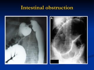
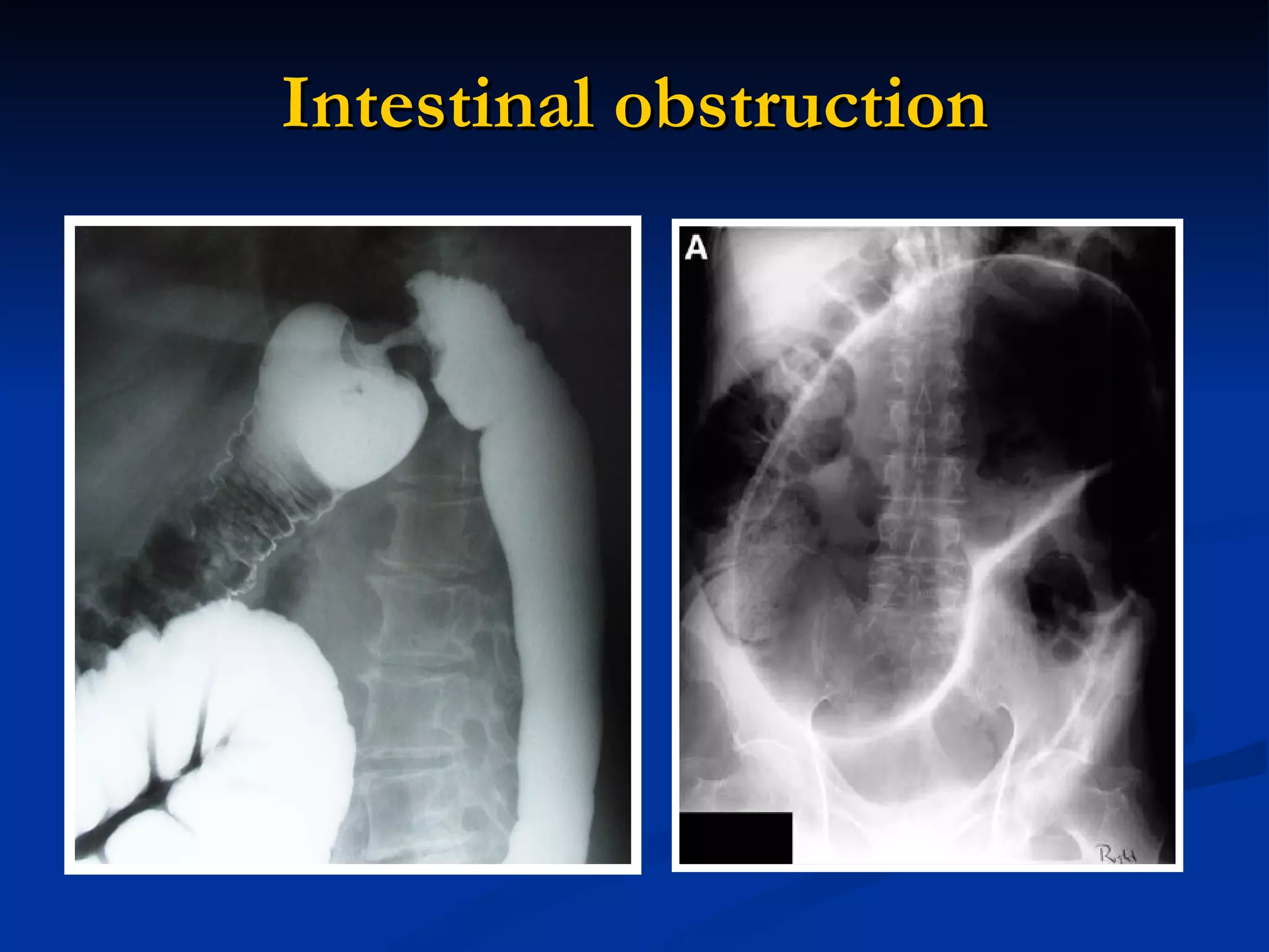
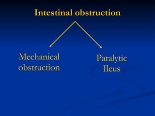
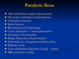
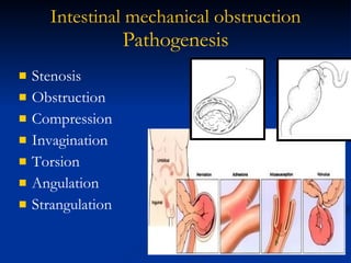
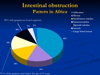
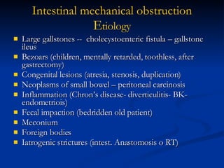
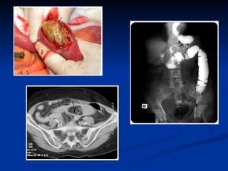
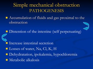
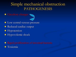
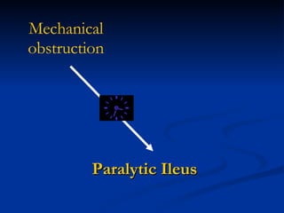
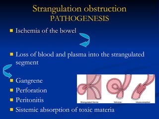
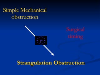
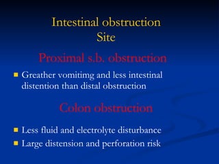
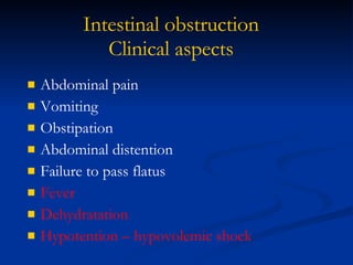
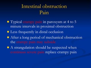
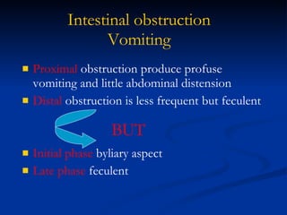
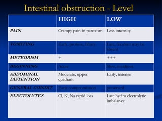
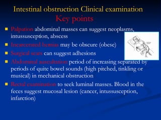
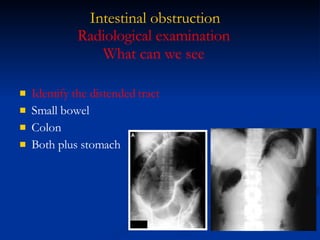
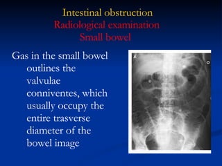
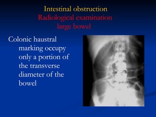
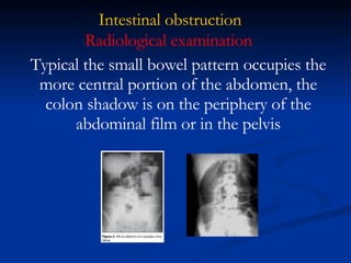
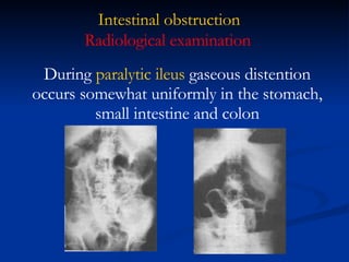
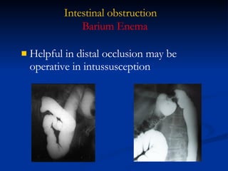
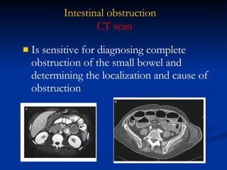
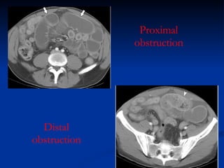
Intestinal   obstruction
Intestinal   obstruction Mechanical   obstruction Paralytic Ileus
Paralytic Ileus After abdominal surgery (laparotomy) Electrolyte imbalances (hypokalemia) Abdominal thrauma Spine fracture Retroperitoneal hemorrhage  Ureter distension – Acute pancreatitis Ischemia of the intestine Drugs (Narcotics, Psychotropics) Peritonitis (ex. Gangrenous cholecystitis) Diabetic coma Extra abdominal infections (Lung) – Sepsis IBD (ulcerative colitis)
Intestinal   mechanical   obstruction Pathogenesis Stenosis Obstruction  Compression Invagination Torsion Angulation Strangulation
Intestinal   obstruction Pattern in Africa 70 % of the patients were below the age of 15 years  80% with gangrenous bowel segments
Large gallstones --  cholecystoenteric fistula – gallstone ileus Bezoars (children, mentally retarded, toothless, after gastrectomy) Congenital lesions (atresia, stenosis, duplication) Neoplasms of small bowel – peritoneal carcinosis Inflammation (Chron’s disease- diverticulitis- BK- endometriois) Fecal impaction (bedridden old patient) Meconium Foreign bodies Iatrogenic strictures (intest. Anastomosis o RT) Intestinal   mechanical   obstruction E tiology
 
 
Accumulation of fluids and gas proximal to the obstruction Simple mechanical   obstruction PATHOGENESIS Distention of the intestine (self perpetuating) Increase intestinal secretion Losses of water, Na, Cl, K, H Dehydratation, ipokalemia, hypochloremia Metabolic alkalosis
Circultory changes Low central venous pressure Reduced cardiac output Hypotention Hypovolemic shock Rapid proliferation of intestinal bacteria   Toxiemia Simple mechanical   obstruction PATHOGENESIS
Paralytic Ileus Mechanical   obstruction
Ischemia of the bowel Strangulation obstruction PATHOGENESIS Loss of blood and plasma into the strangulated segment Gangrene Perforation Peritonitis Sistemic absorption of toxic materia
Strangulation Obstruction Simple Mechanical   obstruction Surgical timing
Intestinal obstruction Site Proximal s.b. obstruction Greather vomitimg and less intestinal distention than distal obstruction Colon obstruction Less fluid and electrolyte disturbance Large distension and perforation risk
Intestinal obstruction Clinical aspects Abdominal pain Vomiting Obstipation Abdominal distention Failure to pass flatus Fever Dehydratation Hypotention – hypovolemic shock
Intestinal obstruction Pain Typical  crampy pain  in paroxysm at 4 to 5 minute intervals in proximal obstruction  Less frequently in distal occlusion After a long period of mechanical obstruction the  crampy pain may subside A strangulation should be suspected when  continuus severe pain  replace crampy pain
Intestinal obstruction Vomiting Proximal  obstruction produce profuse vomiting and little abdominal distension  Distal  obstruction is less frequent but feculent Initial phase  byliary aspect Late phase  feculent BUT
Intestinal obstruction - Level HIGH LOW PAIN Crampy pain in paroxism Less intensity VOMITING Early, profuse, biliary Late, feculent may be absent METEORISM + +++ BEGINNING Acute Slow, insidious ABDOMINAL DISTENTION Moderate, upper quadrant Early, intense GENERAL CONDIT Early compromission preserved ELECTOLYTES Cl, K, Na rapid loss Late hydro electrolytic imbalance
Intestinal obstruction Clinical examination Palpation  abdominal masses can suggest neoplasms, intussusception, abscess  Incarcerated hernias  may be obscure (obese) Surgical scars  can suggest adhesions Abdominal auscultation  period of increasing separated by periods of quite bowel sounds (high pitched, tinkling or musical) in mechanical obstruction Rectal examination  to seek luminal masses. Blood in the feces suggest mucosal lesion (cancer, intussusception, infarction) Key points
Intestinal obstruction Clinical examination Young children and babies  Atresia Volvolus Anal imperforation Meconial ileus Intestinal Duplication Malrotation Intussusception Ascaris infestation Hernia Patient age and sex Adults  Hernia Adhesions Neoplasm Inflammation RT  Endometriosis Gynecological pathology
Intestinal obstruction Abdominal direct X ray exhamination Barium enema CT Endoscopy Ecography (very difficult because of the massive presence of gas) Radiological examination
Intestinal obstruction Gas  abnormally large quantities of gas in the bowel  Multiple gas-fluid levels  in the upright or lateral decubitus position Abdominal direct X ray exhamination
Intestinal obstruction Multiple gas-fluid levels  does not always mean intestinal obstruction Abdominal pain and  diarrhea  can be found in gastroenteritis (cytomegalovirus infection as well as salmonellosis) expecially if profuse watery for 12 or more hours. Abdominal direct X ray exhamination  Remember
Intestinal obstruction Identify the distended tract Small bowel Colon Both plus stomach Radiological examination What can we see
Intestinal obstruction Gas in the small bowel outlines the valvulae conniventes, which usually occupy the entire trasverse diameter of the bowel image Radiological examination Small bowel
Intestinal obstruction Colonic haustral marking occupy only a portion of the transverse diameter of the bowel Radiological examination large bowel
Intestinal obstruction Radiological examination Typical the small bowel pattern occupies the more central portion of the abdomen, the colon shadow is on the periphery of the abdominal film or in the pelvis
Intestinal obstruction Radiological examination During  paralytic ileus  gaseous distention occurs somewhat uniformly in the stomach, small intestine and colon
Intestinal obstruction Helpful in distal occlusion may be operative in intussusception  Barium Enema
Intestinal obstruction Is sensitive for diagnosing complete obstruction of the small bowel and determining the localization and cause of obstruction CT scan
Proximal obstruction Distal obstruction
Intestinal obstruction Hematocrit WBC Electrolytes PCR (C reactive protein) AST -ALT – GGT- LDH Laboratory test
Intestinal obstruction Fluid and electrolytes therapy Intestinal decompression (NG tube) Diuresys monitoring Correct surgical timing for relief of obstruction Treatment
Intestinal obstruction Duration of obstruction Severity of fluid, electrolyte and acid base abnormalities Opportunity to improve vital organ function Consideration of the risk of strangulation Timing of operation depends

Intestinal Obstruction

  • 1.
    Intestinal obstruction
  • 2.
    Intestinal obstruction Mechanical obstruction Paralytic Ileus
  • 3.
    Paralytic Ileus Afterabdominal surgery (laparotomy) Electrolyte imbalances (hypokalemia) Abdominal thrauma Spine fracture Retroperitoneal hemorrhage Ureter distension – Acute pancreatitis Ischemia of the intestine Drugs (Narcotics, Psychotropics) Peritonitis (ex. Gangrenous cholecystitis) Diabetic coma Extra abdominal infections (Lung) – Sepsis IBD (ulcerative colitis)
  • 4.
    Intestinal mechanical obstruction Pathogenesis Stenosis Obstruction Compression Invagination Torsion Angulation Strangulation
  • 5.
    Intestinal obstruction Pattern in Africa 70 % of the patients were below the age of 15 years 80% with gangrenous bowel segments
  • 6.
    Large gallstones -- cholecystoenteric fistula – gallstone ileus Bezoars (children, mentally retarded, toothless, after gastrectomy) Congenital lesions (atresia, stenosis, duplication) Neoplasms of small bowel – peritoneal carcinosis Inflammation (Chron’s disease- diverticulitis- BK- endometriois) Fecal impaction (bedridden old patient) Meconium Foreign bodies Iatrogenic strictures (intest. Anastomosis o RT) Intestinal mechanical obstruction E tiology
  • 7.
  • 8.
  • 9.
    Accumulation of fluidsand gas proximal to the obstruction Simple mechanical obstruction PATHOGENESIS Distention of the intestine (self perpetuating) Increase intestinal secretion Losses of water, Na, Cl, K, H Dehydratation, ipokalemia, hypochloremia Metabolic alkalosis
  • 10.
    Circultory changes Lowcentral venous pressure Reduced cardiac output Hypotention Hypovolemic shock Rapid proliferation of intestinal bacteria Toxiemia Simple mechanical obstruction PATHOGENESIS
  • 11.
  • 12.
    Ischemia of thebowel Strangulation obstruction PATHOGENESIS Loss of blood and plasma into the strangulated segment Gangrene Perforation Peritonitis Sistemic absorption of toxic materia
  • 13.
    Strangulation Obstruction SimpleMechanical obstruction Surgical timing
  • 14.
    Intestinal obstruction SiteProximal s.b. obstruction Greather vomitimg and less intestinal distention than distal obstruction Colon obstruction Less fluid and electrolyte disturbance Large distension and perforation risk
  • 15.
    Intestinal obstruction Clinicalaspects Abdominal pain Vomiting Obstipation Abdominal distention Failure to pass flatus Fever Dehydratation Hypotention – hypovolemic shock
  • 16.
    Intestinal obstruction PainTypical crampy pain in paroxysm at 4 to 5 minute intervals in proximal obstruction Less frequently in distal occlusion After a long period of mechanical obstruction the crampy pain may subside A strangulation should be suspected when continuus severe pain replace crampy pain
  • 17.
    Intestinal obstruction VomitingProximal obstruction produce profuse vomiting and little abdominal distension Distal obstruction is less frequent but feculent Initial phase byliary aspect Late phase feculent BUT
  • 18.
    Intestinal obstruction -Level HIGH LOW PAIN Crampy pain in paroxism Less intensity VOMITING Early, profuse, biliary Late, feculent may be absent METEORISM + +++ BEGINNING Acute Slow, insidious ABDOMINAL DISTENTION Moderate, upper quadrant Early, intense GENERAL CONDIT Early compromission preserved ELECTOLYTES Cl, K, Na rapid loss Late hydro electrolytic imbalance
  • 19.
    Intestinal obstruction Clinicalexamination Palpation abdominal masses can suggest neoplasms, intussusception, abscess Incarcerated hernias may be obscure (obese) Surgical scars can suggest adhesions Abdominal auscultation period of increasing separated by periods of quite bowel sounds (high pitched, tinkling or musical) in mechanical obstruction Rectal examination to seek luminal masses. Blood in the feces suggest mucosal lesion (cancer, intussusception, infarction) Key points
  • 20.
    Intestinal obstruction Clinicalexamination Young children and babies Atresia Volvolus Anal imperforation Meconial ileus Intestinal Duplication Malrotation Intussusception Ascaris infestation Hernia Patient age and sex Adults Hernia Adhesions Neoplasm Inflammation RT Endometriosis Gynecological pathology
  • 21.
    Intestinal obstruction Abdominaldirect X ray exhamination Barium enema CT Endoscopy Ecography (very difficult because of the massive presence of gas) Radiological examination
  • 22.
    Intestinal obstruction Gas abnormally large quantities of gas in the bowel Multiple gas-fluid levels in the upright or lateral decubitus position Abdominal direct X ray exhamination
  • 23.
    Intestinal obstruction Multiplegas-fluid levels does not always mean intestinal obstruction Abdominal pain and diarrhea can be found in gastroenteritis (cytomegalovirus infection as well as salmonellosis) expecially if profuse watery for 12 or more hours. Abdominal direct X ray exhamination Remember
  • 24.
    Intestinal obstruction Identifythe distended tract Small bowel Colon Both plus stomach Radiological examination What can we see
  • 25.
    Intestinal obstruction Gasin the small bowel outlines the valvulae conniventes, which usually occupy the entire trasverse diameter of the bowel image Radiological examination Small bowel
  • 26.
    Intestinal obstruction Colonichaustral marking occupy only a portion of the transverse diameter of the bowel Radiological examination large bowel
  • 27.
    Intestinal obstruction Radiologicalexamination Typical the small bowel pattern occupies the more central portion of the abdomen, the colon shadow is on the periphery of the abdominal film or in the pelvis
  • 28.
    Intestinal obstruction Radiologicalexamination During paralytic ileus gaseous distention occurs somewhat uniformly in the stomach, small intestine and colon
  • 29.
    Intestinal obstruction Helpfulin distal occlusion may be operative in intussusception Barium Enema
  • 30.
    Intestinal obstruction Issensitive for diagnosing complete obstruction of the small bowel and determining the localization and cause of obstruction CT scan
  • 31.
  • 32.
    Intestinal obstruction HematocritWBC Electrolytes PCR (C reactive protein) AST -ALT – GGT- LDH Laboratory test
  • 33.
    Intestinal obstruction Fluidand electrolytes therapy Intestinal decompression (NG tube) Diuresys monitoring Correct surgical timing for relief of obstruction Treatment
  • 34.
    Intestinal obstruction Durationof obstruction Severity of fluid, electrolyte and acid base abnormalities Opportunity to improve vital organ function Consideration of the risk of strangulation Timing of operation depends