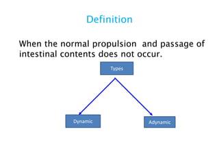
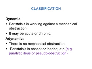
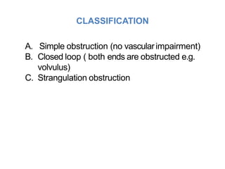
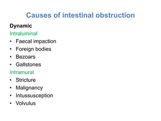
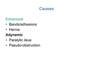
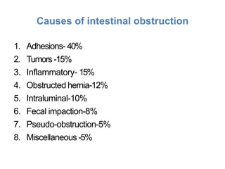
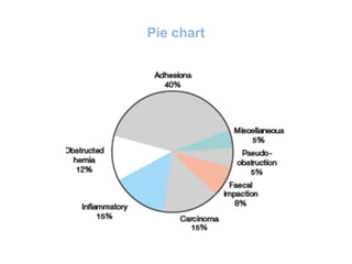
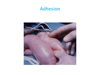
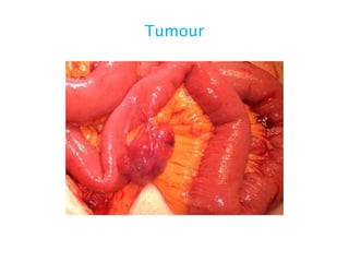
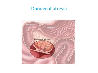
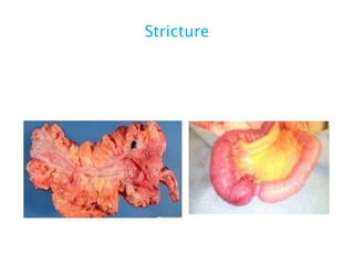
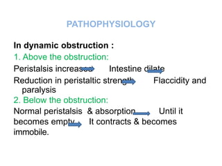
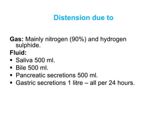
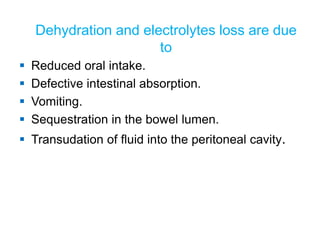
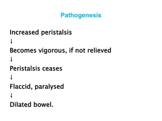
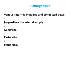
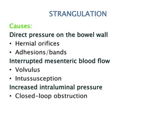
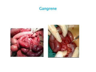
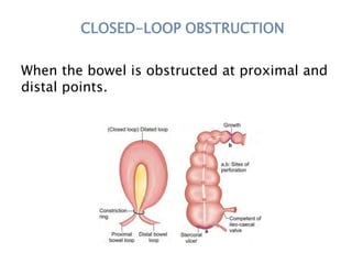
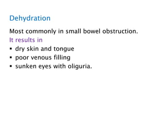
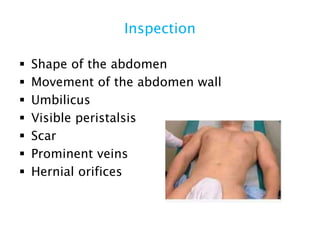
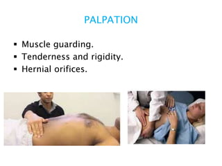
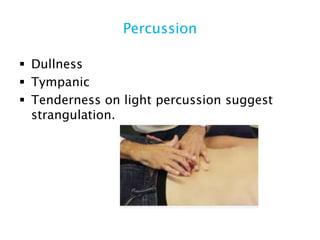
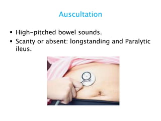
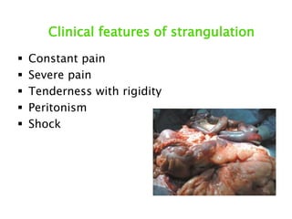
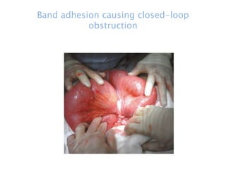
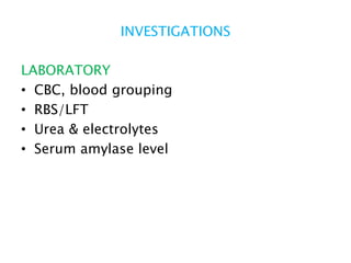
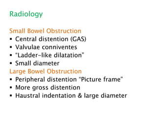
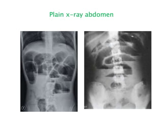
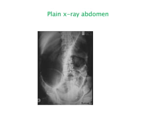
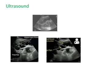
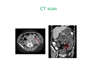
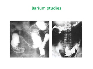
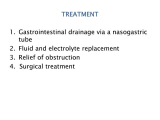
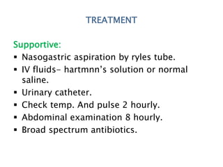
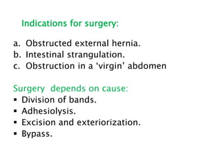
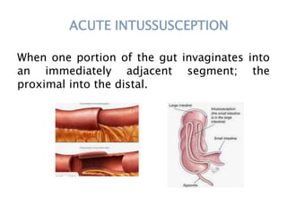
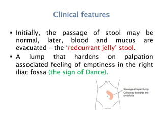
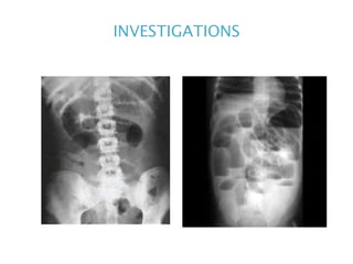
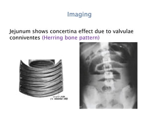
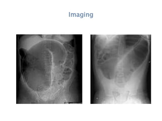
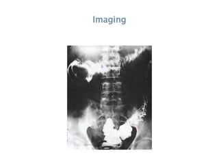
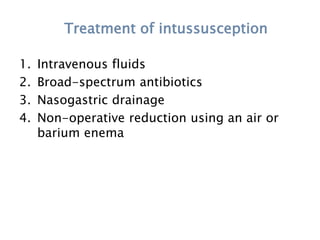
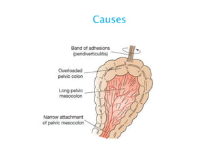
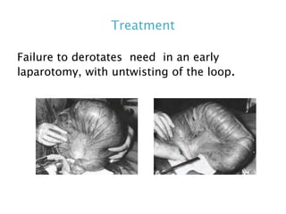
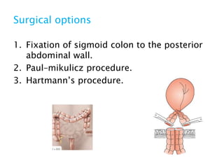
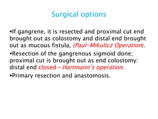
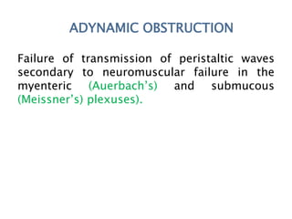
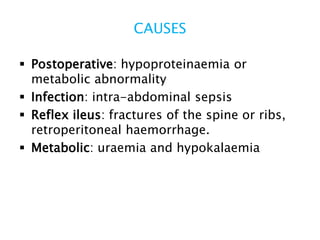
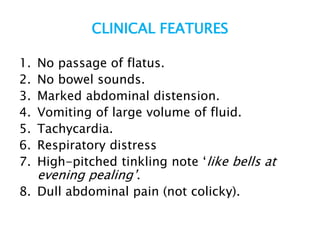
INTESTINAL OBSTRUCTION
LT COL SM SHAHADAT HOSSAIN
MCPS,FCPS(surgery)FCPS(Thoracic Surgery)
Adv Trg on Thoracoscopy CNU, South Korea
Definition
When the normal propulsion and passage of
intestinal contents does not occur.
Dynamic Adynamic
Types
CLASSIFICATION
Dynamic:
 Peristalsis is working against a mechanical
obstruction.
 It may be acute or chronic.
Adynamic:
 There is no mechanical obstruction.
 Peristalsis is absent or inadequate (e.g.
paralytic ileus or pseudo-obstruction).
CLASSIFICATION
A. Simple obstruction (no vascularimpairment)
B. Closed loop ( both ends are obstructed e.g.
volvulus)
C. Strangulation obstruction
Causes of intestinal obstruction
Dynamic
Intraluminal
• Faecal impaction
• Foreign bodies
• Bezoars
• Gallstones
Intramural
• Stricture
• Malignancy
• Intussusception
• Volvulus
Causes
Extramural
• Bands/adhesions
• Hernia
Adynamic
• Paralytic ileus
• Pseudo-obstruction
Causes of intestinal obstruction
1. Adhesions- 40%
2. Tumors -15%
3. Inflammatory- 15%
4. Obstructed hernia-12%
5. Intraluminal-10%
6. Fecal impaction-8%
7. Pseudo-obstruction-5%
8. Miscellaneous -5%
Pie chart
Adhesion
Tumour
Duodenal atresia
Stricture
PATHOPHYSIOLOGY
In dynamic obstruction :
1. Above the obstruction:
Peristalsis increases Intestine dilate
Reduction in peristaltic strength Flaccidity and
paralysis
2. Below the obstruction:
Normal peristalsis & absorption Until it
becomes empty It contracts & becomes
immobile.
Distension due to
Gas: Mainly nitrogen (90%) and hydrogen
sulphide.
Fluid:
 Saliva 500 ml.
 Bile 500 ml.
 Pancreatic secretions 500 ml.
 Gastric secretions 1 litre – all per 24 hours.
Dehydration and electrolytes loss are due
to
 Reduced oral intake.
 Defective intestinal absorption.
 Vomiting.
 Sequestration in the bowel lumen.
 Transudation of fluid into the peritoneal cavity.
Pathogenesis
Increased peristalsis
↓
Becomes vigorous, if not relieved
↓
Peristalsis ceases
↓
Flaccid, paralysed
↓
Dilated bowel.
Pathogenesis
Venous return is impaired and congested bowel
↓
Jeopardizes the arterial supply.
↓
Gangrene.
↓
Perforation.
↓
Peritonitis.
STRANGULATION
Causes:
Direct pressure on the bowel wall
• Hernial orifices
• Adhesions/bands
Interrupted mesenteric blood flow
• Volvulus
• Intussusception
Increased intraluminal pressure
• Closed-loop obstruction
Gangrene
CLOSED-LOOP OBSTRUCTION
When the bowel is obstructed at proximal and
distal points.
SPECIAL TYPES OF
MECHANICAL INTESTINAL
OBSTRUCTION
INTERNAL HERNIA
1. Framen of Winslow
2. Defect in the mesentery
3. Defect in the transverse mesocolon
4. Defects in the broad ligament
5. Diaphragmatic hernia
6. Duodenal retroperitoneal fossae
7. Caecal/appendiceal retroperitoneal
8. Intersigmoid fossa.
OBSTRUCTION FROM ENTERIC
STRICTURES
 Small bowel strictures usually due to
tuberculosis or Crohn’s disease.
 Malignant strictures: lymphoma are
uncommon, whereas carcinoma and
sarcoma are rare.
Stricture
BOLUS OBSTRUCTION
i. Gallstones
ii. Trichobezoars
iii. Phytobezoars
iv. Foreign body
v. Food bolus
vi. Stercoliths
vii. Worms
Gall stone
CLINICAL FEATURES
Vary according to:
● Location of the obstruction;
● Duration of the obstruction;
● Underlying pathology;
● Presence or absence of intestinal
ischaemia.
Pain
 Pain is the first symptom.
 Occurs suddenly and is usually severe.
 Colicky in nature.
 Usually centred on the umbilicus (small
bowel).
 Lower abdomen (large bowel).
Vomiting
More distal the obstruction, longer the
interval between the onset of symptoms and
appearance of nausea and vomiting.
Distension
 In small bowel degree of distension is
dependent on the site of obstruction and
more distal the lesion.
 Distention is much less in high small bowel
obstruction.
 Visible peristalsis may be present
Constipation
This may be
• Absolute (i.e. neither faeces nor flatus is
passed) or
• Relative (where only flatus is passed).
Other manifestations
 Dehydration
 Hypokalaemia
 Pyrexia
 Hypothermia
 Abdominal tenderness
 Bowel sounds:
Dehydration
Most commonly in small bowel obstruction.
It results in
 dry skin and tongue
 poor venous filling
 sunken eyes with oliguria.
PHYSICAL EXAMINATION
 Inspection
 Palpation
 Percussion
 Auscultation
 Rectal examination
Inspection
 Shape of the abdomen
 Movement of the abdomen wall
 Umbilicus
 Visible peristalsis
 Scar
 Prominent veins
 Hernial orifices
PALPATION
 Muscle guarding.
 Tenderness and rigidity.
 Hernial orifices.
Percussion
 Dullness
 Tympanic
 Tenderness on light percussion suggest
strangulation.
Auscultation
 High-pitched bowel sounds.
 Scanty or absent: longstanding and Paralytic
ileus.
RECTAL EXAMINATION
• Presence of mass.e.g.cancer,
Intussuception.
• Feces.
Clinical features of strangulation
 Constant pain
 Severe pain
 Tenderness with rigidity
 Peritonism
 Shock
Band adhesion causing closed-loop
obstruction
INVESTIGATIONS
LABORATORY
• CBC, blood grouping
• RBS/LFT
• Urea & electrolytes
• Serum amylase level
Radiology
Small Bowel Obstruction
 Central distention (GAS)
 Valvulae conniventes
 “Ladder-like dilatation”
 Small diameter
Large Bowel Obstruction
 Peripheral distention “Picture frame”
 More gross distention
 Haustral indentation & large diameter
Plain x-ray abdomen
Plain x-ray abdomen
Ultrasound
CT scan
Barium studies
TREATMENT
1. Gastrointestinal drainage via a nasogastric
tube
2. Fluid and electrolyte replacement
3. Relief of obstruction
4. Surgical treatment
TREATMENT
Supportive:
 Nasogastric aspiration by ryles tube.
 IV fluids- hartmnn’s solution or normal
saline.
 Urinary catheter.
 Check temp. And pulse 2 hourly.
 Abdominal examination 8 hourly.
 Broad spectrum antibiotics.
Indications for surgery:
a. Obstructed external hernia.
b. Intestinal strangulation.
c. Obstruction in a ‘virgin’ abdomen
Surgery depends on cause:
 Division of bands.
 Adhesiolysis.
 Excision and exteriorization.
 Bypass.
Difference between viable and non-viable
intestine
ACUTE INTUSSUSCEPTION
When one portion of the gut invaginates into
an immediately adjacent segment; the
proximal into the distal.
CAUSES
 Most common: children.
 Peak incidence: 5 and 10 months.
Infant:
 90%-idiopathic.
 Upper respiratory tract infection.
 Gastroenteritis.
 Hyperplasia of peyer’s patches.
CAUSES
Children:
 Meckel’s diverticulum
 Polyp, duplication
 Henoch–Schönlein purpura or appendix.
Adult:
 Polyp (e.g. Peutz–jeghers syndrome)
 Submucosal lipoma
 Tumour.
Mechanism of intussusception
Types
Clinical features of intussusception
 Screaming and drawing up of the legs in a
previously well male infant.
 During attacks the child appears pale;
between episodes he may be listless.
 Vomiting may or may not occur but bile-
stained.
Clinical features
 Initially, the passage of stool may be
normal, later, blood and mucus are
evacuated – the ‘redcurrant jelly’ stool.
 A lump that hardens on palpation
associated feeling of emptiness in the right
iliac fossa (the sign of Dance).
Clinical features
On rectal examination:
 Blood-stained mucus
 The apex of intussusception may be
palpable.
DIFFERENTIAL DIAGNOSIS
1. Acute gastroenteritis
Abdominal pain, vomiting with occasional
blood and mucus in the stool.
2. Henoch–schönlein purpura
Characteristic rash and abdominal pain.
3. Rectal prolapse
The projecting mucosa can be felt in
continuity with the perianal skin whereas in
intussusception the finger may pass
indefinitely into the depths of a sulcus.
INVESTIGATIONS
Imaging
Jejunum shows concertina effect due to valvulae
conniventes (Herring bone pattern)
Imaging
Imaging
Triad of small bowel obstruction in plain
x-ray
1. Dilated small bowel loops > 3 cm.
2. Multiple air fluid levels in erect x-ray.
3. Paucity of air in the colon.
Treatment of intussusception
1. Intravenous fluids
2. Broad-spectrum antibiotics
3. Nasogastric drainage
4. Non-operative reduction using an air or
barium enema
SURGERY
 Transverse right sided abdominal incision
 Reduction is achieved by gently
compressing the most distal part of the
intussusception toward its origin not to
pull.
Treatment of sigmoid volvulus
 NG suction
 IV fluids
 Antibiotics
 Catheterisation
 Sigmoidoscopy and insertion of a flatus
tube to allow deflation of the gut.
VOLVULUS
 A volvulus is a twisting or axial rotation of a
portion of bowel about its mesentery.
 The rotation causes obstruction to the
lumen (>180° torsion) and if tight enough
also causes vascular occlusion in the
mesentery (>360° torsion).
Types of Volvulus
Primary:
 Volvulus neonatorum
 Caecal volvulus
 Sigmoid volvulus
Secondary: Common and is due to rotation of
a segment of bowel around an acquired
adhesion or stoma.
SIGMOID VOLVULUS
 Rotation nearly always occurs in the
anticlockwise direction.
Predisposing factors:
 Elderly patient
 High-residue diet
 Constipation
Causes
PRESENTATION
Fulminant:
 Sudden onset
 Severe pain
 Early vomiting, rapidly deteriorating clinical
course.
Indolent:
 Insidious onset,
 Slow progressive course
 Less pain, late vomiting.
Treatment of sigmoid volvulus
 NG suction
 IV fluids
 Antibiotics
 Catheterisation
 Sigmoidoscopy and insertion of a flatus
tube to allow deflation of the gut.
Treatment
Failure to derotates need in an early
laparotomy, with untwisting of the loop.
pic
Surgical options
1. Fixation of sigmoid colon to the posterior
abdominal wall.
2. Paul–mikulicz procedure.
3. Hartmann’s procedure.
Surgical options
A flatus tube or Sigmoidoscope is passed in
operation theatre.
If derotation does not occur, laparotomy
If viable, it can be fixed to the lateral wall of
abdomen or pelvis—sigmoidopexy.
Surgical options
If gangrene, it is resected and proximal cut end
brought out as colostomy and distal end brought
out as mucous fistula, (Paul-Mikulicz Operation).
Resection of the gangrenous sigmoid done;
proximal cut is brought out as end colostomy:
distal end closed—Hartmann’s operation.
Primary resection and anastomosis.
ADYNAMIC OBSTRUCTION
Failure of transmission of peristaltic waves
secondary to neuromuscular failure in the
myenteric (Auerbach’s) and submucous
(Meissner’s) plexuses).
CAUSES
 Postoperative: hypoproteinaemia or
metabolic abnormality
 Infection: intra-abdominal sepsis
 Reflex ileus: fractures of the spine or ribs,
retroperitoneal haemorrhage.
 Metabolic: uraemia and hypokalaemia
CLINICAL FEATURES
1. No passage of flatus.
2. No bowel sounds.
3. Marked abdominal distension.
4. Vomiting of large volume of fluid.
5. Tachycardia.
6. Respiratory distress
7. High-pitched tinkling note ‘like bells at
evening pealing’.
8. Dull abdominal pain (not colicky).
INVESTIGATIONS
 Serum electrolytes
 X-ray abdomen.
 Ultrasound abdomen e.g. sepsis
TREATMENT
a. Nasogastric aspiration.
b. IV fluids.
c. Electrolyte management.
d. Catheterisation and urine output measurement.
e. The primary cause is treated.

INTESTINAL OBSTRUCTION

  • 1.
    INTESTINAL OBSTRUCTION LT COLSM SHAHADAT HOSSAIN MCPS,FCPS(surgery)FCPS(Thoracic Surgery) Adv Trg on Thoracoscopy CNU, South Korea
  • 2.
    Definition When the normalpropulsion and passage of intestinal contents does not occur. Dynamic Adynamic Types
  • 3.
    CLASSIFICATION Dynamic:  Peristalsis isworking against a mechanical obstruction.  It may be acute or chronic. Adynamic:  There is no mechanical obstruction.  Peristalsis is absent or inadequate (e.g. paralytic ileus or pseudo-obstruction).
  • 4.
    CLASSIFICATION A. Simple obstruction(no vascularimpairment) B. Closed loop ( both ends are obstructed e.g. volvulus) C. Strangulation obstruction
  • 5.
    Causes of intestinalobstruction Dynamic Intraluminal • Faecal impaction • Foreign bodies • Bezoars • Gallstones Intramural • Stricture • Malignancy • Intussusception • Volvulus
  • 6.
  • 7.
    Causes of intestinalobstruction 1. Adhesions- 40% 2. Tumors -15% 3. Inflammatory- 15% 4. Obstructed hernia-12% 5. Intraluminal-10% 6. Fecal impaction-8% 7. Pseudo-obstruction-5% 8. Miscellaneous -5%
  • 8.
  • 9.
  • 10.
  • 11.
  • 12.
  • 13.
    PATHOPHYSIOLOGY In dynamic obstruction: 1. Above the obstruction: Peristalsis increases Intestine dilate Reduction in peristaltic strength Flaccidity and paralysis 2. Below the obstruction: Normal peristalsis & absorption Until it becomes empty It contracts & becomes immobile.
  • 14.
    Distension due to Gas:Mainly nitrogen (90%) and hydrogen sulphide. Fluid:  Saliva 500 ml.  Bile 500 ml.  Pancreatic secretions 500 ml.  Gastric secretions 1 litre – all per 24 hours.
  • 15.
    Dehydration and electrolytesloss are due to  Reduced oral intake.  Defective intestinal absorption.  Vomiting.  Sequestration in the bowel lumen.  Transudation of fluid into the peritoneal cavity.
  • 16.
    Pathogenesis Increased peristalsis ↓ Becomes vigorous,if not relieved ↓ Peristalsis ceases ↓ Flaccid, paralysed ↓ Dilated bowel.
  • 17.
    Pathogenesis Venous return isimpaired and congested bowel ↓ Jeopardizes the arterial supply. ↓ Gangrene. ↓ Perforation. ↓ Peritonitis.
  • 18.
    STRANGULATION Causes: Direct pressure onthe bowel wall • Hernial orifices • Adhesions/bands Interrupted mesenteric blood flow • Volvulus • Intussusception Increased intraluminal pressure • Closed-loop obstruction
  • 19.
  • 20.
    CLOSED-LOOP OBSTRUCTION When thebowel is obstructed at proximal and distal points.
  • 21.
    SPECIAL TYPES OF MECHANICALINTESTINAL OBSTRUCTION
  • 22.
    INTERNAL HERNIA 1. Framenof Winslow 2. Defect in the mesentery 3. Defect in the transverse mesocolon 4. Defects in the broad ligament 5. Diaphragmatic hernia 6. Duodenal retroperitoneal fossae 7. Caecal/appendiceal retroperitoneal 8. Intersigmoid fossa.
  • 23.
    OBSTRUCTION FROM ENTERIC STRICTURES Small bowel strictures usually due to tuberculosis or Crohn’s disease.  Malignant strictures: lymphoma are uncommon, whereas carcinoma and sarcoma are rare.
  • 24.
  • 25.
    BOLUS OBSTRUCTION i. Gallstones ii.Trichobezoars iii. Phytobezoars iv. Foreign body v. Food bolus vi. Stercoliths vii. Worms
  • 26.
  • 27.
    CLINICAL FEATURES Vary accordingto: ● Location of the obstruction; ● Duration of the obstruction; ● Underlying pathology; ● Presence or absence of intestinal ischaemia.
  • 28.
    Pain  Pain isthe first symptom.  Occurs suddenly and is usually severe.  Colicky in nature.  Usually centred on the umbilicus (small bowel).  Lower abdomen (large bowel).
  • 29.
    Vomiting More distal theobstruction, longer the interval between the onset of symptoms and appearance of nausea and vomiting.
  • 30.
    Distension  In smallbowel degree of distension is dependent on the site of obstruction and more distal the lesion.  Distention is much less in high small bowel obstruction.  Visible peristalsis may be present
  • 31.
    Constipation This may be •Absolute (i.e. neither faeces nor flatus is passed) or • Relative (where only flatus is passed).
  • 32.
    Other manifestations  Dehydration Hypokalaemia  Pyrexia  Hypothermia  Abdominal tenderness  Bowel sounds:
  • 33.
    Dehydration Most commonly insmall bowel obstruction. It results in  dry skin and tongue  poor venous filling  sunken eyes with oliguria.
  • 34.
    PHYSICAL EXAMINATION  Inspection Palpation  Percussion  Auscultation  Rectal examination
  • 35.
    Inspection  Shape ofthe abdomen  Movement of the abdomen wall  Umbilicus  Visible peristalsis  Scar  Prominent veins  Hernial orifices
  • 36.
    PALPATION  Muscle guarding. Tenderness and rigidity.  Hernial orifices.
  • 37.
    Percussion  Dullness  Tympanic Tenderness on light percussion suggest strangulation.
  • 38.
    Auscultation  High-pitched bowelsounds.  Scanty or absent: longstanding and Paralytic ileus.
  • 39.
    RECTAL EXAMINATION • Presenceof mass.e.g.cancer, Intussuception. • Feces.
  • 40.
    Clinical features ofstrangulation  Constant pain  Severe pain  Tenderness with rigidity  Peritonism  Shock
  • 41.
    Band adhesion causingclosed-loop obstruction
  • 42.
    INVESTIGATIONS LABORATORY • CBC, bloodgrouping • RBS/LFT • Urea & electrolytes • Serum amylase level
  • 43.
    Radiology Small Bowel Obstruction Central distention (GAS)  Valvulae conniventes  “Ladder-like dilatation”  Small diameter Large Bowel Obstruction  Peripheral distention “Picture frame”  More gross distention  Haustral indentation & large diameter
  • 44.
  • 45.
  • 46.
  • 47.
  • 48.
  • 49.
    TREATMENT 1. Gastrointestinal drainagevia a nasogastric tube 2. Fluid and electrolyte replacement 3. Relief of obstruction 4. Surgical treatment
  • 50.
    TREATMENT Supportive:  Nasogastric aspirationby ryles tube.  IV fluids- hartmnn’s solution or normal saline.  Urinary catheter.  Check temp. And pulse 2 hourly.  Abdominal examination 8 hourly.  Broad spectrum antibiotics.
  • 51.
    Indications for surgery: a.Obstructed external hernia. b. Intestinal strangulation. c. Obstruction in a ‘virgin’ abdomen Surgery depends on cause:  Division of bands.  Adhesiolysis.  Excision and exteriorization.  Bypass.
  • 52.
    Difference between viableand non-viable intestine
  • 53.
    ACUTE INTUSSUSCEPTION When oneportion of the gut invaginates into an immediately adjacent segment; the proximal into the distal.
  • 54.
    CAUSES  Most common:children.  Peak incidence: 5 and 10 months. Infant:  90%-idiopathic.  Upper respiratory tract infection.  Gastroenteritis.  Hyperplasia of peyer’s patches.
  • 55.
    CAUSES Children:  Meckel’s diverticulum Polyp, duplication  Henoch–Schönlein purpura or appendix. Adult:  Polyp (e.g. Peutz–jeghers syndrome)  Submucosal lipoma  Tumour.
  • 56.
  • 57.
  • 58.
    Clinical features ofintussusception  Screaming and drawing up of the legs in a previously well male infant.  During attacks the child appears pale; between episodes he may be listless.  Vomiting may or may not occur but bile- stained.
  • 59.
    Clinical features  Initially,the passage of stool may be normal, later, blood and mucus are evacuated – the ‘redcurrant jelly’ stool.  A lump that hardens on palpation associated feeling of emptiness in the right iliac fossa (the sign of Dance).
  • 60.
    Clinical features On rectalexamination:  Blood-stained mucus  The apex of intussusception may be palpable.
  • 61.
    DIFFERENTIAL DIAGNOSIS 1. Acutegastroenteritis Abdominal pain, vomiting with occasional blood and mucus in the stool. 2. Henoch–schönlein purpura Characteristic rash and abdominal pain. 3. Rectal prolapse The projecting mucosa can be felt in continuity with the perianal skin whereas in intussusception the finger may pass indefinitely into the depths of a sulcus.
  • 62.
  • 63.
    Imaging Jejunum shows concertinaeffect due to valvulae conniventes (Herring bone pattern)
  • 64.
  • 65.
  • 66.
    Triad of smallbowel obstruction in plain x-ray 1. Dilated small bowel loops > 3 cm. 2. Multiple air fluid levels in erect x-ray. 3. Paucity of air in the colon.
  • 67.
    Treatment of intussusception 1.Intravenous fluids 2. Broad-spectrum antibiotics 3. Nasogastric drainage 4. Non-operative reduction using an air or barium enema
  • 68.
    SURGERY  Transverse rightsided abdominal incision  Reduction is achieved by gently compressing the most distal part of the intussusception toward its origin not to pull.
  • 69.
    Treatment of sigmoidvolvulus  NG suction  IV fluids  Antibiotics  Catheterisation  Sigmoidoscopy and insertion of a flatus tube to allow deflation of the gut.
  • 70.
    VOLVULUS  A volvulusis a twisting or axial rotation of a portion of bowel about its mesentery.  The rotation causes obstruction to the lumen (>180° torsion) and if tight enough also causes vascular occlusion in the mesentery (>360° torsion).
  • 71.
    Types of Volvulus Primary: Volvulus neonatorum  Caecal volvulus  Sigmoid volvulus Secondary: Common and is due to rotation of a segment of bowel around an acquired adhesion or stoma.
  • 72.
    SIGMOID VOLVULUS  Rotationnearly always occurs in the anticlockwise direction. Predisposing factors:  Elderly patient  High-residue diet  Constipation
  • 73.
  • 74.
    PRESENTATION Fulminant:  Sudden onset Severe pain  Early vomiting, rapidly deteriorating clinical course. Indolent:  Insidious onset,  Slow progressive course  Less pain, late vomiting.
  • 75.
    Treatment of sigmoidvolvulus  NG suction  IV fluids  Antibiotics  Catheterisation  Sigmoidoscopy and insertion of a flatus tube to allow deflation of the gut.
  • 76.
    Treatment Failure to derotatesneed in an early laparotomy, with untwisting of the loop.
  • 77.
  • 78.
    Surgical options 1. Fixationof sigmoid colon to the posterior abdominal wall. 2. Paul–mikulicz procedure. 3. Hartmann’s procedure.
  • 79.
    Surgical options A flatustube or Sigmoidoscope is passed in operation theatre. If derotation does not occur, laparotomy If viable, it can be fixed to the lateral wall of abdomen or pelvis—sigmoidopexy.
  • 80.
    Surgical options If gangrene,it is resected and proximal cut end brought out as colostomy and distal end brought out as mucous fistula, (Paul-Mikulicz Operation). Resection of the gangrenous sigmoid done; proximal cut is brought out as end colostomy: distal end closed—Hartmann’s operation. Primary resection and anastomosis.
  • 81.
    ADYNAMIC OBSTRUCTION Failure oftransmission of peristaltic waves secondary to neuromuscular failure in the myenteric (Auerbach’s) and submucous (Meissner’s) plexuses).
  • 82.
    CAUSES  Postoperative: hypoproteinaemiaor metabolic abnormality  Infection: intra-abdominal sepsis  Reflex ileus: fractures of the spine or ribs, retroperitoneal haemorrhage.  Metabolic: uraemia and hypokalaemia
  • 83.
    CLINICAL FEATURES 1. Nopassage of flatus. 2. No bowel sounds. 3. Marked abdominal distension. 4. Vomiting of large volume of fluid. 5. Tachycardia. 6. Respiratory distress 7. High-pitched tinkling note ‘like bells at evening pealing’. 8. Dull abdominal pain (not colicky).
  • 84.
    INVESTIGATIONS  Serum electrolytes X-ray abdomen.  Ultrasound abdomen e.g. sepsis
  • 85.
    TREATMENT a. Nasogastric aspiration. b.IV fluids. c. Electrolyte management. d. Catheterisation and urine output measurement. e. The primary cause is treated.