INTESTINAL OBSTRUCTION
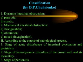
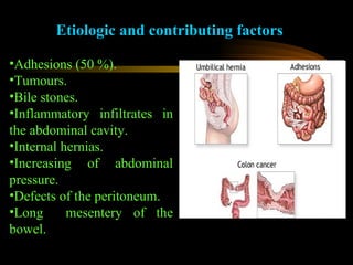
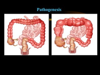
Intestinal obstruction is a complete or partial disturbance of intestinal evacuation and peristalsis resulting from various causes which manifests by specific clinical course and morphologic changes of involved part of the bowel Intestinal obstruction occurs   approximately in 9.4 % among urgent abdominal pathology, consisting 1.2 % of all surgical diseases.
Classification (by D.P.Chuhrienko) 1. Dynamic intestinal obstruction: a) paralytic; b) spastic. 2. Mechanical intestinal obstruction: a) strangulation; b) obturation; c) mixed (invagination). II. According to the course of pathological process. 1. Stage of acute disturbance of intestinal evacuation and peristalsis. 2. Stage of hemodynamic disorders of the bowel wall and its mesentery. 3. Stage of peritonitis.
Etiologic and contributing factors Adhesions (50 %). Tumours. Bile stones. Inflammatory infiltrates in the abdominal cavity. Internal hernias. Increasing of abdominal pressure. Defects of the peritoneum. Long  mesentery of the bowel.
Pathogenesis   More higher the obstruction, more severe the pathologic changes Transudation of intestinal juice Vomiting Intestinal block Accumulation of intestinal contents Distention of intestinal wall Loss of electrolytes, dehydratation, loss of potassium, loss of proteins Necrosis of the bowel Peritonitis  Intoxication, hemodynamic disturbances
Pathogenesis
Peculiarities of strangulated intestinal obstruction Transfusion of large quantity of proteins; Transfusion of erythrocytes and decreasing of  volume of circulating blood; Severe dehydratation, caused by additional edema of the bowel wall; Rapid necrosis of intestinal wall with subsequent peritonitis and intoxication.
Clinical manifestation It depends on the level of the block, type and degree of obstruction and its cause. 1. Acute onset of the disease. 2. Periodic acute diffuse pain   of wavelike character which results in shock. 3. Constant vomiting and nausea without any relief. 4. Signs of dehydratation and intoxication  (The patient looks anxious, with drawn features, hollowed-eyed, his lips and tongue are dry, with brown fur) . 5. Retention of stool and gases.
Objective examination 1. Signs of shock. 2. Distended and asymmetric abdomen. 3. Splashing sound (Sklyarov’s sign). 4. Increased peristalsis in early period with further absence. 5. Wahl’s sign - high  tympanic sound over the distended bowel. 6. PR: empty and distended anus and rectal ampoule - (Grekov’s sign). 7. In intestinal strangulation and advanced cases of obstruction - peritoneal signs.
X-ray examination 1. Kloiber's cups (air-fluid level) 2. Intestinal pneumatisation
 
 
Differential diagnostics of acute intestinal obstruction with perforative peptic ulcer Periodic acute diffuse pain Constant vomiting and nausea without any relief Retention of stool and gases Abdominal distension On X-ray of the   abdomen Kloiber's cups (air-fluid levels) Splashing sound, increased peristalsis   Sharp acute diffuse pain Ulcerative anamnesis Absence of hepatic dullness On X-ray of the abdomen air above the liver (air sickle) Rigidity of anterior abdominal wall
Differential diagnostics of acute intestinal obstruction with acute pancreatitis Periodic acute diffuse pain Constant vomiting and nausea without any relief Retention of stool and gases Abdominal distension On X-ray of the   abdomen Kloiber's cups (air-fluid levels) Splashing sound, increased peristalsis   Sharp acute constant girdle pain with irradiation into left scapule Vomiting and nausea without any relief Mayo-Robson symptom Increased serum amylase Increased urinary diastase No retention of stool and gases Abdominal distension only in advanced cases. No increased peristalsis
Differential diagnostics of acute intestinal obstruction with mesenteric   thrombosis Periodic acute diffuse pain Constant vomiting and nausea without any relief Retention of stool and gases Abdominal distension On X-ray of the   abdomen Kloiber's cups (air-fluid levels) Splashing sound, increased peristalsis   Sharp acute constant pain which results in shock Abdominal distension. Decreased peristalsis   Blood stool Concomitant cardiac pathology Peritoneal signs
Differential diagnostics of acute intestinal obstruction with acute cholecystitis Periodic acute diffuse pain Constant vomiting and nausea without any relief Retention of stool and gases Abdominal distension On X-ray of the   abdomen Kloiber's cups (air-fluid levels) Splashing sound, increased peristalsis   Acute pain in a right hypohondrium with irradiation to the scapula Muscular tenderness in a right hypohondrium  Vomiting by bile and nausea without any relief Ortner's symptom, phrenic symptom, Murphy’s sign Increased serum bilirubin
Conservative treatment Conservative treatment is indicated only in: 1. Adhesive obstruction without signs of strangulation. 2. Initial stages of invagination. 3. Initial stages of low obturation Gastric decompression Siphon enema Paranephral procaine block Ganglio- and sympatholytics Correction of water-electrolyte balance
Surgical treatment is indicated only if no improvement during 3-4 hours of conservative treatment  Wide laparotomy Procaine block of mesenteric root Revision of intestine and detecting of the level and cause of obstruction Decompression of intestine proximal to the obstruction Assessing of viability of the bowel  (peristalsis, colour, vascular pulsation) Removal of the obstruction  (division of adhesions, intestinal resection, collateral anastomosis) External drainage of intestine   (nasogastrointestinal intubation, rectal tube)
 
Strangulation intestinal obstruction 1. Volvulus, (torsion). 2. Nodulus (knots). 3. Hernial strangulation (incarceration). 4.Invagination (refers to mixed forms of intestinal obstruction) Causes Elongation of intestinal loop. Increasing of abdominal pressure. Long  mesentery of the bowel. Cicatrical shortening of mesenteric root. Adhesions Extensive functional overload of the bowel.
Volvulus (torsion) Small intestine volvulus Clinically manifests by high strangulation intestinal obstruction   Cecal volvulus, sigmoid volvulus Clinically: low strangulation intestinal obstruction   asymmetric abdomen    by palpation enlarged and displaced colon (like balloon)   retracted right or left iliac region Treatment:   detorsion, division of adhesions, cecopexia, colon resection
 
Nodulus It is the most severe type of strangulation with manifestation of  a high strangulated obstruction which rapidly results in shock, bowel necrosis and peritonitis  Treatment:   very short preoperative period (less 1 hour) untie of the bowel node (till 4-5 hours from the onset) resection of the bowel
Invagination It is the insertion of one part of the bowel into the lumen of another  Treatment:   desinvagination or resection Clinically:   signs of obturation and in advanced cases the strangulation ileus elastic, painful, tumourlike formation in the abdomen blood in stool (or during rectal examination)
 
Paralytic ileus Abdominal peritonitis mesenteric thromboses hemoperitoneum pancreatitis postoperative ileus Retroperitoneal phlegmone hematoma renal colic spinal trauma Other cranial trauma acidosis diminished potassium hypoproteinemia uremia
Spastic ileus Abdominal hepatic  colic   ascaridosis  Retroperitoneal renal colic Other lead poisonong
Treatment of dynamic ileus 1. Cholynomymetics  (Proserin, ubretid). 2. Intravenous infusion of hypertonic solution  (10 % NaCl). 3. Hypertonic enema. 4. Oil enema. 5. Gastric decompression. 6. Paranephral novocaine block. 7. Ultrasound stimulation.

Intestinal obstruction

  • 1.
  • 2.
    Intestinal obstruction isa complete or partial disturbance of intestinal evacuation and peristalsis resulting from various causes which manifests by specific clinical course and morphologic changes of involved part of the bowel Intestinal obstruction occurs approximately in 9.4 % among urgent abdominal pathology, consisting 1.2 % of all surgical diseases.
  • 3.
    Classification (by D.P.Chuhrienko)1. Dynamic intestinal obstruction: a) paralytic; b) spastic. 2. Mechanical intestinal obstruction: a) strangulation; b) obturation; c) mixed (invagination). II. According to the course of pathological process. 1. Stage of acute disturbance of intestinal evacuation and peristalsis. 2. Stage of hemodynamic disorders of the bowel wall and its mesentery. 3. Stage of peritonitis.
  • 4.
    Etiologic and contributingfactors Adhesions (50 %). Tumours. Bile stones. Inflammatory infiltrates in the abdominal cavity. Internal hernias. Increasing of abdominal pressure. Defects of the peritoneum. Long mesentery of the bowel.
  • 5.
    Pathogenesis More higher the obstruction, more severe the pathologic changes Transudation of intestinal juice Vomiting Intestinal block Accumulation of intestinal contents Distention of intestinal wall Loss of electrolytes, dehydratation, loss of potassium, loss of proteins Necrosis of the bowel Peritonitis Intoxication, hemodynamic disturbances
  • 6.
  • 7.
    Peculiarities of strangulatedintestinal obstruction Transfusion of large quantity of proteins; Transfusion of erythrocytes and decreasing of volume of circulating blood; Severe dehydratation, caused by additional edema of the bowel wall; Rapid necrosis of intestinal wall with subsequent peritonitis and intoxication.
  • 8.
    Clinical manifestation Itdepends on the level of the block, type and degree of obstruction and its cause. 1. Acute onset of the disease. 2. Periodic acute diffuse pain of wavelike character which results in shock. 3. Constant vomiting and nausea without any relief. 4. Signs of dehydratation and intoxication (The patient looks anxious, with drawn features, hollowed-eyed, his lips and tongue are dry, with brown fur) . 5. Retention of stool and gases.
  • 9.
    Objective examination 1.Signs of shock. 2. Distended and asymmetric abdomen. 3. Splashing sound (Sklyarov’s sign). 4. Increased peristalsis in early period with further absence. 5. Wahl’s sign - high tympanic sound over the distended bowel. 6. PR: empty and distended anus and rectal ampoule - (Grekov’s sign). 7. In intestinal strangulation and advanced cases of obstruction - peritoneal signs.
  • 10.
    X-ray examination 1.Kloiber's cups (air-fluid level) 2. Intestinal pneumatisation
  • 11.
  • 12.
  • 13.
    Differential diagnostics ofacute intestinal obstruction with perforative peptic ulcer Periodic acute diffuse pain Constant vomiting and nausea without any relief Retention of stool and gases Abdominal distension On X-ray of the abdomen Kloiber's cups (air-fluid levels) Splashing sound, increased peristalsis Sharp acute diffuse pain Ulcerative anamnesis Absence of hepatic dullness On X-ray of the abdomen air above the liver (air sickle) Rigidity of anterior abdominal wall
  • 14.
    Differential diagnostics ofacute intestinal obstruction with acute pancreatitis Periodic acute diffuse pain Constant vomiting and nausea without any relief Retention of stool and gases Abdominal distension On X-ray of the abdomen Kloiber's cups (air-fluid levels) Splashing sound, increased peristalsis Sharp acute constant girdle pain with irradiation into left scapule Vomiting and nausea without any relief Mayo-Robson symptom Increased serum amylase Increased urinary diastase No retention of stool and gases Abdominal distension only in advanced cases. No increased peristalsis
  • 15.
    Differential diagnostics ofacute intestinal obstruction with mesenteric thrombosis Periodic acute diffuse pain Constant vomiting and nausea without any relief Retention of stool and gases Abdominal distension On X-ray of the abdomen Kloiber's cups (air-fluid levels) Splashing sound, increased peristalsis Sharp acute constant pain which results in shock Abdominal distension. Decreased peristalsis Blood stool Concomitant cardiac pathology Peritoneal signs
  • 16.
    Differential diagnostics ofacute intestinal obstruction with acute cholecystitis Periodic acute diffuse pain Constant vomiting and nausea without any relief Retention of stool and gases Abdominal distension On X-ray of the abdomen Kloiber's cups (air-fluid levels) Splashing sound, increased peristalsis Acute pain in a right hypohondrium with irradiation to the scapula Muscular tenderness in a right hypohondrium Vomiting by bile and nausea without any relief Ortner's symptom, phrenic symptom, Murphy’s sign Increased serum bilirubin
  • 17.
    Conservative treatment Conservativetreatment is indicated only in: 1. Adhesive obstruction without signs of strangulation. 2. Initial stages of invagination. 3. Initial stages of low obturation Gastric decompression Siphon enema Paranephral procaine block Ganglio- and sympatholytics Correction of water-electrolyte balance
  • 18.
    Surgical treatment isindicated only if no improvement during 3-4 hours of conservative treatment Wide laparotomy Procaine block of mesenteric root Revision of intestine and detecting of the level and cause of obstruction Decompression of intestine proximal to the obstruction Assessing of viability of the bowel (peristalsis, colour, vascular pulsation) Removal of the obstruction (division of adhesions, intestinal resection, collateral anastomosis) External drainage of intestine (nasogastrointestinal intubation, rectal tube)
  • 19.
  • 20.
    Strangulation intestinal obstruction1. Volvulus, (torsion). 2. Nodulus (knots). 3. Hernial strangulation (incarceration). 4.Invagination (refers to mixed forms of intestinal obstruction) Causes Elongation of intestinal loop. Increasing of abdominal pressure. Long mesentery of the bowel. Cicatrical shortening of mesenteric root. Adhesions Extensive functional overload of the bowel.
  • 21.
    Volvulus (torsion) Smallintestine volvulus Clinically manifests by high strangulation intestinal obstruction Cecal volvulus, sigmoid volvulus Clinically: low strangulation intestinal obstruction asymmetric abdomen by palpation enlarged and displaced colon (like balloon) retracted right or left iliac region Treatment: detorsion, division of adhesions, cecopexia, colon resection
  • 22.
  • 23.
    Nodulus It isthe most severe type of strangulation with manifestation of a high strangulated obstruction which rapidly results in shock, bowel necrosis and peritonitis Treatment: very short preoperative period (less 1 hour) untie of the bowel node (till 4-5 hours from the onset) resection of the bowel
  • 24.
    Invagination It isthe insertion of one part of the bowel into the lumen of another Treatment: desinvagination or resection Clinically: signs of obturation and in advanced cases the strangulation ileus elastic, painful, tumourlike formation in the abdomen blood in stool (or during rectal examination)
  • 25.
  • 26.
    Paralytic ileus Abdominalperitonitis mesenteric thromboses hemoperitoneum pancreatitis postoperative ileus Retroperitoneal phlegmone hematoma renal colic spinal trauma Other cranial trauma acidosis diminished potassium hypoproteinemia uremia
  • 27.
    Spastic ileus Abdominalhepatic colic ascaridosis Retroperitoneal renal colic Other lead poisonong
  • 28.
    Treatment of dynamicileus 1. Cholynomymetics (Proserin, ubretid). 2. Intravenous infusion of hypertonic solution (10 % NaCl). 3. Hypertonic enema. 4. Oil enema. 5. Gastric decompression. 6. Paranephral novocaine block. 7. Ultrasound stimulation.