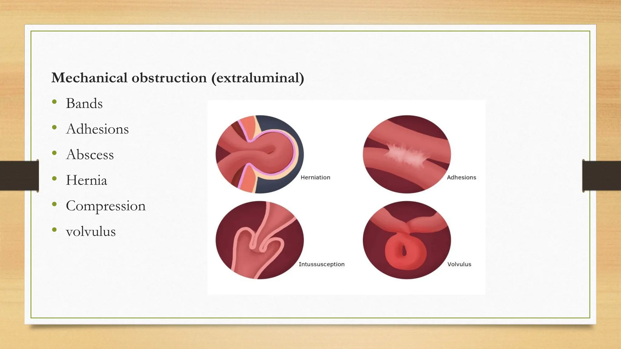
INTESTINAL
OBSTRUCTION
outline
• Introduction
• Epidemiology
• Etiology
• Pathophysiology
• Signs and symptoms
• Diagnosis
• management
Introduction
• Intestinal obstruction, one of the commonest surgical emergencies, is a
condition in which there is stoppage of the onward passage of intestinal
contents - gas, digestive juices and food
• There is either significant impairment or complete impairment of the flow of
materials in the intestines. Obstruction could be mechanical or paralytic
1. Mechanical (or dynamic) ileus, due to mechanical obstruction of the intestinal
canal, is associated with abdominal pain.
2. Paralytic (or adynamic) ileus, due to paralysis of the intestinal musculature, is
characterized by the absence of pain. Could be caused by low potassium or
peritonitis
Mechanical obstruction is of 3 types: ,
• l. Acute, i.e. of sudden onset. Usually occurs in small bowel obstruction
Severe central abdominal pain, distension and early vomiting and constipation
• 2. Chronic, i.e. of slow, progressive severity.
usually seen in large bowel obstruction with lower abdominal colic and absolute
constipation, followed by distention
• 3. Acute-on-chronic, i.e. chronic obstruction suddenly becoming acute by the
obturation of the already narrowed intestinal canal.
Usually occurs in large bowel but gradually involve the small intestine
Early symptoms are pain and constipation, if small intestines are involved, vomiting
and distension
Acute obstruction
1. Site of obstruction. The site of 'obstruction is its position relative 10 the ampulla
of Vater. It may be
(i) High, i.e. relatively near to the ampulla-jejunum and proximal ileum - with rapid
loss of water and electrolytes; or
(ii) Low i.e. relatively distant from the ampulla-distal ileum and colon - with relatively
late onset of fluid and electrolyte imbalances but early onset of distension
2. Nature of obstruction
(i) Simple obstruction. The bowel lumen is occluded with no impairment of the blood
supply to the bowel, e.g. obstruction due to intra-abdominal adhesions which may
improve on conservative treatment alone. Other examples are uncommon causes
such as ball of worms, gallstones and bezoars,
(ii) (ii) Strangulation obstruction, e.g. strangulated inguinal hernia, volvulus or inus
susception. In addition to the occlusion of the lumen, the blood supply to the
segment of bowel involved is also cut off. Pure strangulation without occlusion of
the bowel lumen occurs in mesenteric thrombosis or embolism.
(iii) (iii) Closed loop obstruction. The obstructed loop is "closed" at both ends so that
nothing can escape from it proximally or distally. The blood supply of the bowel wall
may be impaired. Closed loop obstruction occurs in a pure form in obstruction of
the colon with a competent ileocaecal valve
ETIOLOGY
• Intramural(from the wall of the intestines) : congenital, tumor, hematoma, inflammatory
• Extramural(outside the wall): adhesion, volvulus, hernia, abscess, hematoma
• Lumen obstruction(from the wall): stone meconium, foreign body, impaction
Causes of intestinal obstruction
• Adhesions-40%
• Tumors-15%
• Inflammatory-15%
• Obstructed hernia-12%
• Intraluminal-10%
• Others-8%
Mechanical obstruction
intramural
• TB ( ileo-caecal tuberculosis)
• Inflammatory lesions - diverticulitis, Crohn's disease
• Tumors
• Sticture
• Congenital (Atresia, Anorectal anomalies)
• Intussusception
• Aganglionic megacolon
Mechanical obstruction
intraluminal
• Gallstones
• Foreign bodies
• Bezoars
• Worms(Ascariasis)
• Impacted faeces
etiology
Mechanical obstruction (extraluminal)
• Bands
• Adhesions
• Abscess
• Hernia
• Compression
• volvulus
Pathophysiology
1. Simple Obstruction
• In the bowel below the site of obstruction, normal peristalsis and absorption continue until
the contents are absorbed or passed out. The bowel then collapses. Above the obstruction the
bowel becomes distended . Peristalsis is vigorous for several days but if the obstruction is not
relieved, then increasing distension leads ultimately to loss of peristaltic activity
• Distension: The distension results from accumulation of gases and fluids. The gases· nitrogen
(70%), carbon dioxide (6-9%), oxygen (10%), hydrogen (I%), methane (1 %), hydrogen
sulphide (1-10%)- are mostly from swallowed air. But some are products of putrefaction and
fermentation of intestinal contents by bacteria and some result from diffusion from the blood
The fluids are essentially digestive juices - saliva, gastric juice, bile, pancreatic and intestinal
secretions - which accumulate partly due to decreased absorption and increased secretion
• Dehydration And Electrolyte Imbalance
These are due to vomiting, accumulation of gastrointestinal secretions in the bowel
lumen, sequestration of fluid in the bowel wall and peritoneal cavity, diminished oral
intake and decreased absorption. Most of the gastro-intestinal secretions - normally
amounting to 7-10 litres in 24h but increased in obstruction - is lost to the body
through vomiting and/or sequestration in the obstructed gut.
There is also loss of E.C.F. into the peritoneum and the bowel wall. This results in
hypovolaemia - dehydration or shock- hyponatraemia, hypokalaernia, hypochloraemia
and renal failure
2. Strangulation Obstruction
• In addition to the effects of simple occlusion, obstruction of the blood supply has
profound consequences. When the pressure of the occluding band exceeds the venous
pressure, venous engorgement of gut wall occurs. (i) If the strangulated loop is long,
this may lead to sequestration of large quantities of blood from the circulation with
consequent hypovolemia, shock and death. The bowel wall becomes cyanosed
• The venous engorgement leads also to outpouring of fluid, dilatation of intramural
lymph channels that provide a pathway for carrying multiplying bacteria (particularly E.
coli) and their products (exo-and endo-toxins) from the mucosal surface deep into the
muscularis and to the serosa, peritoneal cavity and circulation. This may lead to
endotoxic shock, septicaemia or peritonitis.
• The occluding band may be tight enough to obstruct the arterial supply or the
artery may go into spasm as a reflex response to the venous congestion. Infarction
of the bowel wall then results. Necrosis of tissues is also hastened by thrombosis
of intramural and mesenteric veins resulting from the stasis of venous
engorgement. Hypoxia or anoxia also enhances the growth of anaerobic bacteria
such as Clostridia and Bacteroides
• Distension of the involved loop and the disintegration of mural structures due to
intramural blood accumulation may cause perforation with secondary peritonitis.
(vi) The bowel is at first-congested and bright red later becoming purplish. As
gangrene sets in by about 6h, it becomes black (Fig.34-ld), green or grey due in part
to
3. Closed Loop Obstruction
• The afferent and efferent limbs of the bowel are both obstructed. It is seen
typically in obstruction of the colon. A competent ileocaecal valve allows gases and
liquid faeces to enter the caecum but not to leave it. The rich bacterial flora adds to
the production of a lot of gases. Distension thus increases rapidly and with rising
intraluminal pressure ernbarrasses the circulation. The bowel becomes necrotic,
first in the caecum where distension is usually greatest. Perforation allows free
escape of faeces and bacteria with fulminating peritonitis
Risk factors of bowel obstruction
Previous abdominal or pelvic surgery ( risk for adhesion formation )
Intestinal inflammation ( Crohn disease )
History of or increased risk for neoplasm
History of foreign body ingestion
Physical examination
Abdominal inspection
 auscultation
 percussion
 palpation
Digital rectal examination
Signs and Symptoms
• Abdominal pain(first symptom)
Small bowel obstruction-central pain
Large bowel obstruction- hypogastric pain
• Vomiting
• Absolute constipation
• Distention
• Visible peristalsis
• Hernial orifices and rectum
• Scars(post surgical adhesions)
• Rebound tenderness
• General signs of dehydration
Investigations
Plain abdominal X-ray
Abdominal CT , if suspicion exist despite negative radiography
FBC
Creatinine and Electrolyte ( sodium, potassium and chloride)
Colonoscopy /Sigmoidoscopy
Barium enema
Complications
• : I. Shock (i) Hypovolemic - Loss of E.C.F. and/or sequestration of blood. (ii)
septic shock.
• 2. Dehydration.
• 3. Electrolyte and metabolic imbalance. (i) Hypokalemia (ii) Hyponatremia. (iii)
Alkalosis or acidosis.
• 4. Peritonitis.
• 5. Septicemia
• 6. Renal failure
TREATMENT
• GOALS OF TREATMENT
1. Correction of fluid, electrolyte and metabolic imbalances
Using crystalloids(ringers lactate or DNS and dextrose), KCl is added when urine output
is over 30ml/h.. In severe strangulation, blood transfusion may be necessary
• Nasogastric decompression
• Sedation/analgesia
Pethidine 100mg or morphine 10-15mg
• Antibiotic therapy
Ceftriaxone + metronidazole or cefuroxime or ciprofloxacin
• CONSERVATIVE THERAPY
laparotomy if obstruction continues
• SURGERY
Management is based on the etiology
 Resection and anastomosis (R&A); malignant enteric strictures, diverticulitis, caecal/sigmoid
volvulus
 Palliative gastrojejunostomy ;duodenal lesion not removable
 Laparotomy ;complete SBO
 Enterotomy and cholecystectomy ; gall stone obstruction
 Colostomy with R&A; colon cancers
 Ileostomy
Criteria for immediate surgery
• Bowel ischemia , necrosis or perforation
• Closed-loop obstruction ( including volvulus )
• Acute incarcerated hernia
• Intussusception
• Gallstone ileus
• Foreign body ingestion
• Localized small bowel tumor

A Slide on INTESTINAL or bowel OBSTRUCTION.pptx

  • 1.
  • 2.
    outline • Introduction • Epidemiology •Etiology • Pathophysiology • Signs and symptoms • Diagnosis • management
  • 4.
    Introduction • Intestinal obstruction,one of the commonest surgical emergencies, is a condition in which there is stoppage of the onward passage of intestinal contents - gas, digestive juices and food • There is either significant impairment or complete impairment of the flow of materials in the intestines. Obstruction could be mechanical or paralytic 1. Mechanical (or dynamic) ileus, due to mechanical obstruction of the intestinal canal, is associated with abdominal pain. 2. Paralytic (or adynamic) ileus, due to paralysis of the intestinal musculature, is characterized by the absence of pain. Could be caused by low potassium or peritonitis
  • 5.
    Mechanical obstruction isof 3 types: , • l. Acute, i.e. of sudden onset. Usually occurs in small bowel obstruction Severe central abdominal pain, distension and early vomiting and constipation • 2. Chronic, i.e. of slow, progressive severity. usually seen in large bowel obstruction with lower abdominal colic and absolute constipation, followed by distention • 3. Acute-on-chronic, i.e. chronic obstruction suddenly becoming acute by the obturation of the already narrowed intestinal canal. Usually occurs in large bowel but gradually involve the small intestine Early symptoms are pain and constipation, if small intestines are involved, vomiting and distension
  • 6.
    Acute obstruction 1. Siteof obstruction. The site of 'obstruction is its position relative 10 the ampulla of Vater. It may be (i) High, i.e. relatively near to the ampulla-jejunum and proximal ileum - with rapid loss of water and electrolytes; or (ii) Low i.e. relatively distant from the ampulla-distal ileum and colon - with relatively late onset of fluid and electrolyte imbalances but early onset of distension
  • 7.
    2. Nature ofobstruction (i) Simple obstruction. The bowel lumen is occluded with no impairment of the blood supply to the bowel, e.g. obstruction due to intra-abdominal adhesions which may improve on conservative treatment alone. Other examples are uncommon causes such as ball of worms, gallstones and bezoars, (ii) (ii) Strangulation obstruction, e.g. strangulated inguinal hernia, volvulus or inus susception. In addition to the occlusion of the lumen, the blood supply to the segment of bowel involved is also cut off. Pure strangulation without occlusion of the bowel lumen occurs in mesenteric thrombosis or embolism. (iii) (iii) Closed loop obstruction. The obstructed loop is "closed" at both ends so that nothing can escape from it proximally or distally. The blood supply of the bowel wall may be impaired. Closed loop obstruction occurs in a pure form in obstruction of the colon with a competent ileocaecal valve
  • 8.
    ETIOLOGY • Intramural(from thewall of the intestines) : congenital, tumor, hematoma, inflammatory • Extramural(outside the wall): adhesion, volvulus, hernia, abscess, hematoma • Lumen obstruction(from the wall): stone meconium, foreign body, impaction Causes of intestinal obstruction • Adhesions-40% • Tumors-15% • Inflammatory-15% • Obstructed hernia-12% • Intraluminal-10% • Others-8%
  • 9.
    Mechanical obstruction intramural • TB( ileo-caecal tuberculosis) • Inflammatory lesions - diverticulitis, Crohn's disease • Tumors • Sticture • Congenital (Atresia, Anorectal anomalies) • Intussusception • Aganglionic megacolon Mechanical obstruction intraluminal • Gallstones • Foreign bodies • Bezoars • Worms(Ascariasis) • Impacted faeces etiology
  • 10.
    Mechanical obstruction (extraluminal) •Bands • Adhesions • Abscess • Hernia • Compression • volvulus
  • 11.
    Pathophysiology 1. Simple Obstruction •In the bowel below the site of obstruction, normal peristalsis and absorption continue until the contents are absorbed or passed out. The bowel then collapses. Above the obstruction the bowel becomes distended . Peristalsis is vigorous for several days but if the obstruction is not relieved, then increasing distension leads ultimately to loss of peristaltic activity • Distension: The distension results from accumulation of gases and fluids. The gases· nitrogen (70%), carbon dioxide (6-9%), oxygen (10%), hydrogen (I%), methane (1 %), hydrogen sulphide (1-10%)- are mostly from swallowed air. But some are products of putrefaction and fermentation of intestinal contents by bacteria and some result from diffusion from the blood The fluids are essentially digestive juices - saliva, gastric juice, bile, pancreatic and intestinal secretions - which accumulate partly due to decreased absorption and increased secretion
  • 12.
    • Dehydration AndElectrolyte Imbalance These are due to vomiting, accumulation of gastrointestinal secretions in the bowel lumen, sequestration of fluid in the bowel wall and peritoneal cavity, diminished oral intake and decreased absorption. Most of the gastro-intestinal secretions - normally amounting to 7-10 litres in 24h but increased in obstruction - is lost to the body through vomiting and/or sequestration in the obstructed gut. There is also loss of E.C.F. into the peritoneum and the bowel wall. This results in hypovolaemia - dehydration or shock- hyponatraemia, hypokalaernia, hypochloraemia and renal failure
  • 13.
    2. Strangulation Obstruction •In addition to the effects of simple occlusion, obstruction of the blood supply has profound consequences. When the pressure of the occluding band exceeds the venous pressure, venous engorgement of gut wall occurs. (i) If the strangulated loop is long, this may lead to sequestration of large quantities of blood from the circulation with consequent hypovolemia, shock and death. The bowel wall becomes cyanosed • The venous engorgement leads also to outpouring of fluid, dilatation of intramural lymph channels that provide a pathway for carrying multiplying bacteria (particularly E. coli) and their products (exo-and endo-toxins) from the mucosal surface deep into the muscularis and to the serosa, peritoneal cavity and circulation. This may lead to endotoxic shock, septicaemia or peritonitis.
  • 14.
    • The occludingband may be tight enough to obstruct the arterial supply or the artery may go into spasm as a reflex response to the venous congestion. Infarction of the bowel wall then results. Necrosis of tissues is also hastened by thrombosis of intramural and mesenteric veins resulting from the stasis of venous engorgement. Hypoxia or anoxia also enhances the growth of anaerobic bacteria such as Clostridia and Bacteroides • Distension of the involved loop and the disintegration of mural structures due to intramural blood accumulation may cause perforation with secondary peritonitis. (vi) The bowel is at first-congested and bright red later becoming purplish. As gangrene sets in by about 6h, it becomes black (Fig.34-ld), green or grey due in part to
  • 15.
    3. Closed LoopObstruction • The afferent and efferent limbs of the bowel are both obstructed. It is seen typically in obstruction of the colon. A competent ileocaecal valve allows gases and liquid faeces to enter the caecum but not to leave it. The rich bacterial flora adds to the production of a lot of gases. Distension thus increases rapidly and with rising intraluminal pressure ernbarrasses the circulation. The bowel becomes necrotic, first in the caecum where distension is usually greatest. Perforation allows free escape of faeces and bacteria with fulminating peritonitis
  • 16.
    Risk factors ofbowel obstruction Previous abdominal or pelvic surgery ( risk for adhesion formation ) Intestinal inflammation ( Crohn disease ) History of or increased risk for neoplasm History of foreign body ingestion
  • 17.
    Physical examination Abdominal inspection auscultation  percussion  palpation Digital rectal examination
  • 18.
    Signs and Symptoms •Abdominal pain(first symptom) Small bowel obstruction-central pain Large bowel obstruction- hypogastric pain • Vomiting • Absolute constipation • Distention • Visible peristalsis • Hernial orifices and rectum • Scars(post surgical adhesions) • Rebound tenderness • General signs of dehydration
  • 19.
    Investigations Plain abdominal X-ray AbdominalCT , if suspicion exist despite negative radiography FBC Creatinine and Electrolyte ( sodium, potassium and chloride) Colonoscopy /Sigmoidoscopy Barium enema
  • 20.
    Complications • : I.Shock (i) Hypovolemic - Loss of E.C.F. and/or sequestration of blood. (ii) septic shock. • 2. Dehydration. • 3. Electrolyte and metabolic imbalance. (i) Hypokalemia (ii) Hyponatremia. (iii) Alkalosis or acidosis. • 4. Peritonitis. • 5. Septicemia • 6. Renal failure
  • 21.
    TREATMENT • GOALS OFTREATMENT 1. Correction of fluid, electrolyte and metabolic imbalances Using crystalloids(ringers lactate or DNS and dextrose), KCl is added when urine output is over 30ml/h.. In severe strangulation, blood transfusion may be necessary • Nasogastric decompression • Sedation/analgesia Pethidine 100mg or morphine 10-15mg • Antibiotic therapy Ceftriaxone + metronidazole or cefuroxime or ciprofloxacin
  • 22.
    • CONSERVATIVE THERAPY laparotomyif obstruction continues • SURGERY Management is based on the etiology  Resection and anastomosis (R&A); malignant enteric strictures, diverticulitis, caecal/sigmoid volvulus  Palliative gastrojejunostomy ;duodenal lesion not removable  Laparotomy ;complete SBO  Enterotomy and cholecystectomy ; gall stone obstruction  Colostomy with R&A; colon cancers  Ileostomy
  • 23.
    Criteria for immediatesurgery • Bowel ischemia , necrosis or perforation • Closed-loop obstruction ( including volvulus ) • Acute incarcerated hernia • Intussusception • Gallstone ileus • Foreign body ingestion • Localized small bowel tumor