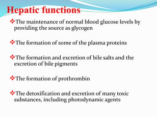
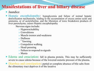
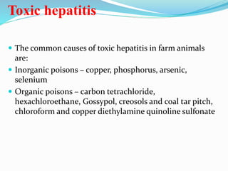
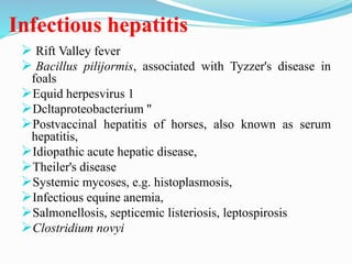
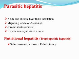
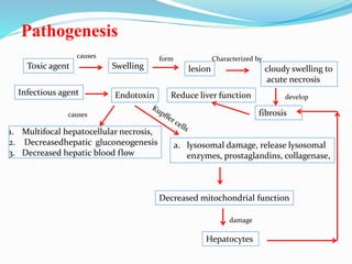
Hepatic functions
The maintenance of normal blood glucose levels by
providing the source as glycogen
The formation of some of the plasma proteins
The formation and excretion of bile salts and the
excretion of bile pigments
The formation of prothrombin
The detoxification and excretion of many toxic
substances, including photodynamic agents
Manifestations of liver and biliary disease
 Jaundice
 Hepatic encephalopathy: hypoglycemia and failure of normal hepatic
detoxification mechanisms, leading to the accumulation of excess amino acids and
ammonia, or of acetylcholine, and the liberation of toxic breakdown products of
liver parenchyma, causes Hepatic encephalopathy
Nervous signs include:
 Hyperexcitability
 Convulsions
 Muscle tremor and weakness
 Dullness
 " Yawning
 Compulsive walking
 Head-pressing
 Failure to respond to signals
 Mania
 Edema and emaciation: fall in plasma protein. This may be sufficiently
severe to cause edema because of the lowered osmotic pressure of the plasma.
 Diarrhea and constipation: partial or complete absence of bile salts from
the alimentary tract deprives it of the laxative
 Abdominal pain: distension of the organ with increased
tension of the capsule, and lesions of the capsule
 Alteration in size of the liver
 Photosensitization: In hepatic or biliary insufficiency
excretion Most photosensitizing substances, including
Phylloerythrin is retarded and photosensitization
occurs
 Haemorrhagic diathesis: deficiency in prothrombin
formation and a consequent prolongation of the
clotting time of the blood
 Displacement of the liver: displaced from its normal
position and protrude into the thoracic cavity through
a diaphragmatic hernia, causing respiratory distress
 Rupture of the liver
Jaundice
Jaundice is the yellowish staining of the
skin and sclerae (the whites of the eyes)
that is caused by high levels in blood of
the chemical bilirubin. The color of the
skin and sclerae vary depending on the
level of bilirubin. When the bilirubin
level is mildly elevated, they are
yellowish. When the bilirubin level is
high, they tend to be brown.
Physiology of jaundice
Obstructive Jaundice
Prehepatic (Hemolytic) jaundice
Occurs in case of hemolytic anemia.
Total bilirubin level is increased due to increased blood indirect
bilirubin level.
The color of urine remains normal, because indirect bilirubin is
bind to albumin, and subsequently unable to pass the glomerular
filter.
Higher level of blood indirect bilirubin, results in higher bilirubin
uptake by the liver and increased the rate of formation of direct
bilirubin, and increased the direct bilirubin that pass to the small
intestine. This results in dark brown color of the feces.
The increased stercobilinogen level in the small intestine results
in increase the formation of urobilinogen, which is excreted in
urine.
The most important changes in pre-hepatic jaundice are
increased total and indirect bilirubin in blood, normal urine color,
dark brown feces and increased urobilinogen in urine.
Hepatic jaundice
Occurs in case of hepatitis.
Serum levels of total bilirubin are increased
because ofretention of direct bilirubin,
passes out in the urine, causing an
elevation of urine levels.
The urobilinogen levels in the urine also
rise.
Dark brown color of urine, as a result of
excretion of direct bilirubin in urine.
Normal color of feces
Post-hepatic (Obstructive) Jaundice
Occurs in case of obstruction of main bile duct.
Total bilirubin increased due to increase blood direct
bilirubin level.
Dark brown color of urine.
Clay color of feces
Absence of urobilinogn from urine
Obstructive jaundice = failure of bilirubin to reach
the gut > light colored stool, dark urine
Principles of treatment
 Oral or intravenous injections of glucose , eg. Dextrose 5%
 Amino acid mixtures, especially those ontaining
methionine, are used with apparently good results.eg.
Hermin
 Diets high in carbohydrate, calcium and protein of high
biological value and a number of specific substances are
known to have a protective effect against hepatotoxic
agents.
 Administration of lipotrophic factors including choline
and maintenance on a diet low in fat and protein may
reduce the compressive effects of fibrous tissue
contraction.
Hepatitis
Diffuse, degenerative and inflammatory diseases that
affect the liver
Only difference between hepatitis and hepatosis
being that the onset of the disease is slower and less
acute than in hepatitis.
Toxic hepatitis
 The common causes of toxic hepatitis in farm animals
are:
 Inorganic poisons – copper, phosphorus, arsenic,
selenium
 Organic poisons – carbon tetrachloride,
hexachloroethane, Gossypol, creosols and coal tar pitch,
chloroform and copper diethylamine quinoline sulfonate
Infectious hepatitis
 Rift Valley fever
 Bacillus pilijormis, associated with Tyzzer's disease in
foals
Equid herpesvirus 1
Dcltaproteobacterium "
Postvaccinal hepatitis of horses, also known as serum
hepatitis,
Idiopathic acute hepatic disease,
Theiler's disease
Systemic mycoses, e.g. histoplasmosis,
Infectious equine anemia,
Salmonellosis, septicemic listeriosis, leptospirosis
Clostridium novyi
Parasitic hepatitis
Acute and chronic liver fluke infestation
Migrating larvae of Ascaris sp.
chronic shistosomiasis1
Hepatic sarcocystosis in a horse
Nutritional hepatitis (Trophopathic hepatitis)
Selenium and vitamin E deficiency
Pathogenesis
Toxic agent Swelling
causes form
lesion
Characterized by
cloudy swelling to
acute necrosis
fibrosis
develop
Infectious agent Endotoxin
1. Multifocal hepatocellular necrosis,
2. Decreasedhepatic gluconeogenesis
3. Decreased hepatic blood flow
causes
a. lysosomal damage, release lysosomal
enzymes, prostaglandins, collagenase,
Hepatocytes
damage
Decreased mitochondrial function
Reduce liver function
Clinical findings
Anorexia
 Mental depression- with excitement
 Muscular weakness
Jaundice
Recumbency
coma
intermittent convulsion.
Photosensitization may also occur
but only when the animals are on a
diet containing green feed and are
exposed to sunlight.
Weight loss,
Tachycardia,
Intennittent fever
 Abdominal pain,
 Ventral body wall edema
Clotting deficiency.
Diarrhea or constipation
Nervous signs
Ataxia and lethargy
Yawning
Coma
Hyperexcitability
Muscle tremor
Mania, including aggressive behavior, and convulsions.
A characteristic syndrome is the dummy syndrome, in which
affected animals push with the head, do not respond to normal
stimuli and may be blind.
There may be subacute abdominal pain
pain on palpation of the liver.
Most common cause of acute hepatic failure in the horse.
administration of tetanus antitoxin.
 Lactating mares appear to be at a higher risk than other horses.
Clinical findings include
Serum Hepatitis (TheHer's disease
Sudden anorexia,
Marked lethargy,
Stiff gait,
 Subcutaneous edema of the distal
aspects of all four limbs and body
wall,
Blindness,
Head pressing
Circling
 Abdominal pain,
Tachycardia,
Icterus
a marked reduction in gastrointestinal
sounds.
Death in a few days
Examination of the liver
 Palpation and percussion
 Biopsy
 Ultrasonography
 Radiography
 ,
Laboratory tests for hepatic disease
and function
 Serum bile acids, arginase and gamma-glutamyl transferase
(GGT) –sensitive indicator of cholestasis and/or hepatocellular
necrosis
 serum activities of sorbitol dehydrogenase (SDH),GGT and
aspartate aminotransferase (AST, formerly known as SGOT),
and the BSP clearance' test – sensitive indicators of
hepatocellular injury in cattle
 Chronic hepatic
 leukocytosis
 neutrophilia,
 hypoalbuminemia,
 hyperbetaglobulinemia,
 increased ALP and GGT
 increases in AST, SDH, total lactate dehydrogenase
Hepatic function
 The sulfobromophthalein sodium clearance- - test has been
used in cattle, sheep and horses.
 The time required by the normal liver to reduce the plasma
concentration of BSP to half the initial concentration is
taken as the standard BSP half-life
 Cattle is 2.5-5.5 minutes,
 Sheep 2.0 minutes
 Normal horses about 2.0 minutes
Icteric index
 Measurement of the icteric index of plasma, by comparing
its color with a standard solution of potassium dichromate
Serum hepatic enzymes
Sorbitol dehydrogenase (also called L-iditol dehydrogenase
(ID) - sheep and cattle Lactate dehydrogenase (LDH)- abundant in
liver, kidney, muscle and myocardium Aspartate aminotransferase
or L-alanine aminotransferase (ALT, previously known as
SGPT)- Dog
Arginase
Gamma-glutamyl transferase- cattle, sheep and horses. GGT is
a sensitive indicator of liver damage in horses
Glutamate dehydrogenase (GD)- ruminants and horses
Ornithine carbamoyl-transferase (OCT)

Liver

  • 2.
    Hepatic functions The maintenanceof normal blood glucose levels by providing the source as glycogen The formation of some of the plasma proteins The formation and excretion of bile salts and the excretion of bile pigments The formation of prothrombin The detoxification and excretion of many toxic substances, including photodynamic agents
  • 3.
    Manifestations of liverand biliary disease  Jaundice  Hepatic encephalopathy: hypoglycemia and failure of normal hepatic detoxification mechanisms, leading to the accumulation of excess amino acids and ammonia, or of acetylcholine, and the liberation of toxic breakdown products of liver parenchyma, causes Hepatic encephalopathy Nervous signs include:  Hyperexcitability  Convulsions  Muscle tremor and weakness  Dullness  " Yawning  Compulsive walking  Head-pressing  Failure to respond to signals  Mania  Edema and emaciation: fall in plasma protein. This may be sufficiently severe to cause edema because of the lowered osmotic pressure of the plasma.  Diarrhea and constipation: partial or complete absence of bile salts from the alimentary tract deprives it of the laxative
  • 4.
     Abdominal pain:distension of the organ with increased tension of the capsule, and lesions of the capsule  Alteration in size of the liver  Photosensitization: In hepatic or biliary insufficiency excretion Most photosensitizing substances, including Phylloerythrin is retarded and photosensitization occurs  Haemorrhagic diathesis: deficiency in prothrombin formation and a consequent prolongation of the clotting time of the blood  Displacement of the liver: displaced from its normal position and protrude into the thoracic cavity through a diaphragmatic hernia, causing respiratory distress  Rupture of the liver
  • 5.
    Jaundice Jaundice is theyellowish staining of the skin and sclerae (the whites of the eyes) that is caused by high levels in blood of the chemical bilirubin. The color of the skin and sclerae vary depending on the level of bilirubin. When the bilirubin level is mildly elevated, they are yellowish. When the bilirubin level is high, they tend to be brown.
  • 6.
  • 7.
    Prehepatic (Hemolytic) jaundice Occursin case of hemolytic anemia. Total bilirubin level is increased due to increased blood indirect bilirubin level. The color of urine remains normal, because indirect bilirubin is bind to albumin, and subsequently unable to pass the glomerular filter. Higher level of blood indirect bilirubin, results in higher bilirubin uptake by the liver and increased the rate of formation of direct bilirubin, and increased the direct bilirubin that pass to the small intestine. This results in dark brown color of the feces. The increased stercobilinogen level in the small intestine results in increase the formation of urobilinogen, which is excreted in urine. The most important changes in pre-hepatic jaundice are increased total and indirect bilirubin in blood, normal urine color, dark brown feces and increased urobilinogen in urine.
  • 8.
    Hepatic jaundice Occurs incase of hepatitis. Serum levels of total bilirubin are increased because ofretention of direct bilirubin, passes out in the urine, causing an elevation of urine levels. The urobilinogen levels in the urine also rise. Dark brown color of urine, as a result of excretion of direct bilirubin in urine. Normal color of feces
  • 9.
    Post-hepatic (Obstructive) Jaundice Occursin case of obstruction of main bile duct. Total bilirubin increased due to increase blood direct bilirubin level. Dark brown color of urine. Clay color of feces Absence of urobilinogn from urine Obstructive jaundice = failure of bilirubin to reach the gut > light colored stool, dark urine
  • 10.
    Principles of treatment Oral or intravenous injections of glucose , eg. Dextrose 5%  Amino acid mixtures, especially those ontaining methionine, are used with apparently good results.eg. Hermin  Diets high in carbohydrate, calcium and protein of high biological value and a number of specific substances are known to have a protective effect against hepatotoxic agents.  Administration of lipotrophic factors including choline and maintenance on a diet low in fat and protein may reduce the compressive effects of fibrous tissue contraction.
  • 11.
    Hepatitis Diffuse, degenerative andinflammatory diseases that affect the liver Only difference between hepatitis and hepatosis being that the onset of the disease is slower and less acute than in hepatitis.
  • 12.
    Toxic hepatitis  Thecommon causes of toxic hepatitis in farm animals are:  Inorganic poisons – copper, phosphorus, arsenic, selenium  Organic poisons – carbon tetrachloride, hexachloroethane, Gossypol, creosols and coal tar pitch, chloroform and copper diethylamine quinoline sulfonate
  • 13.
    Infectious hepatitis  RiftValley fever  Bacillus pilijormis, associated with Tyzzer's disease in foals Equid herpesvirus 1 Dcltaproteobacterium " Postvaccinal hepatitis of horses, also known as serum hepatitis, Idiopathic acute hepatic disease, Theiler's disease Systemic mycoses, e.g. histoplasmosis, Infectious equine anemia, Salmonellosis, septicemic listeriosis, leptospirosis Clostridium novyi
  • 14.
    Parasitic hepatitis Acute andchronic liver fluke infestation Migrating larvae of Ascaris sp. chronic shistosomiasis1 Hepatic sarcocystosis in a horse Nutritional hepatitis (Trophopathic hepatitis) Selenium and vitamin E deficiency
  • 15.
    Pathogenesis Toxic agent Swelling causesform lesion Characterized by cloudy swelling to acute necrosis fibrosis develop Infectious agent Endotoxin 1. Multifocal hepatocellular necrosis, 2. Decreasedhepatic gluconeogenesis 3. Decreased hepatic blood flow causes a. lysosomal damage, release lysosomal enzymes, prostaglandins, collagenase, Hepatocytes damage Decreased mitochondrial function Reduce liver function
  • 16.
    Clinical findings Anorexia  Mentaldepression- with excitement  Muscular weakness Jaundice Recumbency coma intermittent convulsion. Photosensitization may also occur but only when the animals are on a diet containing green feed and are exposed to sunlight. Weight loss, Tachycardia, Intennittent fever  Abdominal pain,  Ventral body wall edema Clotting deficiency. Diarrhea or constipation
  • 17.
    Nervous signs Ataxia andlethargy Yawning Coma Hyperexcitability Muscle tremor Mania, including aggressive behavior, and convulsions. A characteristic syndrome is the dummy syndrome, in which affected animals push with the head, do not respond to normal stimuli and may be blind. There may be subacute abdominal pain pain on palpation of the liver.
  • 18.
    Most common causeof acute hepatic failure in the horse. administration of tetanus antitoxin.  Lactating mares appear to be at a higher risk than other horses. Clinical findings include Serum Hepatitis (TheHer's disease Sudden anorexia, Marked lethargy, Stiff gait,  Subcutaneous edema of the distal aspects of all four limbs and body wall, Blindness, Head pressing Circling  Abdominal pain, Tachycardia, Icterus a marked reduction in gastrointestinal sounds. Death in a few days
  • 19.
    Examination of theliver  Palpation and percussion  Biopsy  Ultrasonography  Radiography  ,
  • 20.
    Laboratory tests forhepatic disease and function  Serum bile acids, arginase and gamma-glutamyl transferase (GGT) –sensitive indicator of cholestasis and/or hepatocellular necrosis  serum activities of sorbitol dehydrogenase (SDH),GGT and aspartate aminotransferase (AST, formerly known as SGOT), and the BSP clearance' test – sensitive indicators of hepatocellular injury in cattle  Chronic hepatic  leukocytosis  neutrophilia,  hypoalbuminemia,  hyperbetaglobulinemia,  increased ALP and GGT  increases in AST, SDH, total lactate dehydrogenase
  • 21.
    Hepatic function  Thesulfobromophthalein sodium clearance- - test has been used in cattle, sheep and horses.  The time required by the normal liver to reduce the plasma concentration of BSP to half the initial concentration is taken as the standard BSP half-life  Cattle is 2.5-5.5 minutes,  Sheep 2.0 minutes  Normal horses about 2.0 minutes
  • 22.
    Icteric index  Measurementof the icteric index of plasma, by comparing its color with a standard solution of potassium dichromate Serum hepatic enzymes Sorbitol dehydrogenase (also called L-iditol dehydrogenase (ID) - sheep and cattle Lactate dehydrogenase (LDH)- abundant in liver, kidney, muscle and myocardium Aspartate aminotransferase or L-alanine aminotransferase (ALT, previously known as SGPT)- Dog Arginase Gamma-glutamyl transferase- cattle, sheep and horses. GGT is a sensitive indicator of liver damage in horses Glutamate dehydrogenase (GD)- ruminants and horses Ornithine carbamoyl-transferase (OCT)

Editor's Notes

  • #7 Obstructive Joundice
  • #16 Reduce liver function