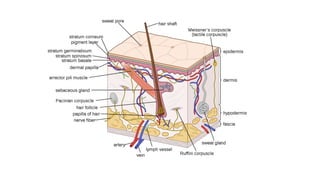
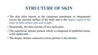
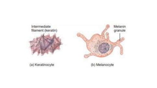
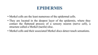
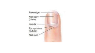
SlideShare a Scribd company logo
INTEGUMENTARY SYSTEM
Presented by:-
HIMIKA RATHI
INTRODUCTION
• The integumentary system is composed of the skin, hair, oil
and sweat glands, nails, and sensory receptors.
• The integumentary system helps maintain a constant body
temperature, protects the body, and provides sensory
information about the surrounding environment.
STRUCTURE OF SKIN
• The skin (also known as the cutaneous membrane or integument)
covers the external surface of the body and is the largest organ of the
body in both surface area and weight.
• Structurally, the skin consists of two main parts.
• The superficial, thinner portion which is composed of epithelial tissue,
is the epidermis.
• The deeper, thicker connective tissue portion is the dermis.
EPIDERMIS
• The epidermis is composed of keratinized stratified squamous
epithelium.
• It contains four principal types of cells: keratinocytes, melanocytes,
Langerhans cells, and Merkel cells
• About 90% of epidermal cells are keratinocytes, which are arranged in
four or five layers and produce the protein keratin.
• keratin is a tough, fibrous protein that helps protect the skin and
underlying tissues from heat, microbes, and chemicals.
EPIDERMIS
• About 8% of the epidermal cells are melanocytes, which produces the
pigment melanin.
• Their long, slender projections extend between the keratinocytes and
transfer melanin granules to them.
• Melanin is a yellow-red or brown-black pigment that contributes to
skin color and absorbs damaging ultraviolet (UV) light.
EPIDERMIS
• Langerhans cells arise from red bone marrow and migrate to the
epidermis , where they constitute a small fraction of the epidermal
cells.
• They participate in immune responses mounted against microbes that
invade the skin, and are easily damaged by UV light.
• Their role in the immune response is to help other cells of the immune
system recognize an invading microbe and destroy it.
EPIDERMIS
• Merkel cells are the least numerous of the epidermal cells.
• They are located in the deepest layer of the epidermis, where they
contact the flattened process of a sensory neuron (nerve cell), a
structure called a Merkel (tactile) disc.
• Merkel cells and their associated Merkel discs detect touch sensations.
EPIDERMIS
• In most regions of the body the epidermis has four strata or layers—
• Stratum basale
• Stratum spinosum
• Stratum granulosum and
• a thin stratum corneum
Stratum basale
• The deepest layer of the epidermis is the stratum basale, composed of
a single row of cuboidal or columnar keratinocytes.
Stratum spinosum
• Superficial to the stratum basale is the stratum spinosum, arranged in 8
to 10 layers of many-sided keratinocytes fitting closely together.
Stratum Granulosum
• At about the middle of the epidermis, the stratum granulosum consists
of three to five layers of flattened keratinocytes.
Stratum Lucidum
• The stratum lucidum is present only in the thick skin of areas such as
the fingertips, palms, and soles.
Stratum Corneum
• The stratum corneum consists on average of 25 to 30 layers of
flattened dead keratinocytes.
Dermis
• The second, deeper part of the skin, the dermis, is composed of a
strong connective tissue containing collagen and elastic fibers.
• The dermis also has the ability to stretch and recoil easily.
• Leather, which we use for belts, shoes, baseball gloves, and
basketballs, is the dried and treated dermis of other animals.
• Based on its tissue structure, the dermis can be divided into a
superficial papillary region and a deeper reticular region.
• The papillary region makes up about one-fifth of the thickness of the
total layer. It consists of areolar connective tissue containing thin
collagen and fine elastic fibers.
• The reticular region, which is attached to the subcutaneous layer,
consists of dense irregular connective tissue containing fibroblasts,
bundles of collagen, and some coarse elastic fibers.
• The combination of collagen and elastic fibers in the reticular region
provides the skin with strength, extensibility (ability to stretch), and
elasticity (ability to return to original shape after stretching).
Accessory structures of the skin
• Accessory structures of the skin—hair, skin glands, and nails—
develop from the embryonic epidermis.
• They have a host of important functions.
• For example, hair and nails protect the body, and sweat glands help
regulate body temperature.
Hair
• Hairs, or pili, are present on most skin surfaces except the palms,
palmar surfaces of the fingers, the soles, and plantar surfaces of the
feet.
• Although the protection it offers is limited, hair on the head guards the
scalp from injury and the sun’s rays.
• It also decreases heat loss from the scalp.
• Eyebrows and eyelashes protect the eyes from foreign particles, as
does hair in the nostrils and in the external ear canal.
Anatomy of a Hair
• The shaft is the superficial portion of the hair, which projects above
the surface of the skin.
• The root is the portion of the hair deep to the shaft that penetrates into
the dermis, and sometimes into the subcutaneous layer.
• The shaft and root of the hair both consist of three concentric layers of
cells: medulla, cortex, and cuticle of the hair.
• Cuticle of the hair, the outermost layer, consists of a single layer of
thin, flat cells that are the most heavily keratinized
Anatomy of a Hair
• Surrounding the root of the hair is the hair follicle, which is made up
of an external root sheath and an internal root sheath, together referred
to as an epithelial root sheath.
• The dense dermis surrounding the hair follicle is called the dermal root
sheath.
• The base of each hair follicle and its surrounding dermal root sheath is
an onion-shaped structure, the bulb.
Nails
• Nails are plates of tightly packed, hard, dead, keratinized epidermal
cells that form a clear, solid covering over the dorsal surfaces of the
distal portions of the digits.
• Each nail consists of a nail body, a free edge, and a nail root.
• The nail body (plate) is the visible portion of the nail.
• The free edge is the part of the nail body that may extend past the
distal end of the digit. The free edge is white because there are no
underlying capillaries.
Nails
• The nail root is the portion of the nail that is buried in a fold of skin.
• The whitish, crescent-shaped area of the proximal end of the nail body
is called the lunula.
• The proximal portion of the epithelium deep to the nail root is the nail
matrix, where cells divide by mitosis to produce growth.
Skin Glands
• Two types of Skin Glands are present:
1) Sebaceous (Oil) Glands
2) Sudoriferous (Sweat) Glands
Sebaceous (Oil) Glands
• Sebaceous glands or oil glands are simple, branched acinar glands.
• They are connected to hair follicles.
• The secreting portion of a sebaceous gland lies in the dermis and
usually opens into the neck of a hair follicle.
• Sebaceous glands secrete an oily substance called sebum, a mixture of
triglycerides, cholesterol, proteins, and inorganic salts.
• Sebum coats the surface of hairs and helps keep them from drying and
becoming brittle. Sebum also prevents excessive evaporation of water
from the skin, keeps the skin soft.
Sudoriferous Glands
• There are three to four million sweat glands, or sudoriferous glands.
• The cells of these glands release sweat, or perspiration, into hair
follicles or onto the skin surface through pores.
• Sweat glands are divided into two main types, eccrine and apocrine,
based on their structure, location, and type of secretion.
• Eccrine sweat glands , also known as merocrine sweat glands, are
simple, coiled tubular glands that are much more common than
apocrine sweat glands
Sudoriferous Glands
• They are distributed throughout the skin of most regions of the body,
especially in the skin of the forehead, palms, and soles.
• Eccrine sweat glands are not present, however, in the margins of the
lips, nail beds of the fingers and toes,and eardrums.
• The secretory portion of eccrine sweat glands is located mostly in the
deep dermis (sometimes in the upper subcutaneous layer)
Sudoriferous Glands
• The sweat produced by eccrine sweat glands (about 600 mL per day)
consists of water, ions (mostly Na and Cl), urea, uric acid, ammonia,
amino acids, glucose, and lactic acid.
• The main function of eccrine sweat glands is to help regulate body
temperature through evaporation.
Sudoriferous Glands
• Apocrine sweat glands are also simple, coiled tubular glands.
• Eccrine sweat glands start to function soon after birth, but apocrine
sweat glands do not begin to function until puberty.
• Apocrine sweat contains the same components as eccrine sweat plus
lipids and proteins.
Ceruminous Glands
• Modified sweat glands in the external ear, called ceruminous glands,
produce a waxy lubricating secretion.
• The combined secretion of the ceruminous and sebaceous glands is a
yellowish material called cerumen, or earwax.
• Cerumen also waterproofs the canal and prevents bacteria and fungi
from entering cells.
Function of Skin
• Thermoregulation
• Blood reservoir
• Protection
• Cutaneous sensations
• Excretion
• Synthesis of vitamin D
32

More Related Content

What's hot

Anatomy and physiologyof skin
Anatomy and physiologyof skinAnatomy and physiologyof skin
Anatomy and physiologyof skin
DR .PALLAVI PATHANIA
 
The integumentary system
The integumentary systemThe integumentary system
The integumentary system
Abhay Rajpoot
 
Skeletal system
Skeletal system Skeletal system
Skeletal system
ambika bagora
 
Skeletal system
Skeletal systemSkeletal system
Skeletal system
abhay joshi
 
Tissue its types, structure and function
Tissue its types, structure and functionTissue its types, structure and function
Tissue its types, structure and function
Amjad Afridi
 
Muscular System.ppt
Muscular System.pptMuscular System.ppt
Muscular System.ppt
Vijay Salvekar
 
Tissues
TissuesTissues
The integumentary system (slide show)
The integumentary system (slide show)The integumentary system (slide show)
The integumentary system (slide show)
William Banaag
 
All about skin
All about skinAll about skin
All about skin
সারন দাস
 
Anatomy and physiology of Lymphatic System
Anatomy and physiology of Lymphatic SystemAnatomy and physiology of Lymphatic System
Anatomy and physiology of Lymphatic System
Indra Mani Mishra
 
Integumentary system
Integumentary systemIntegumentary system
Integumentary system
Ahmed Al-Dawoodi
 
Skin anatomy and physiology
Skin anatomy and physiologySkin anatomy and physiology
Skin anatomy and physiology
DeboraJasmin S
 
Skin structure function and disease
Skin structure function and diseaseSkin structure function and disease
Skin structure function and disease
SUJIT DAS
 
Appendicular skeleton
Appendicular skeletonAppendicular skeleton
Appendicular skeleton
abhay joshi
 
Integumentary System
Integumentary SystemIntegumentary System
Integumentary System
clarot16
 

What's hot (20)

Anatomy and physiologyof skin
Anatomy and physiologyof skinAnatomy and physiologyof skin
Anatomy and physiologyof skin
 
The integumentary system
The integumentary systemThe integumentary system
The integumentary system
 
Skeletal system
Skeletal system Skeletal system
Skeletal system
 
Skeletal system
Skeletal systemSkeletal system
Skeletal system
 
Tissue its types, structure and function
Tissue its types, structure and functionTissue its types, structure and function
Tissue its types, structure and function
 
Muscular System.ppt
Muscular System.pptMuscular System.ppt
Muscular System.ppt
 
Skin
SkinSkin
Skin
 
Tissues
TissuesTissues
Tissues
 
The integumentary system (slide show)
The integumentary system (slide show)The integumentary system (slide show)
The integumentary system (slide show)
 
Sense organs
Sense organsSense organs
Sense organs
 
All about skin
All about skinAll about skin
All about skin
 
Anatomy and physiology of Lymphatic System
Anatomy and physiology of Lymphatic SystemAnatomy and physiology of Lymphatic System
Anatomy and physiology of Lymphatic System
 
Integumentary system
Integumentary systemIntegumentary system
Integumentary system
 
Lymphatic system
Lymphatic systemLymphatic system
Lymphatic system
 
Functions of skin
Functions of skinFunctions of skin
Functions of skin
 
Skin anatomy and physiology
Skin anatomy and physiologySkin anatomy and physiology
Skin anatomy and physiology
 
Skin structure function and disease
Skin structure function and diseaseSkin structure function and disease
Skin structure function and disease
 
Epithelial tissue
Epithelial tissueEpithelial tissue
Epithelial tissue
 
Appendicular skeleton
Appendicular skeletonAppendicular skeleton
Appendicular skeleton
 
Integumentary System
Integumentary SystemIntegumentary System
Integumentary System
 

Similar to Integumentary system

THE INTEGUMENTARY SYSTEM.pptx
THE INTEGUMENTARY SYSTEM.pptxTHE INTEGUMENTARY SYSTEM.pptx
THE INTEGUMENTARY SYSTEM.pptx
RupaSingh83
 
SKIN STRUCTURE AND FUNCTION.pptx
SKIN STRUCTURE AND FUNCTION.pptxSKIN STRUCTURE AND FUNCTION.pptx
SKIN STRUCTURE AND FUNCTION.pptx
DR.P.S SUDHAKAR
 
Anotomy of Skin
Anotomy of SkinAnotomy of Skin
Anotomy of Skin
abhay joshi
 
SKIN AND ITS APPENDAGES.pptx
SKIN AND ITS APPENDAGES.pptxSKIN AND ITS APPENDAGES.pptx
SKIN AND ITS APPENDAGES.pptx
lekhakuriakose1
 
The sense of touch, pain & temperature
The sense of touch, pain & temperatureThe sense of touch, pain & temperature
The sense of touch, pain & temperature
Hashim Ali
 
Dermatology
DermatologyDermatology
Dermatology
ssuser47c7ec1
 
Anatomy of the skin
Anatomy of the skinAnatomy of the skin
Skin
SkinSkin
B pharmacy HAP-1 Sem-1 skin and bones.pptx
B pharmacy HAP-1 Sem-1 skin and bones.pptxB pharmacy HAP-1 Sem-1 skin and bones.pptx
B pharmacy HAP-1 Sem-1 skin and bones.pptx
omkarshindeminecraft
 
SemiSolid Dosage Forms
SemiSolid Dosage FormsSemiSolid Dosage Forms
SemiSolid Dosage Forms
Eknath D. Ahire
 
Integumentary System
Integumentary System Integumentary System
Integumentary System
Dr Asma Lashari
 
Skin presentation
Skin presentationSkin presentation
Unit II, Chapter-1-Integumentary System
Unit II, Chapter-1-Integumentary SystemUnit II, Chapter-1-Integumentary System
Unit II, Chapter-1-Integumentary System
Audumbar Mali
 
Skin and fascia
Skin and fasciaSkin and fascia
Skin and fascia
Aqsa Mushtaq
 
Normal histology of skin with pathological skin conditions.pptx
Normal histology of skin with pathological skin conditions.pptxNormal histology of skin with pathological skin conditions.pptx
Normal histology of skin with pathological skin conditions.pptx
Dr.Sunil mitran
 
Anatomy & physiology of skin
Anatomy & physiology of skinAnatomy & physiology of skin
Anatomy & physiology of skin
Ayshah Hashimi
 
Anatomy of skin S.A.pptx
Anatomy of skin S.A.pptxAnatomy of skin S.A.pptx
Anatomy of skin S.A.pptx
ssuser3ae7cb
 
Powerpoint of integumentary syytem
Powerpoint of  integumentary syytemPowerpoint of  integumentary syytem
Powerpoint of integumentary syytemnone
 
Powerpoint of integumentary system of vertebrates
Powerpoint of  integumentary system of vertebratesPowerpoint of  integumentary system of vertebrates
Powerpoint of integumentary system of vertebratesnone
 

Similar to Integumentary system (20)

THE INTEGUMENTARY SYSTEM.pptx
THE INTEGUMENTARY SYSTEM.pptxTHE INTEGUMENTARY SYSTEM.pptx
THE INTEGUMENTARY SYSTEM.pptx
 
SKIN STRUCTURE AND FUNCTION.pptx
SKIN STRUCTURE AND FUNCTION.pptxSKIN STRUCTURE AND FUNCTION.pptx
SKIN STRUCTURE AND FUNCTION.pptx
 
Layers of skin
Layers of skinLayers of skin
Layers of skin
 
Anotomy of Skin
Anotomy of SkinAnotomy of Skin
Anotomy of Skin
 
SKIN AND ITS APPENDAGES.pptx
SKIN AND ITS APPENDAGES.pptxSKIN AND ITS APPENDAGES.pptx
SKIN AND ITS APPENDAGES.pptx
 
The sense of touch, pain & temperature
The sense of touch, pain & temperatureThe sense of touch, pain & temperature
The sense of touch, pain & temperature
 
Dermatology
DermatologyDermatology
Dermatology
 
Anatomy of the skin
Anatomy of the skinAnatomy of the skin
Anatomy of the skin
 
Skin
SkinSkin
Skin
 
B pharmacy HAP-1 Sem-1 skin and bones.pptx
B pharmacy HAP-1 Sem-1 skin and bones.pptxB pharmacy HAP-1 Sem-1 skin and bones.pptx
B pharmacy HAP-1 Sem-1 skin and bones.pptx
 
SemiSolid Dosage Forms
SemiSolid Dosage FormsSemiSolid Dosage Forms
SemiSolid Dosage Forms
 
Integumentary System
Integumentary System Integumentary System
Integumentary System
 
Skin presentation
Skin presentationSkin presentation
Skin presentation
 
Unit II, Chapter-1-Integumentary System
Unit II, Chapter-1-Integumentary SystemUnit II, Chapter-1-Integumentary System
Unit II, Chapter-1-Integumentary System
 
Skin and fascia
Skin and fasciaSkin and fascia
Skin and fascia
 
Normal histology of skin with pathological skin conditions.pptx
Normal histology of skin with pathological skin conditions.pptxNormal histology of skin with pathological skin conditions.pptx
Normal histology of skin with pathological skin conditions.pptx
 
Anatomy & physiology of skin
Anatomy & physiology of skinAnatomy & physiology of skin
Anatomy & physiology of skin
 
Anatomy of skin S.A.pptx
Anatomy of skin S.A.pptxAnatomy of skin S.A.pptx
Anatomy of skin S.A.pptx
 
Powerpoint of integumentary syytem
Powerpoint of  integumentary syytemPowerpoint of  integumentary syytem
Powerpoint of integumentary syytem
 
Powerpoint of integumentary system of vertebrates
Powerpoint of  integumentary system of vertebratesPowerpoint of  integumentary system of vertebrates
Powerpoint of integumentary system of vertebrates
 

More from HimikaRathi

National health intervention programme for mother and child
National health intervention programme for mother and childNational health intervention programme for mother and child
National health intervention programme for mother and child
HimikaRathi
 
Antiallergic activity by mast cell stabilization assay
Antiallergic activity by mast cell stabilization assayAntiallergic activity by mast cell stabilization assay
Antiallergic activity by mast cell stabilization assay
HimikaRathi
 
Epilepsy
EpilepsyEpilepsy
Epilepsy
HimikaRathi
 
Non randomized controlled trial
Non randomized controlled trial Non randomized controlled trial
Non randomized controlled trial
HimikaRathi
 
Novel antibiotics
Novel antibioticsNovel antibiotics
Novel antibiotics
HimikaRathi
 
Regulatory guidelines for conducting toxicity studies
Regulatory guidelines for conducting toxicity studiesRegulatory guidelines for conducting toxicity studies
Regulatory guidelines for conducting toxicity studies
HimikaRathi
 
Antiischemics
AntiischemicsAntiischemics
Antiischemics
HimikaRathi
 
Screening models of antiepileptic and nootropic drugs
Screening models of antiepileptic and nootropic drugsScreening models of antiepileptic and nootropic drugs
Screening models of antiepileptic and nootropic drugs
HimikaRathi
 

More from HimikaRathi (8)

National health intervention programme for mother and child
National health intervention programme for mother and childNational health intervention programme for mother and child
National health intervention programme for mother and child
 
Antiallergic activity by mast cell stabilization assay
Antiallergic activity by mast cell stabilization assayAntiallergic activity by mast cell stabilization assay
Antiallergic activity by mast cell stabilization assay
 
Epilepsy
EpilepsyEpilepsy
Epilepsy
 
Non randomized controlled trial
Non randomized controlled trial Non randomized controlled trial
Non randomized controlled trial
 
Novel antibiotics
Novel antibioticsNovel antibiotics
Novel antibiotics
 
Regulatory guidelines for conducting toxicity studies
Regulatory guidelines for conducting toxicity studiesRegulatory guidelines for conducting toxicity studies
Regulatory guidelines for conducting toxicity studies
 
Antiischemics
AntiischemicsAntiischemics
Antiischemics
 
Screening models of antiepileptic and nootropic drugs
Screening models of antiepileptic and nootropic drugsScreening models of antiepileptic and nootropic drugs
Screening models of antiepileptic and nootropic drugs
 

Recently uploaded

Surgical Site Infections, pathophysiology, and prevention.pptx
Surgical Site Infections, pathophysiology, and prevention.pptxSurgical Site Infections, pathophysiology, and prevention.pptx
Surgical Site Infections, pathophysiology, and prevention.pptx
jval Landero
 
Phone Us ❤85270-49040❤ #ℂall #gIRLS In Surat By Surat @ℂall @Girls Hotel With...
Phone Us ❤85270-49040❤ #ℂall #gIRLS In Surat By Surat @ℂall @Girls Hotel With...Phone Us ❤85270-49040❤ #ℂall #gIRLS In Surat By Surat @ℂall @Girls Hotel With...
Phone Us ❤85270-49040❤ #ℂall #gIRLS In Surat By Surat @ℂall @Girls Hotel With...
Savita Shen $i11
 
THOA 2.ppt Human Organ Transplantation Act
THOA 2.ppt Human Organ Transplantation ActTHOA 2.ppt Human Organ Transplantation Act
THOA 2.ppt Human Organ Transplantation Act
DrSathishMS1
 
Hemodialysis: Chapter 3, Dialysis Water Unit - Dr.Gawad
Hemodialysis: Chapter 3, Dialysis Water Unit - Dr.GawadHemodialysis: Chapter 3, Dialysis Water Unit - Dr.Gawad
Hemodialysis: Chapter 3, Dialysis Water Unit - Dr.Gawad
NephroTube - Dr.Gawad
 
Ophthalmology Clinical Tests for OSCE exam
Ophthalmology Clinical Tests for OSCE examOphthalmology Clinical Tests for OSCE exam
Ophthalmology Clinical Tests for OSCE exam
KafrELShiekh University
 
heat stroke and heat exhaustion in children
heat stroke and heat exhaustion in childrenheat stroke and heat exhaustion in children
heat stroke and heat exhaustion in children
SumeraAhmad5
 
Non-respiratory Functions of the Lungs.pdf
Non-respiratory Functions of the Lungs.pdfNon-respiratory Functions of the Lungs.pdf
Non-respiratory Functions of the Lungs.pdf
MedicoseAcademics
 
The POPPY STUDY (Preconception to post-partum cardiovascular function in prim...
The POPPY STUDY (Preconception to post-partum cardiovascular function in prim...The POPPY STUDY (Preconception to post-partum cardiovascular function in prim...
The POPPY STUDY (Preconception to post-partum cardiovascular function in prim...
Catherine Liao
 
24 Upakrama.pptx class ppt useful in all
24 Upakrama.pptx class ppt useful in all24 Upakrama.pptx class ppt useful in all
24 Upakrama.pptx class ppt useful in all
DrSathishMS1
 
ARTIFICIAL INTELLIGENCE IN HEALTHCARE.pdf
ARTIFICIAL INTELLIGENCE IN  HEALTHCARE.pdfARTIFICIAL INTELLIGENCE IN  HEALTHCARE.pdf
ARTIFICIAL INTELLIGENCE IN HEALTHCARE.pdf
Anujkumaranit
 
BRACHYTHERAPY OVERVIEW AND APPLICATORS
BRACHYTHERAPY OVERVIEW  AND  APPLICATORSBRACHYTHERAPY OVERVIEW  AND  APPLICATORS
BRACHYTHERAPY OVERVIEW AND APPLICATORS
Krishan Murari
 
Physiology of Chemical Sensation of smell.pdf
Physiology of Chemical Sensation of smell.pdfPhysiology of Chemical Sensation of smell.pdf
Physiology of Chemical Sensation of smell.pdf
MedicoseAcademics
 
Are There Any Natural Remedies To Treat Syphilis.pdf
Are There Any Natural Remedies To Treat Syphilis.pdfAre There Any Natural Remedies To Treat Syphilis.pdf
Are There Any Natural Remedies To Treat Syphilis.pdf
Little Cross Family Clinic
 
New Directions in Targeted Therapeutic Approaches for Older Adults With Mantl...
New Directions in Targeted Therapeutic Approaches for Older Adults With Mantl...New Directions in Targeted Therapeutic Approaches for Older Adults With Mantl...
New Directions in Targeted Therapeutic Approaches for Older Adults With Mantl...
i3 Health
 
Prix Galien International 2024 Forum Program
Prix Galien International 2024 Forum ProgramPrix Galien International 2024 Forum Program
Prix Galien International 2024 Forum Program
Levi Shapiro
 
Factory Supply Best Quality Pmk Oil CAS 28578–16–7 PMK Powder in Stock
Factory Supply Best Quality Pmk Oil CAS 28578–16–7 PMK Powder in StockFactory Supply Best Quality Pmk Oil CAS 28578–16–7 PMK Powder in Stock
Factory Supply Best Quality Pmk Oil CAS 28578–16–7 PMK Powder in Stock
rebeccabio
 
The Normal Electrocardiogram - Part I of II
The Normal Electrocardiogram - Part I of IIThe Normal Electrocardiogram - Part I of II
The Normal Electrocardiogram - Part I of II
MedicoseAcademics
 
Charaka Samhita Sutra sthana Chapter 15 Upakalpaniyaadhyaya
Charaka Samhita Sutra sthana Chapter 15 UpakalpaniyaadhyayaCharaka Samhita Sutra sthana Chapter 15 Upakalpaniyaadhyaya
Charaka Samhita Sutra sthana Chapter 15 Upakalpaniyaadhyaya
Dr KHALID B.M
 
ANATOMY AND PHYSIOLOGY OF URINARY SYSTEM.pptx
ANATOMY AND PHYSIOLOGY OF URINARY SYSTEM.pptxANATOMY AND PHYSIOLOGY OF URINARY SYSTEM.pptx
ANATOMY AND PHYSIOLOGY OF URINARY SYSTEM.pptx
Swetaba Besh
 
TEST BANK for Operations Management, 14th Edition by William J. Stevenson, Ve...
TEST BANK for Operations Management, 14th Edition by William J. Stevenson, Ve...TEST BANK for Operations Management, 14th Edition by William J. Stevenson, Ve...
TEST BANK for Operations Management, 14th Edition by William J. Stevenson, Ve...
kevinkariuki227
 

Recently uploaded (20)

Surgical Site Infections, pathophysiology, and prevention.pptx
Surgical Site Infections, pathophysiology, and prevention.pptxSurgical Site Infections, pathophysiology, and prevention.pptx
Surgical Site Infections, pathophysiology, and prevention.pptx
 
Phone Us ❤85270-49040❤ #ℂall #gIRLS In Surat By Surat @ℂall @Girls Hotel With...
Phone Us ❤85270-49040❤ #ℂall #gIRLS In Surat By Surat @ℂall @Girls Hotel With...Phone Us ❤85270-49040❤ #ℂall #gIRLS In Surat By Surat @ℂall @Girls Hotel With...
Phone Us ❤85270-49040❤ #ℂall #gIRLS In Surat By Surat @ℂall @Girls Hotel With...
 
THOA 2.ppt Human Organ Transplantation Act
THOA 2.ppt Human Organ Transplantation ActTHOA 2.ppt Human Organ Transplantation Act
THOA 2.ppt Human Organ Transplantation Act
 
Hemodialysis: Chapter 3, Dialysis Water Unit - Dr.Gawad
Hemodialysis: Chapter 3, Dialysis Water Unit - Dr.GawadHemodialysis: Chapter 3, Dialysis Water Unit - Dr.Gawad
Hemodialysis: Chapter 3, Dialysis Water Unit - Dr.Gawad
 
Ophthalmology Clinical Tests for OSCE exam
Ophthalmology Clinical Tests for OSCE examOphthalmology Clinical Tests for OSCE exam
Ophthalmology Clinical Tests for OSCE exam
 
heat stroke and heat exhaustion in children
heat stroke and heat exhaustion in childrenheat stroke and heat exhaustion in children
heat stroke and heat exhaustion in children
 
Non-respiratory Functions of the Lungs.pdf
Non-respiratory Functions of the Lungs.pdfNon-respiratory Functions of the Lungs.pdf
Non-respiratory Functions of the Lungs.pdf
 
The POPPY STUDY (Preconception to post-partum cardiovascular function in prim...
The POPPY STUDY (Preconception to post-partum cardiovascular function in prim...The POPPY STUDY (Preconception to post-partum cardiovascular function in prim...
The POPPY STUDY (Preconception to post-partum cardiovascular function in prim...
 
24 Upakrama.pptx class ppt useful in all
24 Upakrama.pptx class ppt useful in all24 Upakrama.pptx class ppt useful in all
24 Upakrama.pptx class ppt useful in all
 
ARTIFICIAL INTELLIGENCE IN HEALTHCARE.pdf
ARTIFICIAL INTELLIGENCE IN  HEALTHCARE.pdfARTIFICIAL INTELLIGENCE IN  HEALTHCARE.pdf
ARTIFICIAL INTELLIGENCE IN HEALTHCARE.pdf
 
BRACHYTHERAPY OVERVIEW AND APPLICATORS
BRACHYTHERAPY OVERVIEW  AND  APPLICATORSBRACHYTHERAPY OVERVIEW  AND  APPLICATORS
BRACHYTHERAPY OVERVIEW AND APPLICATORS
 
Physiology of Chemical Sensation of smell.pdf
Physiology of Chemical Sensation of smell.pdfPhysiology of Chemical Sensation of smell.pdf
Physiology of Chemical Sensation of smell.pdf
 
Are There Any Natural Remedies To Treat Syphilis.pdf
Are There Any Natural Remedies To Treat Syphilis.pdfAre There Any Natural Remedies To Treat Syphilis.pdf
Are There Any Natural Remedies To Treat Syphilis.pdf
 
New Directions in Targeted Therapeutic Approaches for Older Adults With Mantl...
New Directions in Targeted Therapeutic Approaches for Older Adults With Mantl...New Directions in Targeted Therapeutic Approaches for Older Adults With Mantl...
New Directions in Targeted Therapeutic Approaches for Older Adults With Mantl...
 
Prix Galien International 2024 Forum Program
Prix Galien International 2024 Forum ProgramPrix Galien International 2024 Forum Program
Prix Galien International 2024 Forum Program
 
Factory Supply Best Quality Pmk Oil CAS 28578–16–7 PMK Powder in Stock
Factory Supply Best Quality Pmk Oil CAS 28578–16–7 PMK Powder in StockFactory Supply Best Quality Pmk Oil CAS 28578–16–7 PMK Powder in Stock
Factory Supply Best Quality Pmk Oil CAS 28578–16–7 PMK Powder in Stock
 
The Normal Electrocardiogram - Part I of II
The Normal Electrocardiogram - Part I of IIThe Normal Electrocardiogram - Part I of II
The Normal Electrocardiogram - Part I of II
 
Charaka Samhita Sutra sthana Chapter 15 Upakalpaniyaadhyaya
Charaka Samhita Sutra sthana Chapter 15 UpakalpaniyaadhyayaCharaka Samhita Sutra sthana Chapter 15 Upakalpaniyaadhyaya
Charaka Samhita Sutra sthana Chapter 15 Upakalpaniyaadhyaya
 
ANATOMY AND PHYSIOLOGY OF URINARY SYSTEM.pptx
ANATOMY AND PHYSIOLOGY OF URINARY SYSTEM.pptxANATOMY AND PHYSIOLOGY OF URINARY SYSTEM.pptx
ANATOMY AND PHYSIOLOGY OF URINARY SYSTEM.pptx
 
TEST BANK for Operations Management, 14th Edition by William J. Stevenson, Ve...
TEST BANK for Operations Management, 14th Edition by William J. Stevenson, Ve...TEST BANK for Operations Management, 14th Edition by William J. Stevenson, Ve...
TEST BANK for Operations Management, 14th Edition by William J. Stevenson, Ve...
 

Integumentary system

  • 2. INTRODUCTION • The integumentary system is composed of the skin, hair, oil and sweat glands, nails, and sensory receptors. • The integumentary system helps maintain a constant body temperature, protects the body, and provides sensory information about the surrounding environment.
  • 3.
  • 4. STRUCTURE OF SKIN • The skin (also known as the cutaneous membrane or integument) covers the external surface of the body and is the largest organ of the body in both surface area and weight. • Structurally, the skin consists of two main parts. • The superficial, thinner portion which is composed of epithelial tissue, is the epidermis. • The deeper, thicker connective tissue portion is the dermis.
  • 5. EPIDERMIS • The epidermis is composed of keratinized stratified squamous epithelium. • It contains four principal types of cells: keratinocytes, melanocytes, Langerhans cells, and Merkel cells • About 90% of epidermal cells are keratinocytes, which are arranged in four or five layers and produce the protein keratin. • keratin is a tough, fibrous protein that helps protect the skin and underlying tissues from heat, microbes, and chemicals.
  • 6. EPIDERMIS • About 8% of the epidermal cells are melanocytes, which produces the pigment melanin. • Their long, slender projections extend between the keratinocytes and transfer melanin granules to them. • Melanin is a yellow-red or brown-black pigment that contributes to skin color and absorbs damaging ultraviolet (UV) light.
  • 7.
  • 8. EPIDERMIS • Langerhans cells arise from red bone marrow and migrate to the epidermis , where they constitute a small fraction of the epidermal cells. • They participate in immune responses mounted against microbes that invade the skin, and are easily damaged by UV light. • Their role in the immune response is to help other cells of the immune system recognize an invading microbe and destroy it.
  • 9. EPIDERMIS • Merkel cells are the least numerous of the epidermal cells. • They are located in the deepest layer of the epidermis, where they contact the flattened process of a sensory neuron (nerve cell), a structure called a Merkel (tactile) disc. • Merkel cells and their associated Merkel discs detect touch sensations.
  • 10.
  • 11. EPIDERMIS • In most regions of the body the epidermis has four strata or layers— • Stratum basale • Stratum spinosum • Stratum granulosum and • a thin stratum corneum
  • 12.
  • 13. Stratum basale • The deepest layer of the epidermis is the stratum basale, composed of a single row of cuboidal or columnar keratinocytes. Stratum spinosum • Superficial to the stratum basale is the stratum spinosum, arranged in 8 to 10 layers of many-sided keratinocytes fitting closely together. Stratum Granulosum • At about the middle of the epidermis, the stratum granulosum consists of three to five layers of flattened keratinocytes.
  • 14. Stratum Lucidum • The stratum lucidum is present only in the thick skin of areas such as the fingertips, palms, and soles. Stratum Corneum • The stratum corneum consists on average of 25 to 30 layers of flattened dead keratinocytes.
  • 15. Dermis • The second, deeper part of the skin, the dermis, is composed of a strong connective tissue containing collagen and elastic fibers. • The dermis also has the ability to stretch and recoil easily. • Leather, which we use for belts, shoes, baseball gloves, and basketballs, is the dried and treated dermis of other animals. • Based on its tissue structure, the dermis can be divided into a superficial papillary region and a deeper reticular region.
  • 16. • The papillary region makes up about one-fifth of the thickness of the total layer. It consists of areolar connective tissue containing thin collagen and fine elastic fibers. • The reticular region, which is attached to the subcutaneous layer, consists of dense irregular connective tissue containing fibroblasts, bundles of collagen, and some coarse elastic fibers. • The combination of collagen and elastic fibers in the reticular region provides the skin with strength, extensibility (ability to stretch), and elasticity (ability to return to original shape after stretching).
  • 17. Accessory structures of the skin • Accessory structures of the skin—hair, skin glands, and nails— develop from the embryonic epidermis. • They have a host of important functions. • For example, hair and nails protect the body, and sweat glands help regulate body temperature.
  • 18. Hair • Hairs, or pili, are present on most skin surfaces except the palms, palmar surfaces of the fingers, the soles, and plantar surfaces of the feet. • Although the protection it offers is limited, hair on the head guards the scalp from injury and the sun’s rays. • It also decreases heat loss from the scalp. • Eyebrows and eyelashes protect the eyes from foreign particles, as does hair in the nostrils and in the external ear canal.
  • 19. Anatomy of a Hair • The shaft is the superficial portion of the hair, which projects above the surface of the skin. • The root is the portion of the hair deep to the shaft that penetrates into the dermis, and sometimes into the subcutaneous layer. • The shaft and root of the hair both consist of three concentric layers of cells: medulla, cortex, and cuticle of the hair. • Cuticle of the hair, the outermost layer, consists of a single layer of thin, flat cells that are the most heavily keratinized
  • 20. Anatomy of a Hair • Surrounding the root of the hair is the hair follicle, which is made up of an external root sheath and an internal root sheath, together referred to as an epithelial root sheath. • The dense dermis surrounding the hair follicle is called the dermal root sheath. • The base of each hair follicle and its surrounding dermal root sheath is an onion-shaped structure, the bulb.
  • 21. Nails • Nails are plates of tightly packed, hard, dead, keratinized epidermal cells that form a clear, solid covering over the dorsal surfaces of the distal portions of the digits. • Each nail consists of a nail body, a free edge, and a nail root. • The nail body (plate) is the visible portion of the nail. • The free edge is the part of the nail body that may extend past the distal end of the digit. The free edge is white because there are no underlying capillaries.
  • 22. Nails • The nail root is the portion of the nail that is buried in a fold of skin. • The whitish, crescent-shaped area of the proximal end of the nail body is called the lunula. • The proximal portion of the epithelium deep to the nail root is the nail matrix, where cells divide by mitosis to produce growth.
  • 23.
  • 24. Skin Glands • Two types of Skin Glands are present: 1) Sebaceous (Oil) Glands 2) Sudoriferous (Sweat) Glands
  • 25. Sebaceous (Oil) Glands • Sebaceous glands or oil glands are simple, branched acinar glands. • They are connected to hair follicles. • The secreting portion of a sebaceous gland lies in the dermis and usually opens into the neck of a hair follicle. • Sebaceous glands secrete an oily substance called sebum, a mixture of triglycerides, cholesterol, proteins, and inorganic salts. • Sebum coats the surface of hairs and helps keep them from drying and becoming brittle. Sebum also prevents excessive evaporation of water from the skin, keeps the skin soft.
  • 26. Sudoriferous Glands • There are three to four million sweat glands, or sudoriferous glands. • The cells of these glands release sweat, or perspiration, into hair follicles or onto the skin surface through pores. • Sweat glands are divided into two main types, eccrine and apocrine, based on their structure, location, and type of secretion. • Eccrine sweat glands , also known as merocrine sweat glands, are simple, coiled tubular glands that are much more common than apocrine sweat glands
  • 27. Sudoriferous Glands • They are distributed throughout the skin of most regions of the body, especially in the skin of the forehead, palms, and soles. • Eccrine sweat glands are not present, however, in the margins of the lips, nail beds of the fingers and toes,and eardrums. • The secretory portion of eccrine sweat glands is located mostly in the deep dermis (sometimes in the upper subcutaneous layer)
  • 28. Sudoriferous Glands • The sweat produced by eccrine sweat glands (about 600 mL per day) consists of water, ions (mostly Na and Cl), urea, uric acid, ammonia, amino acids, glucose, and lactic acid. • The main function of eccrine sweat glands is to help regulate body temperature through evaporation.
  • 29. Sudoriferous Glands • Apocrine sweat glands are also simple, coiled tubular glands. • Eccrine sweat glands start to function soon after birth, but apocrine sweat glands do not begin to function until puberty. • Apocrine sweat contains the same components as eccrine sweat plus lipids and proteins.
  • 30. Ceruminous Glands • Modified sweat glands in the external ear, called ceruminous glands, produce a waxy lubricating secretion. • The combined secretion of the ceruminous and sebaceous glands is a yellowish material called cerumen, or earwax. • Cerumen also waterproofs the canal and prevents bacteria and fungi from entering cells.
  • 31. Function of Skin • Thermoregulation • Blood reservoir • Protection • Cutaneous sensations • Excretion • Synthesis of vitamin D
  • 32. 32