Downloaded 478 times
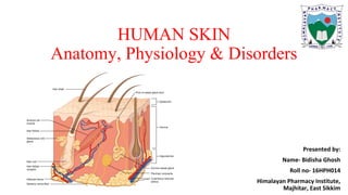
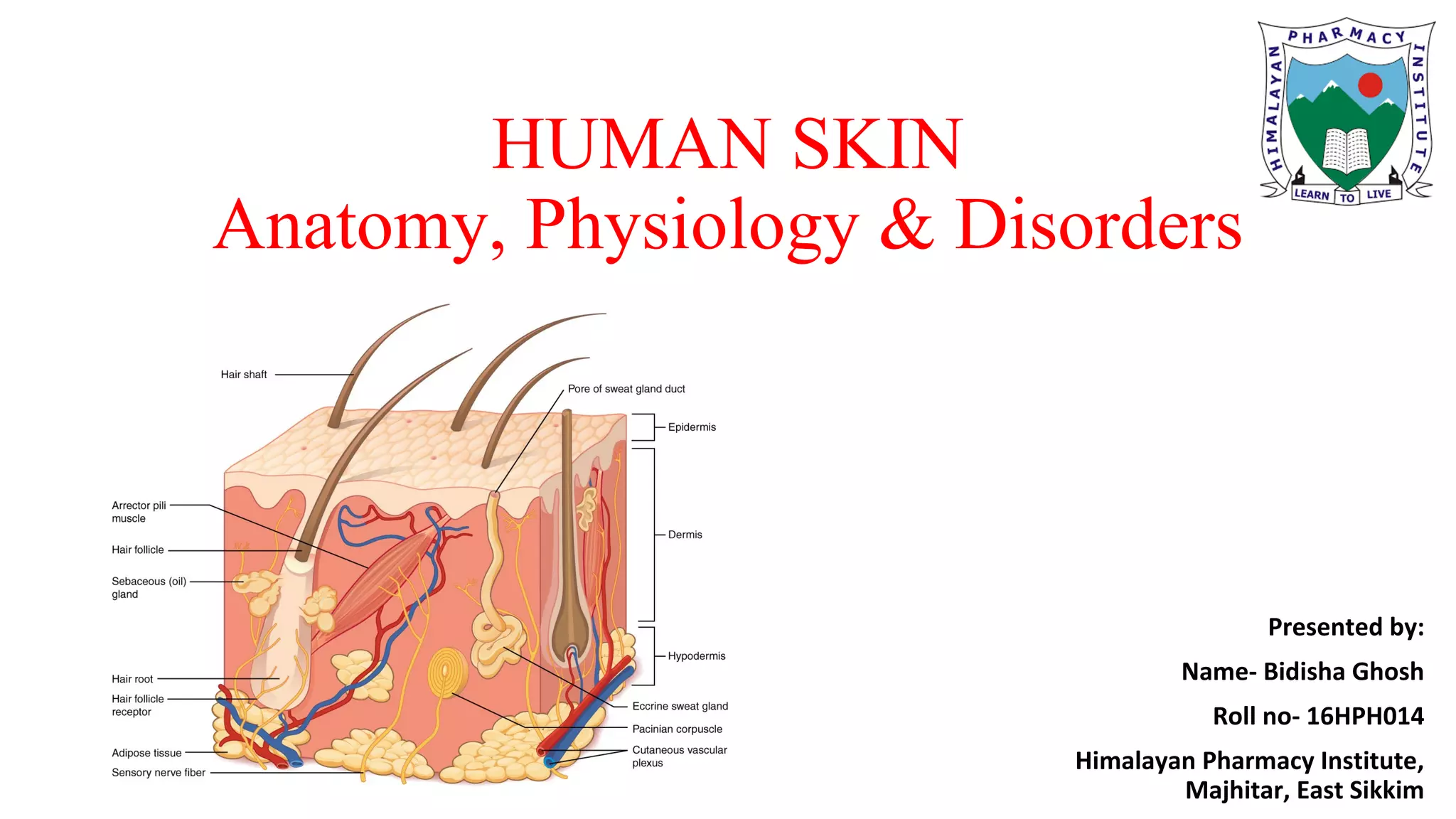

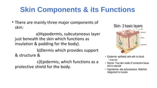
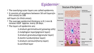


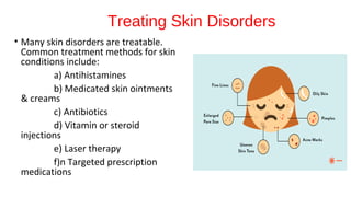


This document provides an overview of human skin anatomy, physiology, and common disorders. It describes the three main layers of skin - epidermis, dermis, and hypodermis - and their functions. Fifteen common skin disorders are then outlined, including acne, cold sores, hives, psoriasis, and skin cancers. Finally, it lists several treatment methods for skin conditions, such as antihistamines, medicated creams, antibiotics, and laser therapy.