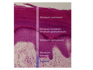
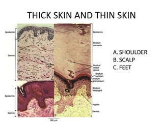
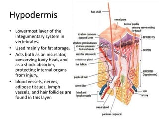
The epidermis is the outermost layer of skin composed of keratinocytes and other cell types. It forms a protective barrier against pathogens and regulates water loss. The epidermis consists of several layers - the stratum basale, stratum spinosum, stratum granulosum, stratum lucidum, and stratum corneum. The dermis lies beneath the epidermis and contains structures like hair follicles, sweat and oil glands, blood vessels, nerves. The hypodermis is the lowest layer and stores fat. Together these layers comprise the integumentary system which acts as a protective barrier for the body.
The skin is the largest organ of the body, with a total area of about 20 square feet. ... Skin has three layers: The epidermis, the outermost layer of skin, provides a waterproof barrier and creates our skin tone. The dermis, beneath the epidermis, contains tough connective tissue, hair follicles, and sweat glands.
basics of skin, review of skin, Integumentary system, the structure of the skin, Functions of skin, skin appendages, Hair, sweat glands, sebaceous glands, Nails, dermis, epidermis,
subcutaneous tissue. anatomy and physiology
This presentation includes structure and functions of sweat glands i.e. eccrine, apocrine and apoeccrine glands. mechanism of sweat secretion and role of sweat in thermoregulation is included.
Brief Anatomy of Skin and Skin GraftingRishi Gupta
Brief Anatomy of Skin and Skin Grafting.
Anatomy of Skin
History of skin grafting.
Recent Advances in Skin Grafting.
Dermal Substitutes.
Cell cultures in skin grafting.
Anatomy and physiology of skin. Explanation on epidermis, dermis and hypodermis. The various mechanoreceptors, pigments and glands of skin. Physiologic function of the skin
Integumentary system
-The organs of the integumentary system include the skin and its accessory structures including hair, nails, and glands, as well as blood vessels, muscles and nerves.
-Dermatology is the medical specialty for the diagnosis and treatment of disorders of the integumentary system.
Structure Of The Skin
The skin (cutaneous membrane) covers the body and is the largest organ of the body by surface area and weight.
Its area is about 2 square meters (22 square feet) and weighs 4.5-5kg (10-11 lb), about 7% of body weight.
It is 0.5 – 4 mm thick, thinnest on the eyelids, thickest on the heels, the average thickness is 1 – 2 mm.
It consists of two major layers:
Outer, thinner layer called the epidermis, consists of epithelial tissue. Inner, thicker layer called the dermis.
Beneath the dermis is a subcutaneous layer (also called hypodermis) which attaches the skin to the underlying tissues and organs.
1. EPIDERMIS
-Covers, protects and waterproofs.
Contains five main layers:
- Stratum Basale
- Stratum Spinosum
- Stratum Granulosum
- Stratum lucidum
- Stratum corneum
The epidermis has a number of important characteristics:
The epidermis is composed of keratinized stratified squamous epithelium.
-It contains four major types of cells:
Keratinocytes (90% of the cells): It produce keratin which is a tough fibrous protein that provides protection.
-Melanocytes: which produce the pigment melanin that protects against damage by ultraviolet radiation.
-Langerhans cells: involved in immune responses, arise from red bone marrow.
-Merkel cells: which function in the sensation of touch along with the adjacent tactile discs.
2. DERMIS
-It is a deeper layer of skin, composed of connective tissue containing collagen and elastic fibers.
-It contains blood and lymph vessels, nerves, and other structures, such as hair follicles and sweat glands.
-The epidermis is avascular and cells of this layer get their oxygen and nutrients from capillaries in the dermis.
-The dermis can be divided into papillary layer and reticular layer.
Hypodermis :
- The hypodermis (also called the subcutaneous layer) is a layer directly below the dermis and serves to connect the skin to the underlying fascia (fibrous tissue) of the bones and muscles.
--Two types of glands are present in the skin over most of the body
These are sweat glands and sebaceous glands.
--Function Of Skin
Protection:
Sensation:
Heat regulation:
Storage and synthesis:
Synthesis of vitamin D:
Excretion and homeostasis:
Secretion:
Absorption:
Water resistance:
Colour :
Healing of wounds:
Aids in the diagnosis:
The integumentary system consists of the skin and its accessory structures, including the hair, nails, sebaceous glands, and sweat glands.
The skin is largest organ and the exterior covering of the body.
It is a protective covering for the skeletal system and vital organs.
For the adult human the average surface area of the skin is between 1.5 – 2.0 square meters.
The thickness of the skin varies over all parts of the body and between men (1.3 mm) and women (1.26 mm).
The skin is composed of three primary layers, the epidermis, the dermis and the hypodermis.
The ph of the varies from 4.5 to 6. The epidermis is the outer or top layer and composed of stratified keratinised squamous epithelium.
There are several layers (strata) of cells in the epidermis -Stratum Corneum; Stratum Lucidum; Stratum Granulosum; Stratum Mucosum; Stratum Germinativum. The dermis is a connective tissue layer sandwiched between the epidermis and subcutaneous tissue. sometimes called the true skin. The hypodermis, also known as the subcutaneous layer, is the layer of tissue beneath the skin that is mostly composed of fat. The hypodermis also helps to regulate body temperature and stores energy.
GraphRAG is All You need? LLM & Knowledge GraphGuy Korland
Guy Korland, CEO and Co-founder of FalkorDB, will review two articles on the integration of language models with knowledge graphs.
1. Unifying Large Language Models and Knowledge Graphs: A Roadmap.
https://arxiv.org/abs/2306.08302
2. Microsoft Research's GraphRAG paper and a review paper on various uses of knowledge graphs:
https://www.microsoft.com/en-us/research/blog/graphrag-unlocking-llm-discovery-on-narrative-private-data/
Elevating Tactical DDD Patterns Through Object CalisthenicsDorra BARTAGUIZ
After immersing yourself in the blue book and its red counterpart, attending DDD-focused conferences, and applying tactical patterns, you're left with a crucial question: How do I ensure my design is effective? Tactical patterns within Domain-Driven Design (DDD) serve as guiding principles for creating clear and manageable domain models. However, achieving success with these patterns requires additional guidance. Interestingly, we've observed that a set of constraints initially designed for training purposes remarkably aligns with effective pattern implementation, offering a more ‘mechanical’ approach. Let's explore together how Object Calisthenics can elevate the design of your tactical DDD patterns, offering concrete help for those venturing into DDD for the first time!
Essentials of Automations: Optimizing FME Workflows with ParametersSafe Software
Are you looking to streamline your workflows and boost your projects’ efficiency? Do you find yourself searching for ways to add flexibility and control over your FME workflows? If so, you’re in the right place.
Join us for an insightful dive into the world of FME parameters, a critical element in optimizing workflow efficiency. This webinar marks the beginning of our three-part “Essentials of Automation” series. This first webinar is designed to equip you with the knowledge and skills to utilize parameters effectively: enhancing the flexibility, maintainability, and user control of your FME projects.
Here’s what you’ll gain:
- Essentials of FME Parameters: Understand the pivotal role of parameters, including Reader/Writer, Transformer, User, and FME Flow categories. Discover how they are the key to unlocking automation and optimization within your workflows.
- Practical Applications in FME Form: Delve into key user parameter types including choice, connections, and file URLs. Allow users to control how a workflow runs, making your workflows more reusable. Learn to import values and deliver the best user experience for your workflows while enhancing accuracy.
- Optimization Strategies in FME Flow: Explore the creation and strategic deployment of parameters in FME Flow, including the use of deployment and geometry parameters, to maximize workflow efficiency.
- Pro Tips for Success: Gain insights on parameterizing connections and leveraging new features like Conditional Visibility for clarity and simplicity.
We’ll wrap up with a glimpse into future webinars, followed by a Q&A session to address your specific questions surrounding this topic.
Don’t miss this opportunity to elevate your FME expertise and drive your projects to new heights of efficiency.
Connector Corner: Automate dynamic content and events by pushing a buttonDianaGray10
Here is something new! In our next Connector Corner webinar, we will demonstrate how you can use a single workflow to:
Create a campaign using Mailchimp with merge tags/fields
Send an interactive Slack channel message (using buttons)
Have the message received by managers and peers along with a test email for review
But there’s more:
In a second workflow supporting the same use case, you’ll see:
Your campaign sent to target colleagues for approval
If the “Approve” button is clicked, a Jira/Zendesk ticket is created for the marketing design team
But—if the “Reject” button is pushed, colleagues will be alerted via Slack message
Join us to learn more about this new, human-in-the-loop capability, brought to you by Integration Service connectors.
And...
Speakers:
Akshay Agnihotri, Product Manager
Charlie Greenberg, Host
LF Energy Webinar: Electrical Grid Modelling and Simulation Through PowSyBl -...DanBrown980551
Do you want to learn how to model and simulate an electrical network from scratch in under an hour?
Then welcome to this PowSyBl workshop, hosted by Rte, the French Transmission System Operator (TSO)!
During the webinar, you will discover the PowSyBl ecosystem as well as handle and study an electrical network through an interactive Python notebook.
PowSyBl is an open source project hosted by LF Energy, which offers a comprehensive set of features for electrical grid modelling and simulation. Among other advanced features, PowSyBl provides:
- A fully editable and extendable library for grid component modelling;
- Visualization tools to display your network;
- Grid simulation tools, such as power flows, security analyses (with or without remedial actions) and sensitivity analyses;
The framework is mostly written in Java, with a Python binding so that Python developers can access PowSyBl functionalities as well.
What you will learn during the webinar:
- For beginners: discover PowSyBl's functionalities through a quick general presentation and the notebook, without needing any expert coding skills;
- For advanced developers: master the skills to efficiently apply PowSyBl functionalities to your real-world scenarios.
Kubernetes & AI - Beauty and the Beast !?! @KCD Istanbul 2024Tobias Schneck
As AI technology is pushing into IT I was wondering myself, as an “infrastructure container kubernetes guy”, how get this fancy AI technology get managed from an infrastructure operational view? Is it possible to apply our lovely cloud native principals as well? What benefit’s both technologies could bring to each other?
Let me take this questions and provide you a short journey through existing deployment models and use cases for AI software. On practical examples, we discuss what cloud/on-premise strategy we may need for applying it to our own infrastructure to get it to work from an enterprise perspective. I want to give an overview about infrastructure requirements and technologies, what could be beneficial or limiting your AI use cases in an enterprise environment. An interactive Demo will give you some insides, what approaches I got already working for real.
Dev Dives: Train smarter, not harder – active learning and UiPath LLMs for do...UiPathCommunity
💥 Speed, accuracy, and scaling – discover the superpowers of GenAI in action with UiPath Document Understanding and Communications Mining™:
See how to accelerate model training and optimize model performance with active learning
Learn about the latest enhancements to out-of-the-box document processing – with little to no training required
Get an exclusive demo of the new family of UiPath LLMs – GenAI models specialized for processing different types of documents and messages
This is a hands-on session specifically designed for automation developers and AI enthusiasts seeking to enhance their knowledge in leveraging the latest intelligent document processing capabilities offered by UiPath.
Speakers:
👨🏫 Andras Palfi, Senior Product Manager, UiPath
👩🏫 Lenka Dulovicova, Product Program Manager, UiPath
Accelerate your Kubernetes clusters with Varnish CachingThijs Feryn
A presentation about the usage and availability of Varnish on Kubernetes. This talk explores the capabilities of Varnish caching and shows how to use the Varnish Helm chart to deploy it to Kubernetes.
This presentation was delivered at K8SUG Singapore. See https://feryn.eu/presentations/accelerate-your-kubernetes-clusters-with-varnish-caching-k8sug-singapore-28-2024 for more details.
Neuro-symbolic is not enough, we need neuro-*semantic*Frank van Harmelen
Neuro-symbolic (NeSy) AI is on the rise. However, simply machine learning on just any symbolic structure is not sufficient to really harvest the gains of NeSy. These will only be gained when the symbolic structures have an actual semantics. I give an operational definition of semantics as “predictable inference”.
All of this illustrated with link prediction over knowledge graphs, but the argument is general.
DevOps and Testing slides at DASA ConnectKari Kakkonen
My and Rik Marselis slides at 30.5.2024 DASA Connect conference. We discuss about what is testing, then what is agile testing and finally what is Testing in DevOps. Finally we had lovely workshop with the participants trying to find out different ways to think about quality and testing in different parts of the DevOps infinity loop.
3. • The epidermis as the body's major barrier
against an inhospitable environment, by
preventing pathogens from entering, making
the skin a natural barrier to infection.
• regulates the amount of water released from
the body into the atmosphere through
transepidermal water loss
5. • Keratinocyte is the predominant cell type in
the epidermis, the outermost layer of
the skin. Those keratinocytes found in the
basal layer of the skin are sometimes referred
to as "basal cells“. The primary function of
keratinocytes is the formation of a barrier
against environmental damage
6. • Melanocytes are melanin
producing cells located in the bottom layer
(the stratum basale) of the skin's epidermis,
the middle layer of the eye (the uvea),
7. • Langerhans cells are dendritic cells (antigen-
presenting immune cells) of the skin and
mucosa, and contain large granules
called Birbeck granules
• In skin infections, the local Langerhans cells
take up and process microbial antigens to
become fully functional antigen-presenting
cells.
8. • Merkel cells are oval receptor cells found in
the skin of vertebrates that have synaptic
contacts with somatosensory afferents. They
are associated with the sense of light touch
discrimination of shapes and textures.
9. LAYERS OF THE EPIDERMIS
• Stratum Corneum
• Stratum Lucidum
• Stratum Granulosum
• Stratum Spinosum
• Stratum Basale/Germinativum
10.
11. STRATUM GERMINATIVUM
• is a continuous layer of cells. It is often described
as one cell thick, though it may in fact be two to
three cells thick in glabrous (hairless) skin.
• is primarily made up of basal keratinocyte cells,
which can be considered the stem cells of the
epidermis. They divide to form the keratinocytes
of the stratum spinosum, which migrate
superficially
12. STRATUM SPINOSUM
• This layer is also referred to as the "spinous"
or "prickle-cell" layer. This appearance is due
to desmosomal connections of adjacent cells.
• Keratinization begins.
13. STRATUM GRANULOSUM
• thin layer of cells in
the epidermis. Keratinocytes migrating from
the underlying stratum spinosum become
known as granular cells in this layer. These
cells contain keratohyalin granules, protein
structures that promote hydration and cross
linking of keratin.
14. STRATUM LUCIDUM
• The stratum lucidum is a thin, clear layer of
dead skin cells in the epidermis named for its
translucent appearance under a microscope. It
is found only in areas of thick skin.
• The keratinocytes of the stratum lucidum do
not feature distinct boundaries and are filled
with eleidin, an intermediate form of keratin.
15. STRATUM CORNEUM
• The purpose of the stratum corneum is to
form a barrier to protect underlying tissue
from infection, dehydration, chemicals and
mechanical stress.
• Desquamation, the process of cell shedding
from the surface of the stratum corneum,
balances proliferating keratinocytes that form
in the stratum basale.
16. • During cornification, the process whereby
living keratinocytes are transformed into non-
living corneocytes, the cell membrane is
replaced by a layer of ceramides which
become covalently linked to an envelope of
structural proteins.
• Cells of the stratum corneum contain a dense
network of keratin
19. • Characteristics of the barrier
• Physical barrier through keratinocytes attached together via cell–
cell junctions and associated to cytoskeletal proteins, which gives
the epidermis its mechanical strength.
• Chemical barrier through the presence of highly
organized lipids, acids, hydrolytic enzymes and antimicrobial
peptides.
• Immunologically active barrier
through humoral and cellular constituents of the immune system.
• Water content of the stratum corneum drops towards the surface,
creating hostile conditions for pathogenic microorganism growth.
• An acidic pH (around 5.0) and low amounts of water make it hostile
to many micro organic pathogens.
• The presence of non-pathogenic microorganism on the epidermis
surface help defend against pathogenic one by
limiting food availability and through chemical secretions.
21. • lies beneath epidermis
• the thickest of the three layers of the skin.
• home to most of the skin’s structures,
including sweat and oil glands (which secrete
substances through openings in the skin called
pores, or comedos), hair follicles, nerve
endings, and blood and lymph ves-sels.
• the main components of the dermis are
connective tissues (collagenous fibers, elastic
fibers, reticular fibers)
22. Dermal Papillae
• Blood vessels in the
dermal papillae
nourish all hair
follicles and bring
oxygen and nutrients
to the lower layers of
epidermal cells.
• Responsible for
“fingerprints”
23. Arrector Pili
• small muscles
attached to hair
follicles in mammals
. Contraction of
these muscles
causes the hairs to
stand on end -
known colloquially
as goose bumps.
24. Nerve Fibers
• responsible for the
sense of touch,
relaying information
to the brain for
interpretation
• Thermoreceptors –
trigger shivering
25. Connective
Tissues
• Collagen - tough,
insoluble protein found
throughout the body in
the connective tissues
that hold muscles and
organs in place. In the
skin, collagen supports
the epidermis, lending
it its durability.
• Elas-tin, a similar
protein, keeps the skin
flexible.
27. • The eccrine glands are the true sweat glands.
Found over the entire body, these glands
regulate body temperature by bringing water
via the pores to the surface of the skin, where
it evaporates and releases heat.
• respond to heat, exercise, and fever, and some
eccrine glands, such as those on the palms,
respond to emo-tional stress as well. It’s these
glands that give you clammy hands when
you’re nervous.
28. • Apocrine glands are specialized sweat glands
that can be found only in the armpits and
pubic region.
• these glands secrete a milky sweat that
encourages the growth of bacteria responsible
for body odor. These glands are activated at
puberty when stimulated by hormones.
30. Apocrine vs. Eccrine
• Unlike apocrine glands, eccrine glands function
from child-hood, though they do increase their
activity at puberty.
• Though eccrine glands can produce up to two
liters of sweat an hour when they’re working at
their full potential, they’re not usually to blame
for body odor.
• Eccrine glands secrete mostly water, which
doesn’t encourage the growth of odor-producing
bacteria.
31. Sebaceous glands
• attached to hair follicles, cylindrical
structures that house the roots of the
hair
• found everywhere on the body except
for the palms of the hands and the
soles of the feet.
• Usually called into action by hormones
during puberty, these glands secrete
oil (sebum) that helps keep the skin
smooth and supple.
• The oil also helps keep skin waterproof
and protects against an overgrowth of
bacteria and fungi on the skin.
• At times, these glands overproduce
and cause acne, a condition in which
pores become clogged and inflamed.
32. blood and lymph
vessels
• blood vessels bring nutrients and oxygen
to the skin and remove cell waste and cell
products. The blood vessels also carry the
vitamin D produced in the skin back to the
rest of the body.
• Enlarged vessels that can be seen through
the skin are known as spider veins or
vari-cose veins. Broken blood vessels
appear as bruises.
• The lymph vessels bathe the tissues of the
skin with lymph, a milky substance that
contains infection-fighting immune
system cells. The cells work to destroy any
infection or invading organisms as the
lymph gradually circulates back through
the body’s tissues to the lymph nodes
33. Hypodermis
• Lowermost layer of the
integumentary system in
vertebrates.
• Used mainly for fat storage.
• Acts both as an insu-lator,
conserving body heat, and
as a shock absorber,
protecting internal organs
from injury.
• blood vessels, nerves,
adipose tissues, lymph
vessels, and hair follicles are
found in this layer.
34. Hair
• The hair can be divided into two parts, the
root and shaft.
• Root - the hair root is contained in a tube-
like structure called the hair follicle.
• New cells are created in the hair root.
• Papilla
– Large structure at the base of the hair follicle
• Matrix
– Surrounds the papilla composed of epithelial cells.
• Hair Fiber
– composed of keratin.
35. Hair
• Inner root sheath
– sheath formed of several
layers of cells.
• Hair follicle
– sac-like anatomical structure
from which hair grows.
36. Hair
– Shaft – 3 layers
• Cuticle -outer layer, the cuticle is
made up of hard, transparent
cells.
• It is the layer giving elasticity
and resiliency to the hair.
• Said to be water resistant
– Cortex
• layer between cuticle and
medulla.
• This contains the pigment and
keratin.
• Cortex determines the bulk and
strength of hair.
– Core/Medulla
• Innermost layer composed of
large cells.
• Supporting structure for a strand
of hair.