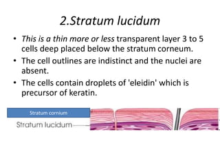
SlideShare a Scribd company logo
SKIN STRUCTURE AND FUNCTION
DR POTNURU SRINIVASA SUDHAKAR
M.D(HOMOEO)
SKIN
• Skin acts as a protective covering for the body
minimizing loss of water from the body
tissues.
• Various sensory nerve endings on the skin
help to protect the body from injury by
invoking appropriate response to noxious
stimuli.
• The skin also plays an important role in
temperature regulation.
Structure
• Two Main Layers
1. Epidermis
2. Dermis
EPIDERMIS
EPIDERMIS
• Epidermis is the most superficial layer and is
composed of stratified squamous type of
epithelmm.
• From outside inward stratified epithelium may be
divided into 5 layers:
l. Stratum corneum
2. Stratum lucidum
3. Stratum granulosum
4. Stratum spinosum
5. Stratum germinativum
l. Stratum corneum
• Stratum corneum is most superficially placed.
The cells are keratinised.
• The cell outlines are indistinct and the nuclei
are absent.
• This layer is thickest at the sole and the palm
and thinnest at the lip.
• Hairs, loops, nails, feathers, scales, etc. are
special outgrowth of this layer.
2.Stratum lucidum
• This is a thin more or less transparent layer 3 to 5
cells deep placed below the stratum corneum.
• The cell outlines are indistinct and the nuclei are
absent.
• The cells contain droplets of 'eleidin' which is
precursor of keratin.
Stratum cornium
3. Stratum granulosum
• Stratum granulosum is situated below the
stratum lucidum and consists of 3 to 5 layers of
flattened polyhedral cells filled with keratohyalin
granules which take a deep stain with
haematoxylin.
4. Stratum spinosum
• This is a broad layer of variable thickness and is made up of
polyhedral cells.
• The surface of these cells is apparently covered with
minute spines, which interdigitate with similar spines of
adjacent cells.
• These are consequently known as "prickle cells".
PRICKLE CELLS
4. Stratum spinosum
• As the cells move towards the surface, keratin
is synthesized within them.
• Microscopic studies indicate that the prickle
cells are in fact cytoplasmic protrusions and
the branches from two cells actually do not
have cytoplasmic continuity, but attached by
well-developed cytoplasmic nodes called
desmosomes.
4. Stratum spinosum
• These cells are basophilic and are supported by a
network of cytoplasmic fibrils.
• Scattered irregularly throughout this layer are
branched star-shaped cells known as Langerhans
cells.
• The functions of these cells are not known and
they are capable of active synthesis of DNA.
5. Stratum germinativum
• Stratum germinativum (stratum malpighi):
• This growing layer is composed of a single layer
of columnar epithelium which has got transverse,
thin, short cytoplasmic processes on its basal
lamina by means of which they anchor the
epithelium to the underlying dermis.
Stratum germinativum
5. Stratum germinativum
• These cuboidal to columnar cells with oblong
nuclei, placed perpendicularly on the basement
membrane, produce new cells to replace those of
the above layers by the process of mitosis.
• Mitotic activity of the epidermis occurs in
rhythmic diurnal cycles, the greatest activity of
the epidermis occurs in rhythmic diurnal cycles,
the greatest activity in humans occurring during
the hours of sleep.
5. Stratum germinativum
• At the junction of the epidermis with the
dermis there occur number of melanin-
containing branched cells, known as
melanocytes*.
• They send processes among the malpighian
cells and the melanin present in the deeper
cells of the stratum.
5. Stratum germinativum
• Malpighi is produced by these melanocytes.
Cytocrine secretion transfers the fully-formed
melanin granules from melanocytes to the
malpighian cells.
• The ratio of melanocytes to basal epidermal
cells varies between 1 to 4 and 1 to 10.
DERMIS (CUTIS VERA OR TRUE SKIN)
• The true skin is made up of connective tissue
and lies below the epidermal layer which it
supports and binds to the underlying tissues.
• It is made up chiefly of collagenous and elastic
fibres which provide it with a tensile strength
equal to that of a skin steel wire.
• This layer is utilized for the production of
leather after chemical processing.
• From the structural point of view the
superficial part of the dermis is compact and
forms the papillary layer because it sends
innumerable finger-like projections (papillae)
into the prickle cells layer of epidermis.
• The deeper part of the dermis is composed of
rather loose connective tissue and is
infiltrated with fat.
• This layer constitutes what is called reticular
layer of dermis.
• The reticular layer of the dermis merges
imperceptibly into the subcutaneous layer of
fat.
Cells of the Dermis
• These are fibroblasts from which the fibrous
tissue of the dermis develops.
• These cells are belonging to the reticulo-
endothelial system which protects the body
from invading bacteria.
Cells of the Dermis
• Some of these cells are loaded with melanin
pigments and are known as melanophores.
• Besides the above two types of cells-dermis
also contains other types of cells which are
found in areolar tissue of other regions of the
body.
Blood Vessels of the Dermis
• Though the epidermis is devoid of blood
vessels, the dermis has got a luxuriant supply
of capillary blood vessels which form a
network from which "hairpin“ loops supply
the tip of the dermal papillae.
• The blood vessels play an important role in
temperature regulation and have got rich
vasomotor innervations.
There is also a well-organised
lymphatic system in the dermis.
Nerves of the Dermis
• Besides the vasomotor nerves mentioned
above the dermis is provided with sensory
nerve endings of various types, which keep
the individual informed about the
surroundings.
Glands of the Dermis
• The lower ends of the sweat glands reach deep
part of the dermis and act as islands for re-
growth of epidermis in case of injury or bums
involving loss of epidermis.
• Sebaceous glands located in the dermis open into
the hair follicles and secrete oily material called
sebum.
Muscles of the Dermis (Corium)
• 1. Arrectores pilorum: A small bundle of
involuntary muscles attached to the hair in
such a way that they cause the hair to stand
on end.
• 2. Tunica dartos and other involuntary muscles
of the skin of the penis, nipple and areola are
located deep in the dermis.
Pigmentation of the Skin
• Five pigments are known to influence skin
colour:
1. All individuals except albinos have some
melanin pigment in their skin.
2. Melanin is a yellow to black pigment which is
found mainly in the stratum Malpighi, and is
formed on a specific cell particle, the
melanosome within the melanocytes.
Pigmentation of the Skin
3. Melanoid is supposed to be a degradation
product of melanin and is diffused through
the epidermis.
Melanoid has a different absorption band of
visible light.
4. Carotene is a yellow-orange pigment and
present in lipid-rich areas (i.e. the stratum
corneum and the fat of the corium and
subcutaneous tissue).
Pigmentation of the Skin
• Oxyhaemoglobin imparts a reddish hue to the
skin colour and is evident to areas where
there is rich arterial supply (i.e. face, neck,
palms, soles and nipples).
FUNCTIONS OF SKIN
1. Protection
• Stratum corneum which is the outermost
layer is horny and formed by the keratinised
stratified epithelial cells resist the action of
external agencies.
• It protects the internal individual injury and
bacterial invasion.
• The nails (hoops, horns, etc. in animals) are
also defensive appendages of the skin.
2. Regulation of body temperature
• Cutaneous vasoconstriction diverts the blood
to the interior of the body and so diminishes
heat loss.
• This is an important mechanism of protection
against cold environment.
2. Regulation of body temperature
• Vasodilatation of the skin helps in elimination
of heat from the body.
• Sweating--evaporation of sweat cools the
body, the latent heat of vaporisation being
0.56 cal/ gm of H20 vaporised.
2. Regulation of body temperature
• Hairs of the skin-in lower animals entangle a
layer of air in its meshes.
• This layer of stagnant air being poor conductor
of heat intervenes between the warm air in
contact with the skin and odd atmospheric air
and helps to preserve body temperature.
3. General sensation
• The skin serves as the medium for receiving the
general sensation.
• Touch, pain, temperature, etc. are sub-served by
the respective nerve endings present in the skin.
• The hair roots are richly supplied with nerves.
• Consequently, slight movement of the hair, such
as by a blast of wind arouses sensation.
• In this way, hairs help the sensory functions of
the skin.
4. Excretion:
• Through sweat and insensible perspiration,
salts and metabolites are excreted to some
extent.
• Synthetic function: Vitamin D is synthesized by
ultra-violet rays of the sun acting upon the
ergosterole present in the skin and
subcutaneous tissue.
• For this reason exposure to the sun ensures
supply of vitamin D.
5. Secretion:
• Sebum: The sebaceous glands secrete a fatty
substance which is rich in cholesterol, called
sebum.
• It helps to keep the skin greasy and prevents
drying.
• In cold climates the secretion is depressed and
the skin becomes dry and scaly.
• Sweat which is the active secretion of sweat
glands of skin plays a vital role in keeping the
body cool in hot environment.
5. Secretion
• Milk, the secretion of the mammary glands,
which are modified sweat glands. [In the
toads, certain poisonous glands are present in
the skin. They secrete a highly irritant fluid
and in this way act as a defensive weapon for
the animal.
6. Absorption
• Waxy layer hinders water absorption through
the skin.
• But the skin is not completely waterproof and
on prolonged exposure to water, there is
water absorption causing swelling of the
stratum corneum.
• Lipids are easily permeable through the skin.
• Lipid-soluble substances like vitamins are
easily absorbed through the skin.
7. Water balance
• Formation and evaporation of sweat is an
important factor in the regulation of water
balance of the body.
8. Acid-base equilibrium
• Sweat being acid in reaction, a good amount of acid is
excreted through it. In acidosis it becomes more acidic
and in this way helps to maintain a constant reaction in
the body fluids.
Storage Junctions:
• The dermis as well as the subcutaneous tissue can
store (a) fats, (b) water, (c) salts, and (d) glucose and
such other substances.
• It is found that when blood sugar level suddenly rises,
considerable amount of sugar may be temporarily
stored in the subcutaneous tissue and in the skin.
8. Acid-base equilibrium
• As sugar is gradually used up, the skin sugar is
slowly drawn in.
• The areolar tissue of the skin and subcutaneous
tissue have great affinity for water and any excess
of water in the body is stored mainly inside the
skin and subcutaneous tissue.
• After haemorrhage, the lost blood volume is
quickly replenished by drawing fluid from the
tissue and half of it comes from the skin.
8. Acid-base equilibrium
• Similarly, a large quantity of NaCl may remain
stored in the skin.
• After a chloride-rich diet or after an
intravenous saline injection, the heaviest load
of NaCl is found in the skin and subcutaneous
tissue.
• Where as during salt deprivation; blood
chlorides are maintained by drawing upon the
chlorides stored in the skin.
9. Gaseous exchange
• Absorption of oxygen and excretion of CO2 may
go onto a considerable extent through the skin in
those animals whose skin is thin and moist, e.g.
frogs.
• It is said that it can be carried to such an extent
that these animals may live even after the
extirpation of the lungs or in the hibernating
period when the lungs do not function.
• In man, this effect is negligible.
9. Gaseous exchange
• No oxygen is absorbed but a small amount of
CO2 is eliminated through the skin (probably it
passes out being dissolved in the sweat).
• Skin also exhibits signs of vitamin deficiency,
malnutrition and advancement of aging
(appearance of wrinkles on the skin).
GLANDS IN THE SKIN
Sweat Glands
• Modified smooth muscle cells, known as
myoepithelial cells invest the base of the glands
and wind round their ducts in such a way that by
their contraction they help in elimination of
sweat.
• There are about 3 million of active sweat glands
in the body.
• They are divided into two groups:
1. Eccrine and
2. Apocrine
Eccrine sweat gland
• The eccrine glands constitute the majority and
are generally found throughout the surface.
• They are most numerous on the palms and soles,
than on the head, but much less on the trunk and
the extremities.
• The glands on palms and soles do not respond to
temperature but secrete at time of emotional
stress.
• They secrete dilute sweat-containing NaCl, urea,
lactates, creatinine, uric acid, ammonia, amino
acids, glucose, water-soluble vitamins B and C.
NaCl content is variable.
Apocrine sweat gland
• The apocrine glands are larger sweat glands.
• They are derived from the hair follicles and include the
ceruminal glands of the ear, those of the eyelid, and
the mammary glands.
• The distribution of these large glands varies very much
from individual to individual and from race to race and
are found only in special regions, such as axilla, areola
of the nipples, mons pubis, labia majora, etc.
• They do not become active until puberty their
secretion is viscid, milky and odourless at first.
• After puberty their secretions vary in composition and
possess a characteristic odour.
• The odour is due to indoxyl, volatile fatty
acids, hydroxy acids, ammonia, etc.
• Bacterial activity in the presence of the
secretions contributes to the intensity and
quality of the odour.
• The mammary glands which eject milk are
form of the apocrine sweat glands and an
interesting study in evolution.
THANKYOU

More Related Content

Similar to SKIN STRUCTURE AND FUNCTION.pptx

Histology of Integumentary System for medical school.pdf
Histology of Integumentary System for medical school.pdfHistology of Integumentary System for medical school.pdf
Histology of Integumentary System for medical school.pdf
edwardkisembo46
 
SKIN AND ITS APPENDAGES.pptx
SKIN AND ITS APPENDAGES.pptxSKIN AND ITS APPENDAGES.pptx
SKIN AND ITS APPENDAGES.pptx
lekhakuriakose1
 
Pdf |Histology| - The skin -
Pdf |Histology| - The skin -Pdf |Histology| - The skin -
Pdf |Histology| - The skin -
Ameen ALzaidy
 
Brief Anatomy of Skin and Skin Grafting
Brief Anatomy of Skin and Skin GraftingBrief Anatomy of Skin and Skin Grafting
Brief Anatomy of Skin and Skin Grafting
Rishi Gupta
 
Integumentary System.pptx
Integumentary System.pptxIntegumentary System.pptx
Integumentary System.pptx
Paramanand Gopie
 
SKIN Final.pptx
SKIN Final.pptxSKIN Final.pptx
SKIN Final.pptx
Prarthana Sharma
 
Anatomy of the skin
Anatomy of the skinAnatomy of the skin
Integumentary System.pptx
Integumentary System.pptxIntegumentary System.pptx
Integumentary System.pptx
Sheetal Patil
 
SemiSolid Dosage Forms
SemiSolid Dosage FormsSemiSolid Dosage Forms
SemiSolid Dosage Forms
Eknath D. Ahire
 
Anatomy of skin S.A.pptx
Anatomy of skin S.A.pptxAnatomy of skin S.A.pptx
Anatomy of skin S.A.pptx
ssuser3ae7cb
 
2015 a&p integumentary handout
2015 a&p integumentary handout2015 a&p integumentary handout
2015 a&p integumentary handout
DINESH sharma
 
The integumentary system special class lec
The integumentary system special class lecThe integumentary system special class lec
The integumentary system special class lecBENZ BUNGGAY
 
Anatomy & physiology of skin
Anatomy & physiology of skinAnatomy & physiology of skin
Anatomy & physiology of skin
Ayshah Hashimi
 
Skin basic structure
Skin basic structureSkin basic structure
Skin basic structure
raviddv
 
ANATOMY AND PHYSIOLOGY OF SKIN.pptx
ANATOMY AND PHYSIOLOGY OF SKIN.pptxANATOMY AND PHYSIOLOGY OF SKIN.pptx
ANATOMY AND PHYSIOLOGY OF SKIN.pptx
Saili Gaude
 
skin.pptx
skin.pptxskin.pptx
skin.pptx
monagajbhiye
 
Skin presentation
Skin presentationSkin presentation
Structure of Skin.fxtxrxtcyftftfyfyfftfpptx
Structure of Skin.fxtxrxtcyftftfyfyfftfpptxStructure of Skin.fxtxrxtcyftftfyfyfftfpptx
Structure of Skin.fxtxrxtcyftftfyfyfftfpptx
hyyhelo226
 
Structure of Skin.pptx
Structure of Skin.pptxStructure of Skin.pptx
Structure of Skin.pptx
Shakti College Of Pharmacy
 
Anotomy of Skin
Anotomy of SkinAnotomy of Skin
Anotomy of Skin
abhay joshi
 

Similar to SKIN STRUCTURE AND FUNCTION.pptx (20)

Histology of Integumentary System for medical school.pdf
Histology of Integumentary System for medical school.pdfHistology of Integumentary System for medical school.pdf
Histology of Integumentary System for medical school.pdf
 
SKIN AND ITS APPENDAGES.pptx
SKIN AND ITS APPENDAGES.pptxSKIN AND ITS APPENDAGES.pptx
SKIN AND ITS APPENDAGES.pptx
 
Pdf |Histology| - The skin -
Pdf |Histology| - The skin -Pdf |Histology| - The skin -
Pdf |Histology| - The skin -
 
Brief Anatomy of Skin and Skin Grafting
Brief Anatomy of Skin and Skin GraftingBrief Anatomy of Skin and Skin Grafting
Brief Anatomy of Skin and Skin Grafting
 
Integumentary System.pptx
Integumentary System.pptxIntegumentary System.pptx
Integumentary System.pptx
 
SKIN Final.pptx
SKIN Final.pptxSKIN Final.pptx
SKIN Final.pptx
 
Anatomy of the skin
Anatomy of the skinAnatomy of the skin
Anatomy of the skin
 
Integumentary System.pptx
Integumentary System.pptxIntegumentary System.pptx
Integumentary System.pptx
 
SemiSolid Dosage Forms
SemiSolid Dosage FormsSemiSolid Dosage Forms
SemiSolid Dosage Forms
 
Anatomy of skin S.A.pptx
Anatomy of skin S.A.pptxAnatomy of skin S.A.pptx
Anatomy of skin S.A.pptx
 
2015 a&p integumentary handout
2015 a&p integumentary handout2015 a&p integumentary handout
2015 a&p integumentary handout
 
The integumentary system special class lec
The integumentary system special class lecThe integumentary system special class lec
The integumentary system special class lec
 
Anatomy & physiology of skin
Anatomy & physiology of skinAnatomy & physiology of skin
Anatomy & physiology of skin
 
Skin basic structure
Skin basic structureSkin basic structure
Skin basic structure
 
ANATOMY AND PHYSIOLOGY OF SKIN.pptx
ANATOMY AND PHYSIOLOGY OF SKIN.pptxANATOMY AND PHYSIOLOGY OF SKIN.pptx
ANATOMY AND PHYSIOLOGY OF SKIN.pptx
 
skin.pptx
skin.pptxskin.pptx
skin.pptx
 
Skin presentation
Skin presentationSkin presentation
Skin presentation
 
Structure of Skin.fxtxrxtcyftftfyfyfftfpptx
Structure of Skin.fxtxrxtcyftftfyfyfftfpptxStructure of Skin.fxtxrxtcyftftfyfyfftfpptx
Structure of Skin.fxtxrxtcyftftfyfyfftfpptx
 
Structure of Skin.pptx
Structure of Skin.pptxStructure of Skin.pptx
Structure of Skin.pptx
 
Anotomy of Skin
Anotomy of SkinAnotomy of Skin
Anotomy of Skin
 

More from DR.P.S SUDHAKAR

Cardiac Output.pptx FOR STUDENTS OF BHMS
Cardiac Output.pptx FOR STUDENTS OF BHMSCardiac Output.pptx FOR STUDENTS OF BHMS
Cardiac Output.pptx FOR STUDENTS OF BHMS
DR.P.S SUDHAKAR
 
BURNS FOR HOMOEOPATHIC STUDIENTS SURGERY
BURNS FOR HOMOEOPATHIC STUDIENTS SURGERYBURNS FOR HOMOEOPATHIC STUDIENTS SURGERY
BURNS FOR HOMOEOPATHIC STUDIENTS SURGERY
DR.P.S SUDHAKAR
 
SHOULDER DISLOCTION FOR HOMOEOPATHIC (SURGERY)
SHOULDER DISLOCTION FOR HOMOEOPATHIC (SURGERY)SHOULDER DISLOCTION FOR HOMOEOPATHIC (SURGERY)
SHOULDER DISLOCTION FOR HOMOEOPATHIC (SURGERY)
DR.P.S SUDHAKAR
 
RHEUMATOID ARTHRITIS FOR BHMS(HOMOEOPATHIC)
RHEUMATOID ARTHRITIS FOR BHMS(HOMOEOPATHIC)RHEUMATOID ARTHRITIS FOR BHMS(HOMOEOPATHIC)
RHEUMATOID ARTHRITIS FOR BHMS(HOMOEOPATHIC)
DR.P.S SUDHAKAR
 
FRACTURES FOR HOMOEOPATHIC STUDIENTS SURGERY
FRACTURES FOR HOMOEOPATHIC STUDIENTS SURGERYFRACTURES FOR HOMOEOPATHIC STUDIENTS SURGERY
FRACTURES FOR HOMOEOPATHIC STUDIENTS SURGERY
DR.P.S SUDHAKAR
 
FRACTURE AND DISLOCATION FOR BHMS STUDIENTS
FRACTURE AND DISLOCATION FOR BHMS STUDIENTSFRACTURE AND DISLOCATION FOR BHMS STUDIENTS
FRACTURE AND DISLOCATION FOR BHMS STUDIENTS
DR.P.S SUDHAKAR
 
DISEASES OF BONE FOR HOMOEOPATHIC STUDIENTS
DISEASES OF BONE FOR HOMOEOPATHIC STUDIENTSDISEASES OF BONE FOR HOMOEOPATHIC STUDIENTS
DISEASES OF BONE FOR HOMOEOPATHIC STUDIENTS
DR.P.S SUDHAKAR
 
COMPLICATIONS OF FRACTURES AND DISLOCATIONS.pptx
COMPLICATIONS OF FRACTURES AND DISLOCATIONS.pptxCOMPLICATIONS OF FRACTURES AND DISLOCATIONS.pptx
COMPLICATIONS OF FRACTURES AND DISLOCATIONS.pptx
DR.P.S SUDHAKAR
 
Colles ’ f racture surgery theory ppt bhms
Colles ’ f racture surgery theory  ppt bhmsColles ’ f racture surgery theory  ppt bhms
Colles ’ f racture surgery theory ppt bhms
DR.P.S SUDHAKAR
 
CARDIAC CYCLE AND EVENTS IN CARDIAC CYCLE
CARDIAC CYCLE AND EVENTS IN CARDIAC CYCLECARDIAC CYCLE AND EVENTS IN CARDIAC CYCLE
CARDIAC CYCLE AND EVENTS IN CARDIAC CYCLE
DR.P.S SUDHAKAR
 
BODY LANGUAGE AND USES TO READ THE PEOPLE
BODY LANGUAGE AND USES TO READ THE PEOPLEBODY LANGUAGE AND USES TO READ THE PEOPLE
BODY LANGUAGE AND USES TO READ THE PEOPLE
DR.P.S SUDHAKAR
 
PYREXIA
PYREXIAPYREXIA
EFFECTS OF EXPOSURE TO HIGH AND LOW
EFFECTS OF EXPOSURE TO HIGH AND LOWEFFECTS OF EXPOSURE TO HIGH AND LOW
EFFECTS OF EXPOSURE TO HIGH AND LOW
DR.P.S SUDHAKAR
 
BODY TEMPERATURE AND ITS REGULATION.pptx
BODY TEMPERATURE AND ITS REGULATION.pptxBODY TEMPERATURE AND ITS REGULATION.pptx
BODY TEMPERATURE AND ITS REGULATION.pptx
DR.P.S SUDHAKAR
 
BODY TEMPERATURE AND ITS REGULATION.pptx
BODY TEMPERATURE AND ITS REGULATION.pptxBODY TEMPERATURE AND ITS REGULATION.pptx
BODY TEMPERATURE AND ITS REGULATION.pptx
DR.P.S SUDHAKAR
 
Trichotillomania.pptx
Trichotillomania.pptxTrichotillomania.pptx
Trichotillomania.pptx
DR.P.S SUDHAKAR
 
CYSTITIS.pptx
CYSTITIS.pptxCYSTITIS.pptx
CYSTITIS.pptx
DR.P.S SUDHAKAR
 
MEDORRHINUM.pptx
MEDORRHINUM.pptxMEDORRHINUM.pptx
MEDORRHINUM.pptx
DR.P.S SUDHAKAR
 
E C G.pptx
E C G.pptxE C G.pptx
E C G.pptx
DR.P.S SUDHAKAR
 
HERNIAS.pptx
HERNIAS.pptxHERNIAS.pptx
HERNIAS.pptx
DR.P.S SUDHAKAR
 

More from DR.P.S SUDHAKAR (20)

Cardiac Output.pptx FOR STUDENTS OF BHMS
Cardiac Output.pptx FOR STUDENTS OF BHMSCardiac Output.pptx FOR STUDENTS OF BHMS
Cardiac Output.pptx FOR STUDENTS OF BHMS
 
BURNS FOR HOMOEOPATHIC STUDIENTS SURGERY
BURNS FOR HOMOEOPATHIC STUDIENTS SURGERYBURNS FOR HOMOEOPATHIC STUDIENTS SURGERY
BURNS FOR HOMOEOPATHIC STUDIENTS SURGERY
 
SHOULDER DISLOCTION FOR HOMOEOPATHIC (SURGERY)
SHOULDER DISLOCTION FOR HOMOEOPATHIC (SURGERY)SHOULDER DISLOCTION FOR HOMOEOPATHIC (SURGERY)
SHOULDER DISLOCTION FOR HOMOEOPATHIC (SURGERY)
 
RHEUMATOID ARTHRITIS FOR BHMS(HOMOEOPATHIC)
RHEUMATOID ARTHRITIS FOR BHMS(HOMOEOPATHIC)RHEUMATOID ARTHRITIS FOR BHMS(HOMOEOPATHIC)
RHEUMATOID ARTHRITIS FOR BHMS(HOMOEOPATHIC)
 
FRACTURES FOR HOMOEOPATHIC STUDIENTS SURGERY
FRACTURES FOR HOMOEOPATHIC STUDIENTS SURGERYFRACTURES FOR HOMOEOPATHIC STUDIENTS SURGERY
FRACTURES FOR HOMOEOPATHIC STUDIENTS SURGERY
 
FRACTURE AND DISLOCATION FOR BHMS STUDIENTS
FRACTURE AND DISLOCATION FOR BHMS STUDIENTSFRACTURE AND DISLOCATION FOR BHMS STUDIENTS
FRACTURE AND DISLOCATION FOR BHMS STUDIENTS
 
DISEASES OF BONE FOR HOMOEOPATHIC STUDIENTS
DISEASES OF BONE FOR HOMOEOPATHIC STUDIENTSDISEASES OF BONE FOR HOMOEOPATHIC STUDIENTS
DISEASES OF BONE FOR HOMOEOPATHIC STUDIENTS
 
COMPLICATIONS OF FRACTURES AND DISLOCATIONS.pptx
COMPLICATIONS OF FRACTURES AND DISLOCATIONS.pptxCOMPLICATIONS OF FRACTURES AND DISLOCATIONS.pptx
COMPLICATIONS OF FRACTURES AND DISLOCATIONS.pptx
 
Colles ’ f racture surgery theory ppt bhms
Colles ’ f racture surgery theory  ppt bhmsColles ’ f racture surgery theory  ppt bhms
Colles ’ f racture surgery theory ppt bhms
 
CARDIAC CYCLE AND EVENTS IN CARDIAC CYCLE
CARDIAC CYCLE AND EVENTS IN CARDIAC CYCLECARDIAC CYCLE AND EVENTS IN CARDIAC CYCLE
CARDIAC CYCLE AND EVENTS IN CARDIAC CYCLE
 
BODY LANGUAGE AND USES TO READ THE PEOPLE
BODY LANGUAGE AND USES TO READ THE PEOPLEBODY LANGUAGE AND USES TO READ THE PEOPLE
BODY LANGUAGE AND USES TO READ THE PEOPLE
 
PYREXIA
PYREXIAPYREXIA
PYREXIA
 
EFFECTS OF EXPOSURE TO HIGH AND LOW
EFFECTS OF EXPOSURE TO HIGH AND LOWEFFECTS OF EXPOSURE TO HIGH AND LOW
EFFECTS OF EXPOSURE TO HIGH AND LOW
 
BODY TEMPERATURE AND ITS REGULATION.pptx
BODY TEMPERATURE AND ITS REGULATION.pptxBODY TEMPERATURE AND ITS REGULATION.pptx
BODY TEMPERATURE AND ITS REGULATION.pptx
 
BODY TEMPERATURE AND ITS REGULATION.pptx
BODY TEMPERATURE AND ITS REGULATION.pptxBODY TEMPERATURE AND ITS REGULATION.pptx
BODY TEMPERATURE AND ITS REGULATION.pptx
 
Trichotillomania.pptx
Trichotillomania.pptxTrichotillomania.pptx
Trichotillomania.pptx
 
CYSTITIS.pptx
CYSTITIS.pptxCYSTITIS.pptx
CYSTITIS.pptx
 
MEDORRHINUM.pptx
MEDORRHINUM.pptxMEDORRHINUM.pptx
MEDORRHINUM.pptx
 
E C G.pptx
E C G.pptxE C G.pptx
E C G.pptx
 
HERNIAS.pptx
HERNIAS.pptxHERNIAS.pptx
HERNIAS.pptx
 

Recently uploaded

ANATOMY AND PHYSIOLOGY OF URINARY SYSTEM.pptx
ANATOMY AND PHYSIOLOGY OF URINARY SYSTEM.pptxANATOMY AND PHYSIOLOGY OF URINARY SYSTEM.pptx
ANATOMY AND PHYSIOLOGY OF URINARY SYSTEM.pptx
Swetaba Besh
 
New Directions in Targeted Therapeutic Approaches for Older Adults With Mantl...
New Directions in Targeted Therapeutic Approaches for Older Adults With Mantl...New Directions in Targeted Therapeutic Approaches for Older Adults With Mantl...
New Directions in Targeted Therapeutic Approaches for Older Adults With Mantl...
i3 Health
 
Physiology of Chemical Sensation of smell.pdf
Physiology of Chemical Sensation of smell.pdfPhysiology of Chemical Sensation of smell.pdf
Physiology of Chemical Sensation of smell.pdf
MedicoseAcademics
 
Charaka Samhita Sutra sthana Chapter 15 Upakalpaniyaadhyaya
Charaka Samhita Sutra sthana Chapter 15 UpakalpaniyaadhyayaCharaka Samhita Sutra sthana Chapter 15 Upakalpaniyaadhyaya
Charaka Samhita Sutra sthana Chapter 15 Upakalpaniyaadhyaya
Dr KHALID B.M
 
Superficial & Deep Fascia of the NECK.pptx
Superficial & Deep Fascia of the NECK.pptxSuperficial & Deep Fascia of the NECK.pptx
Superficial & Deep Fascia of the NECK.pptx
Dr. Rabia Inam Gandapore
 
Report Back from SGO 2024: What’s the Latest in Cervical Cancer?
Report Back from SGO 2024: What’s the Latest in Cervical Cancer?Report Back from SGO 2024: What’s the Latest in Cervical Cancer?
Report Back from SGO 2024: What’s the Latest in Cervical Cancer?
bkling
 
Evaluation of antidepressant activity of clitoris ternatea in animals
Evaluation of antidepressant activity of clitoris ternatea in animalsEvaluation of antidepressant activity of clitoris ternatea in animals
Evaluation of antidepressant activity of clitoris ternatea in animals
Shweta
 
Factory Supply Best Quality Pmk Oil CAS 28578–16–7 PMK Powder in Stock
Factory Supply Best Quality Pmk Oil CAS 28578–16–7 PMK Powder in StockFactory Supply Best Quality Pmk Oil CAS 28578–16–7 PMK Powder in Stock
Factory Supply Best Quality Pmk Oil CAS 28578–16–7 PMK Powder in Stock
rebeccabio
 
ARTIFICIAL INTELLIGENCE IN HEALTHCARE.pdf
ARTIFICIAL INTELLIGENCE IN  HEALTHCARE.pdfARTIFICIAL INTELLIGENCE IN  HEALTHCARE.pdf
ARTIFICIAL INTELLIGENCE IN HEALTHCARE.pdf
Anujkumaranit
 
Flu Vaccine Alert in Bangalore Karnataka
Flu Vaccine Alert in Bangalore KarnatakaFlu Vaccine Alert in Bangalore Karnataka
Flu Vaccine Alert in Bangalore Karnataka
addon Scans
 
Tom Selleck Health: A Comprehensive Look at the Iconic Actor’s Wellness Journey
Tom Selleck Health: A Comprehensive Look at the Iconic Actor’s Wellness JourneyTom Selleck Health: A Comprehensive Look at the Iconic Actor’s Wellness Journey
Tom Selleck Health: A Comprehensive Look at the Iconic Actor’s Wellness Journey
greendigital
 
Pulmonary Thromboembolism - etilogy, types, medical- Surgical and nursing man...
Pulmonary Thromboembolism - etilogy, types, medical- Surgical and nursing man...Pulmonary Thromboembolism - etilogy, types, medical- Surgical and nursing man...
Pulmonary Thromboembolism - etilogy, types, medical- Surgical and nursing man...
VarunMahajani
 
Maxilla, Mandible & Hyoid Bone & Clinical Correlations by Dr. RIG.pptx
Maxilla, Mandible & Hyoid Bone & Clinical Correlations by Dr. RIG.pptxMaxilla, Mandible & Hyoid Bone & Clinical Correlations by Dr. RIG.pptx
Maxilla, Mandible & Hyoid Bone & Clinical Correlations by Dr. RIG.pptx
Dr. Rabia Inam Gandapore
 
Hemodialysis: Chapter 3, Dialysis Water Unit - Dr.Gawad
Hemodialysis: Chapter 3, Dialysis Water Unit - Dr.GawadHemodialysis: Chapter 3, Dialysis Water Unit - Dr.Gawad
Hemodialysis: Chapter 3, Dialysis Water Unit - Dr.Gawad
NephroTube - Dr.Gawad
 
Non-respiratory Functions of the Lungs.pdf
Non-respiratory Functions of the Lungs.pdfNon-respiratory Functions of the Lungs.pdf
Non-respiratory Functions of the Lungs.pdf
MedicoseAcademics
 
24 Upakrama.pptx class ppt useful in all
24 Upakrama.pptx class ppt useful in all24 Upakrama.pptx class ppt useful in all
24 Upakrama.pptx class ppt useful in all
DrSathishMS1
 
Lung Cancer: Artificial Intelligence, Synergetics, Complex System Analysis, S...
Lung Cancer: Artificial Intelligence, Synergetics, Complex System Analysis, S...Lung Cancer: Artificial Intelligence, Synergetics, Complex System Analysis, S...
Lung Cancer: Artificial Intelligence, Synergetics, Complex System Analysis, S...
Oleg Kshivets
 
The POPPY STUDY (Preconception to post-partum cardiovascular function in prim...
The POPPY STUDY (Preconception to post-partum cardiovascular function in prim...The POPPY STUDY (Preconception to post-partum cardiovascular function in prim...
The POPPY STUDY (Preconception to post-partum cardiovascular function in prim...
Catherine Liao
 
Physiology of Special Chemical Sensation of Taste
Physiology of Special Chemical Sensation of TastePhysiology of Special Chemical Sensation of Taste
Physiology of Special Chemical Sensation of Taste
MedicoseAcademics
 
Charaka Samhita Sutra Sthana 9 Chapter khuddakachatuspadadhyaya
Charaka Samhita Sutra Sthana 9 Chapter khuddakachatuspadadhyayaCharaka Samhita Sutra Sthana 9 Chapter khuddakachatuspadadhyaya
Charaka Samhita Sutra Sthana 9 Chapter khuddakachatuspadadhyaya
Dr KHALID B.M
 

Recently uploaded (20)

ANATOMY AND PHYSIOLOGY OF URINARY SYSTEM.pptx
ANATOMY AND PHYSIOLOGY OF URINARY SYSTEM.pptxANATOMY AND PHYSIOLOGY OF URINARY SYSTEM.pptx
ANATOMY AND PHYSIOLOGY OF URINARY SYSTEM.pptx
 
New Directions in Targeted Therapeutic Approaches for Older Adults With Mantl...
New Directions in Targeted Therapeutic Approaches for Older Adults With Mantl...New Directions in Targeted Therapeutic Approaches for Older Adults With Mantl...
New Directions in Targeted Therapeutic Approaches for Older Adults With Mantl...
 
Physiology of Chemical Sensation of smell.pdf
Physiology of Chemical Sensation of smell.pdfPhysiology of Chemical Sensation of smell.pdf
Physiology of Chemical Sensation of smell.pdf
 
Charaka Samhita Sutra sthana Chapter 15 Upakalpaniyaadhyaya
Charaka Samhita Sutra sthana Chapter 15 UpakalpaniyaadhyayaCharaka Samhita Sutra sthana Chapter 15 Upakalpaniyaadhyaya
Charaka Samhita Sutra sthana Chapter 15 Upakalpaniyaadhyaya
 
Superficial & Deep Fascia of the NECK.pptx
Superficial & Deep Fascia of the NECK.pptxSuperficial & Deep Fascia of the NECK.pptx
Superficial & Deep Fascia of the NECK.pptx
 
Report Back from SGO 2024: What’s the Latest in Cervical Cancer?
Report Back from SGO 2024: What’s the Latest in Cervical Cancer?Report Back from SGO 2024: What’s the Latest in Cervical Cancer?
Report Back from SGO 2024: What’s the Latest in Cervical Cancer?
 
Evaluation of antidepressant activity of clitoris ternatea in animals
Evaluation of antidepressant activity of clitoris ternatea in animalsEvaluation of antidepressant activity of clitoris ternatea in animals
Evaluation of antidepressant activity of clitoris ternatea in animals
 
Factory Supply Best Quality Pmk Oil CAS 28578–16–7 PMK Powder in Stock
Factory Supply Best Quality Pmk Oil CAS 28578–16–7 PMK Powder in StockFactory Supply Best Quality Pmk Oil CAS 28578–16–7 PMK Powder in Stock
Factory Supply Best Quality Pmk Oil CAS 28578–16–7 PMK Powder in Stock
 
ARTIFICIAL INTELLIGENCE IN HEALTHCARE.pdf
ARTIFICIAL INTELLIGENCE IN  HEALTHCARE.pdfARTIFICIAL INTELLIGENCE IN  HEALTHCARE.pdf
ARTIFICIAL INTELLIGENCE IN HEALTHCARE.pdf
 
Flu Vaccine Alert in Bangalore Karnataka
Flu Vaccine Alert in Bangalore KarnatakaFlu Vaccine Alert in Bangalore Karnataka
Flu Vaccine Alert in Bangalore Karnataka
 
Tom Selleck Health: A Comprehensive Look at the Iconic Actor’s Wellness Journey
Tom Selleck Health: A Comprehensive Look at the Iconic Actor’s Wellness JourneyTom Selleck Health: A Comprehensive Look at the Iconic Actor’s Wellness Journey
Tom Selleck Health: A Comprehensive Look at the Iconic Actor’s Wellness Journey
 
Pulmonary Thromboembolism - etilogy, types, medical- Surgical and nursing man...
Pulmonary Thromboembolism - etilogy, types, medical- Surgical and nursing man...Pulmonary Thromboembolism - etilogy, types, medical- Surgical and nursing man...
Pulmonary Thromboembolism - etilogy, types, medical- Surgical and nursing man...
 
Maxilla, Mandible & Hyoid Bone & Clinical Correlations by Dr. RIG.pptx
Maxilla, Mandible & Hyoid Bone & Clinical Correlations by Dr. RIG.pptxMaxilla, Mandible & Hyoid Bone & Clinical Correlations by Dr. RIG.pptx
Maxilla, Mandible & Hyoid Bone & Clinical Correlations by Dr. RIG.pptx
 
Hemodialysis: Chapter 3, Dialysis Water Unit - Dr.Gawad
Hemodialysis: Chapter 3, Dialysis Water Unit - Dr.GawadHemodialysis: Chapter 3, Dialysis Water Unit - Dr.Gawad
Hemodialysis: Chapter 3, Dialysis Water Unit - Dr.Gawad
 
Non-respiratory Functions of the Lungs.pdf
Non-respiratory Functions of the Lungs.pdfNon-respiratory Functions of the Lungs.pdf
Non-respiratory Functions of the Lungs.pdf
 
24 Upakrama.pptx class ppt useful in all
24 Upakrama.pptx class ppt useful in all24 Upakrama.pptx class ppt useful in all
24 Upakrama.pptx class ppt useful in all
 
Lung Cancer: Artificial Intelligence, Synergetics, Complex System Analysis, S...
Lung Cancer: Artificial Intelligence, Synergetics, Complex System Analysis, S...Lung Cancer: Artificial Intelligence, Synergetics, Complex System Analysis, S...
Lung Cancer: Artificial Intelligence, Synergetics, Complex System Analysis, S...
 
The POPPY STUDY (Preconception to post-partum cardiovascular function in prim...
The POPPY STUDY (Preconception to post-partum cardiovascular function in prim...The POPPY STUDY (Preconception to post-partum cardiovascular function in prim...
The POPPY STUDY (Preconception to post-partum cardiovascular function in prim...
 
Physiology of Special Chemical Sensation of Taste
Physiology of Special Chemical Sensation of TastePhysiology of Special Chemical Sensation of Taste
Physiology of Special Chemical Sensation of Taste
 
Charaka Samhita Sutra Sthana 9 Chapter khuddakachatuspadadhyaya
Charaka Samhita Sutra Sthana 9 Chapter khuddakachatuspadadhyayaCharaka Samhita Sutra Sthana 9 Chapter khuddakachatuspadadhyaya
Charaka Samhita Sutra Sthana 9 Chapter khuddakachatuspadadhyaya
 

SKIN STRUCTURE AND FUNCTION.pptx

  • 1. SKIN STRUCTURE AND FUNCTION DR POTNURU SRINIVASA SUDHAKAR M.D(HOMOEO)
  • 3. • Skin acts as a protective covering for the body minimizing loss of water from the body tissues. • Various sensory nerve endings on the skin help to protect the body from injury by invoking appropriate response to noxious stimuli. • The skin also plays an important role in temperature regulation.
  • 4. Structure • Two Main Layers 1. Epidermis 2. Dermis
  • 5.
  • 7. EPIDERMIS • Epidermis is the most superficial layer and is composed of stratified squamous type of epithelmm. • From outside inward stratified epithelium may be divided into 5 layers: l. Stratum corneum 2. Stratum lucidum 3. Stratum granulosum 4. Stratum spinosum 5. Stratum germinativum
  • 8. l. Stratum corneum • Stratum corneum is most superficially placed. The cells are keratinised. • The cell outlines are indistinct and the nuclei are absent. • This layer is thickest at the sole and the palm and thinnest at the lip. • Hairs, loops, nails, feathers, scales, etc. are special outgrowth of this layer.
  • 9. 2.Stratum lucidum • This is a thin more or less transparent layer 3 to 5 cells deep placed below the stratum corneum. • The cell outlines are indistinct and the nuclei are absent. • The cells contain droplets of 'eleidin' which is precursor of keratin. Stratum cornium
  • 10. 3. Stratum granulosum • Stratum granulosum is situated below the stratum lucidum and consists of 3 to 5 layers of flattened polyhedral cells filled with keratohyalin granules which take a deep stain with haematoxylin.
  • 11. 4. Stratum spinosum • This is a broad layer of variable thickness and is made up of polyhedral cells. • The surface of these cells is apparently covered with minute spines, which interdigitate with similar spines of adjacent cells. • These are consequently known as "prickle cells". PRICKLE CELLS
  • 12. 4. Stratum spinosum • As the cells move towards the surface, keratin is synthesized within them. • Microscopic studies indicate that the prickle cells are in fact cytoplasmic protrusions and the branches from two cells actually do not have cytoplasmic continuity, but attached by well-developed cytoplasmic nodes called desmosomes.
  • 13. 4. Stratum spinosum • These cells are basophilic and are supported by a network of cytoplasmic fibrils. • Scattered irregularly throughout this layer are branched star-shaped cells known as Langerhans cells. • The functions of these cells are not known and they are capable of active synthesis of DNA.
  • 14. 5. Stratum germinativum • Stratum germinativum (stratum malpighi): • This growing layer is composed of a single layer of columnar epithelium which has got transverse, thin, short cytoplasmic processes on its basal lamina by means of which they anchor the epithelium to the underlying dermis. Stratum germinativum
  • 15. 5. Stratum germinativum • These cuboidal to columnar cells with oblong nuclei, placed perpendicularly on the basement membrane, produce new cells to replace those of the above layers by the process of mitosis. • Mitotic activity of the epidermis occurs in rhythmic diurnal cycles, the greatest activity of the epidermis occurs in rhythmic diurnal cycles, the greatest activity in humans occurring during the hours of sleep.
  • 16. 5. Stratum germinativum • At the junction of the epidermis with the dermis there occur number of melanin- containing branched cells, known as melanocytes*. • They send processes among the malpighian cells and the melanin present in the deeper cells of the stratum.
  • 17. 5. Stratum germinativum • Malpighi is produced by these melanocytes. Cytocrine secretion transfers the fully-formed melanin granules from melanocytes to the malpighian cells. • The ratio of melanocytes to basal epidermal cells varies between 1 to 4 and 1 to 10.
  • 18. DERMIS (CUTIS VERA OR TRUE SKIN)
  • 19. • The true skin is made up of connective tissue and lies below the epidermal layer which it supports and binds to the underlying tissues. • It is made up chiefly of collagenous and elastic fibres which provide it with a tensile strength equal to that of a skin steel wire. • This layer is utilized for the production of leather after chemical processing.
  • 20.
  • 21. • From the structural point of view the superficial part of the dermis is compact and forms the papillary layer because it sends innumerable finger-like projections (papillae) into the prickle cells layer of epidermis.
  • 22. • The deeper part of the dermis is composed of rather loose connective tissue and is infiltrated with fat. • This layer constitutes what is called reticular layer of dermis. • The reticular layer of the dermis merges imperceptibly into the subcutaneous layer of fat.
  • 23.
  • 24. Cells of the Dermis • These are fibroblasts from which the fibrous tissue of the dermis develops. • These cells are belonging to the reticulo- endothelial system which protects the body from invading bacteria.
  • 25. Cells of the Dermis • Some of these cells are loaded with melanin pigments and are known as melanophores. • Besides the above two types of cells-dermis also contains other types of cells which are found in areolar tissue of other regions of the body.
  • 26. Blood Vessels of the Dermis • Though the epidermis is devoid of blood vessels, the dermis has got a luxuriant supply of capillary blood vessels which form a network from which "hairpin“ loops supply the tip of the dermal papillae. • The blood vessels play an important role in temperature regulation and have got rich vasomotor innervations.
  • 27. There is also a well-organised lymphatic system in the dermis.
  • 28. Nerves of the Dermis • Besides the vasomotor nerves mentioned above the dermis is provided with sensory nerve endings of various types, which keep the individual informed about the surroundings.
  • 29. Glands of the Dermis • The lower ends of the sweat glands reach deep part of the dermis and act as islands for re- growth of epidermis in case of injury or bums involving loss of epidermis. • Sebaceous glands located in the dermis open into the hair follicles and secrete oily material called sebum.
  • 30. Muscles of the Dermis (Corium) • 1. Arrectores pilorum: A small bundle of involuntary muscles attached to the hair in such a way that they cause the hair to stand on end. • 2. Tunica dartos and other involuntary muscles of the skin of the penis, nipple and areola are located deep in the dermis.
  • 31. Pigmentation of the Skin • Five pigments are known to influence skin colour: 1. All individuals except albinos have some melanin pigment in their skin. 2. Melanin is a yellow to black pigment which is found mainly in the stratum Malpighi, and is formed on a specific cell particle, the melanosome within the melanocytes.
  • 32. Pigmentation of the Skin 3. Melanoid is supposed to be a degradation product of melanin and is diffused through the epidermis. Melanoid has a different absorption band of visible light. 4. Carotene is a yellow-orange pigment and present in lipid-rich areas (i.e. the stratum corneum and the fat of the corium and subcutaneous tissue).
  • 33. Pigmentation of the Skin • Oxyhaemoglobin imparts a reddish hue to the skin colour and is evident to areas where there is rich arterial supply (i.e. face, neck, palms, soles and nipples).
  • 35. 1. Protection • Stratum corneum which is the outermost layer is horny and formed by the keratinised stratified epithelial cells resist the action of external agencies. • It protects the internal individual injury and bacterial invasion. • The nails (hoops, horns, etc. in animals) are also defensive appendages of the skin.
  • 36. 2. Regulation of body temperature • Cutaneous vasoconstriction diverts the blood to the interior of the body and so diminishes heat loss. • This is an important mechanism of protection against cold environment.
  • 37. 2. Regulation of body temperature • Vasodilatation of the skin helps in elimination of heat from the body. • Sweating--evaporation of sweat cools the body, the latent heat of vaporisation being 0.56 cal/ gm of H20 vaporised.
  • 38. 2. Regulation of body temperature • Hairs of the skin-in lower animals entangle a layer of air in its meshes. • This layer of stagnant air being poor conductor of heat intervenes between the warm air in contact with the skin and odd atmospheric air and helps to preserve body temperature.
  • 39. 3. General sensation • The skin serves as the medium for receiving the general sensation. • Touch, pain, temperature, etc. are sub-served by the respective nerve endings present in the skin. • The hair roots are richly supplied with nerves. • Consequently, slight movement of the hair, such as by a blast of wind arouses sensation. • In this way, hairs help the sensory functions of the skin.
  • 40. 4. Excretion: • Through sweat and insensible perspiration, salts and metabolites are excreted to some extent. • Synthetic function: Vitamin D is synthesized by ultra-violet rays of the sun acting upon the ergosterole present in the skin and subcutaneous tissue. • For this reason exposure to the sun ensures supply of vitamin D.
  • 41. 5. Secretion: • Sebum: The sebaceous glands secrete a fatty substance which is rich in cholesterol, called sebum. • It helps to keep the skin greasy and prevents drying. • In cold climates the secretion is depressed and the skin becomes dry and scaly. • Sweat which is the active secretion of sweat glands of skin plays a vital role in keeping the body cool in hot environment.
  • 42. 5. Secretion • Milk, the secretion of the mammary glands, which are modified sweat glands. [In the toads, certain poisonous glands are present in the skin. They secrete a highly irritant fluid and in this way act as a defensive weapon for the animal.
  • 43. 6. Absorption • Waxy layer hinders water absorption through the skin. • But the skin is not completely waterproof and on prolonged exposure to water, there is water absorption causing swelling of the stratum corneum. • Lipids are easily permeable through the skin. • Lipid-soluble substances like vitamins are easily absorbed through the skin.
  • 44. 7. Water balance • Formation and evaporation of sweat is an important factor in the regulation of water balance of the body.
  • 45. 8. Acid-base equilibrium • Sweat being acid in reaction, a good amount of acid is excreted through it. In acidosis it becomes more acidic and in this way helps to maintain a constant reaction in the body fluids. Storage Junctions: • The dermis as well as the subcutaneous tissue can store (a) fats, (b) water, (c) salts, and (d) glucose and such other substances. • It is found that when blood sugar level suddenly rises, considerable amount of sugar may be temporarily stored in the subcutaneous tissue and in the skin.
  • 46. 8. Acid-base equilibrium • As sugar is gradually used up, the skin sugar is slowly drawn in. • The areolar tissue of the skin and subcutaneous tissue have great affinity for water and any excess of water in the body is stored mainly inside the skin and subcutaneous tissue. • After haemorrhage, the lost blood volume is quickly replenished by drawing fluid from the tissue and half of it comes from the skin.
  • 47. 8. Acid-base equilibrium • Similarly, a large quantity of NaCl may remain stored in the skin. • After a chloride-rich diet or after an intravenous saline injection, the heaviest load of NaCl is found in the skin and subcutaneous tissue. • Where as during salt deprivation; blood chlorides are maintained by drawing upon the chlorides stored in the skin.
  • 48. 9. Gaseous exchange • Absorption of oxygen and excretion of CO2 may go onto a considerable extent through the skin in those animals whose skin is thin and moist, e.g. frogs. • It is said that it can be carried to such an extent that these animals may live even after the extirpation of the lungs or in the hibernating period when the lungs do not function. • In man, this effect is negligible.
  • 49. 9. Gaseous exchange • No oxygen is absorbed but a small amount of CO2 is eliminated through the skin (probably it passes out being dissolved in the sweat). • Skin also exhibits signs of vitamin deficiency, malnutrition and advancement of aging (appearance of wrinkles on the skin).
  • 51. Sweat Glands • Modified smooth muscle cells, known as myoepithelial cells invest the base of the glands and wind round their ducts in such a way that by their contraction they help in elimination of sweat. • There are about 3 million of active sweat glands in the body. • They are divided into two groups: 1. Eccrine and 2. Apocrine
  • 52. Eccrine sweat gland • The eccrine glands constitute the majority and are generally found throughout the surface. • They are most numerous on the palms and soles, than on the head, but much less on the trunk and the extremities. • The glands on palms and soles do not respond to temperature but secrete at time of emotional stress. • They secrete dilute sweat-containing NaCl, urea, lactates, creatinine, uric acid, ammonia, amino acids, glucose, water-soluble vitamins B and C. NaCl content is variable.
  • 53. Apocrine sweat gland • The apocrine glands are larger sweat glands. • They are derived from the hair follicles and include the ceruminal glands of the ear, those of the eyelid, and the mammary glands. • The distribution of these large glands varies very much from individual to individual and from race to race and are found only in special regions, such as axilla, areola of the nipples, mons pubis, labia majora, etc. • They do not become active until puberty their secretion is viscid, milky and odourless at first. • After puberty their secretions vary in composition and possess a characteristic odour.
  • 54.
  • 55. • The odour is due to indoxyl, volatile fatty acids, hydroxy acids, ammonia, etc. • Bacterial activity in the presence of the secretions contributes to the intensity and quality of the odour. • The mammary glands which eject milk are form of the apocrine sweat glands and an interesting study in evolution.