Download to read offline

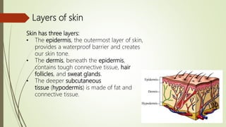

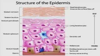
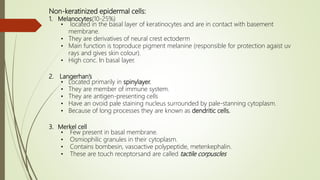
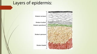
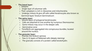
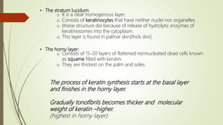

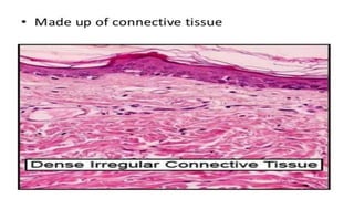
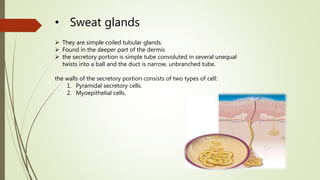
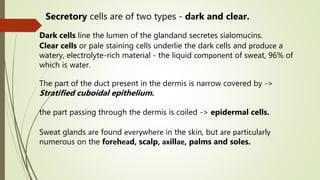

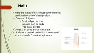
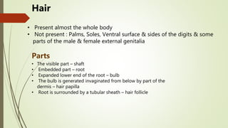

The skin consists of three layers - the epidermis, dermis, and subcutaneous tissue. The epidermis provides protection and contains melanocytes, Langerhans cells, and Merkel cells. The dermis contains tough connective tissue, hair follicles, and sweat glands. Sweat glands help regulate body temperature and excrete waste. Skin also contains sebaceous glands associated with hair follicles and nails composed of keratinized epithelial cells.


