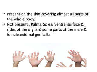
The skin and its appendages are summarized as follows:
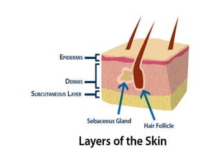
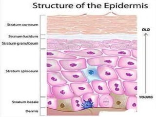
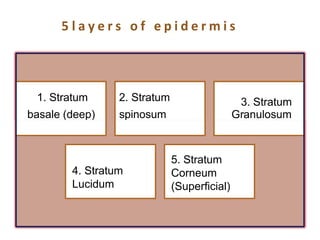
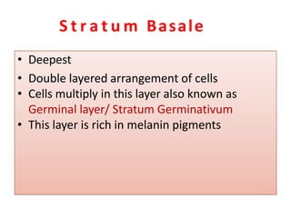
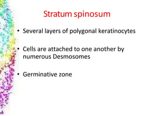
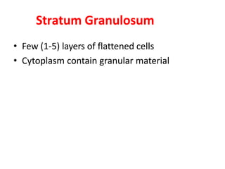
1. The skin is composed of two main layers - the epidermis and dermis. The epidermis contains stratified squamous epithelium in 5 layers that helps protect underlying tissues.
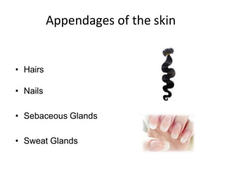
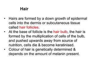
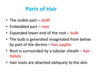
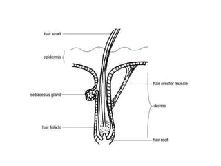
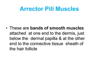
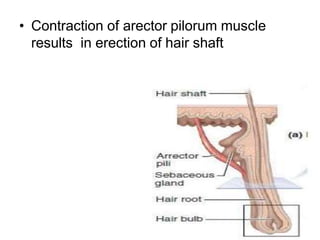
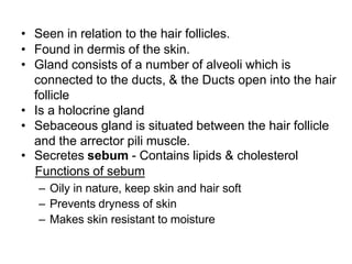
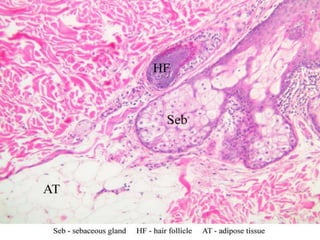
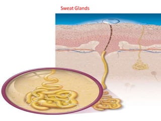
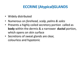
2. Hair, sebaceous glands, sweat glands, and nails are appendages of the skin. Hair grows from hair follicles which contain sebaceous and arrector pili muscles. Sebaceous glands secrete an oily substance while sweat glands secrete sweat to cool the body.
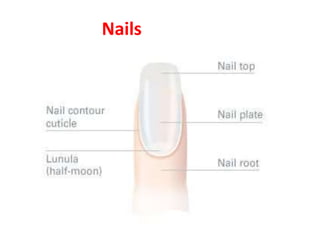
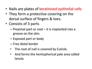
3. Nails provide protection to fingers and toes, consisting of three parts: the root, body,