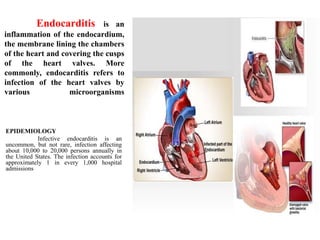
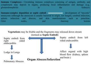
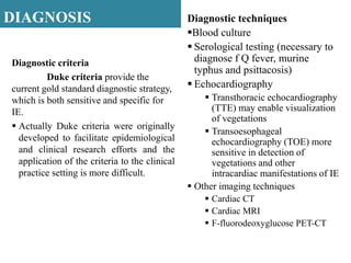
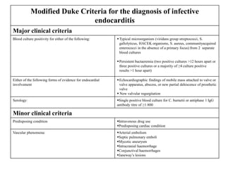
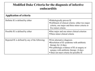
ENDOCARDITIS
Dr. Niranjana ES, Pharm D.,
Assistant Professor,
SSM College of Pharmacy, Erode Tamil Nadu
Endocarditis is an
inflammation of the endocardium,
the membrane lining the chambers
of the heart and covering the cusps
of the heart valves. More
commonly, endocarditis refers to
infection of the heart valves by
various microorganisms
EPIDEMIOLOGY
Infective endocarditis is an
uncommon, but not rare, infection affecting
about 10,000 to 20,000 persons annually in
the United States. The infection accounts for
approximately 1 in every 1,000 hospital
admissions
TYPES
Endocarditis is often referred to as; (depending on the pace and
severity of the clinical presentation)
 Acute
 Subacute.
Acute infective endocarditis - Associated with high fevers and
systemic toxicity. Virulent bacteria, such as Staphylococcus
aureus, frequently cause this syndrome, and if untreated, death
may occur within days to weeks.
Subacute infective endocarditis - More indolent and it is
caused by less-invasive organisms, such as viridans streptococci,
usually occurring in pre existing valvular heart disease.
*Although infective endocarditis is
often referred to as acute or subacute,
it is best classified based on the
etiologic organism, the anatomic site
of infection, and pathogenic risk
factors. Infection also may follow
surgical insertion of a prosthetic heart
valve, resulting in prosthetic-valve
endocarditis (PVE).
RISK FACTORS
Many types of structural heart disease
result in turbulent blood flow that increases the risk
for infective endocarditis
ETIOLOGIC ORGANISMS IN
INFECTIVE ENDOCARDITIS
Three groups of organisms
result in a majority of cases:
 Streptococci (Viridans streptococci/
α-haemolytic streptococci, S. mitis, S. sanguis,
S. Oralis, S. bovis)
 Staphylococci (commonly
s.aureus),
 Enterococci (E. faecalis, E. faecium )
 Others
 Fungi - Candida species, Aspergillus
species
 Presence of a prosthetic valve
(highest risk)
 Previous endocarditis (highest
risk)
 Complex cyanotic congenital
heart disease (e.g., single-
ventricle states)
 Surgically constructed
systemic pulmonary shunts or
conduits
 Acquired valvular dysfunction
(e.g., rheumatic heart disease)
 Hypertrophic cardiomyopathy
 Mitral valve prolapse with
regurgitation
 IVDA
CLINICAL PRESENTATION
Common sign and symptoms
• Fever (most common finding
and is often accompanied by
other vague symptoms )
• Heart murmurs
• Other nonspecific findings, such
as chills, weakness, dyspnea,
night sweats, weight loss, or
malaise
• classic signs of sepsis (those
with a history of IVDA and S.
aureus infective endocarditis)
• Splenomegaly (prolonged
endocarditis)
The clinical presentation of infective endocarditis is highly
variable and nonspecific
Peripheral manifestations
(“stigmata”) of endocarditis
 Osler nodes
 Janeway lesions
 Splinter hemorrhages
 Petechiae
 Clubbing of the fingers
 Roth spots
Osler nodes on palm and finger
Purplish or erythematous subcutaneous papules or nodules on
the pads of the fingers and toes. These lesions are 2 to 15 mm in size
and are painful and tender. These nodes are not specific for infective
endocarditis and may be the result of embolism, immunologic
phenomena, or both
Janeway lesions
Hemorrhagic, painless plaques on
the palms of the hands or soles of
the feet. These lesions are
believed to be embolic in origin.
Splinter hemorrhages
Thin, linear hemorrhages found
under the nail beds of the fingers
or toes. These lesions are not
specific for infective endocarditis
and more commonly are the result
of traumatic injuries.
Clubbing of the fingers—Proliferative
changes in the soft tissues about the
terminal phalanges observed in long-
standing endocarditis.
Petechiae -Small (usually
1 to 2 mm in diameter),
erythematous, painless,
hemorrhagic lesions.
These lesions appear
anywhere on the skin but
more frequently on the
anterior trunk, buccal
mucosa and palate, and
conjunctivae. Petechiae
are nonblanching and
resolve after a few days
Roth spots - Retinal infarct
with central pallor and
surrounding hemorrhage
Emboli
Embolic phenomena occur in up
to one-third of cases and may result in
significant complications. Left-sided
endocarditis can result in renal artery
emboli causing flank pain with
hematuria, splenic artery emboli
causing abdominal pain, and cerebral
emboli, which may result in
hemiplegia or alteration in mental
status. Right-sided endocarditis may
result in pulmonary emboli, causing
pleuritic pain with hemoptysis
LABORATORY ABNORMALITIES ASSOCIATED WITH ENDOCARDITIS
Haematology
Anemia(normocytic, normochromic),
leukocytosis, and thrombocytopenia
The white blood cell count is often
normal or only slightly elevated,.
Elevated erythrocyte sedimentation
rate (90% to 100% of patients)
Elevated C-reactive protein level
Urinary analysis
Proteinuria
Microscopic hematuria
(approximately 50% of
individuals)
Site of infection
Aortic valve more common than mitral
Aortic:
 Vegetation usually on ventricular aspect, all 3 cusps
usually affected
 Perforation or dysfunction of valve
 Root abscess
Mitral:
Dysfunction by rupture of chordae tendineae
PATHOPHYSIOLOGY
The normal valvular endothelium is resistant to bacterial
colonization upon intravascular challenge. Thus, the development of IE
requires the simultaneous occurrence of several independent factors:
Alteration of the cardiac valve surface to produce a suitable site for
bacterial attachment and colonization;
Bacteraemia with an organism capable of attaching to and colonizing valve
tissue;
Survival of bacteria on surface and creation of the infected mass or
‘vegetation’ by ‘burying’ of the proliferating organism within a protective
matrix of serum molecules (for example, fibrin and platelets)
PATHOPHYSIOLOGY
The common route for development of infective endocarditis is Hematogenic spread;
Turbulent blood flow related to primary valvular and
endothelial damage; mechanical injury by catheters or electrodes, or
injury arising from repeated injections of solid particles in IDU
This endothelial damage prompts the formation of
fibrin-platelet deposits overlying interstitial oedema; “nonbacterial
thrombotic endocarditis” (NBTE)
Transient Bacteraemia
(Bloodstream infection is a prerequisite for development of native valve
IE)
Readily detectible in a majority of patients after dental, gastrointestinal,
urologic and gynaecologic procedures
Adherence of the organism to the fibrin-platelet matrices of NBTE
Bacterial colonization of the valve
(stimulation of platelet aggregation and fibrin deposition)
Vegetation
(enlarges by further cycles of platelet-fibrin deposition and bacterial
proliferation)
Bacteria within vegetation grows slowly and are protected from
antibiotics and host defences
After internalization by endothelial cells, microbes such as S. aureus
evoke a potent proinflammatory chemokine response, including
increased expression of IL-6, IL-8 and monocyte chemotactic peptide.
Monocytes are drawn into the endothelial cell microenvironment,
where circulating bacteria may then bind directly to their surface,
inducing the release of tissue thromboplastin (tissue factor). This
release amplifies the procoagulant cascade leading to progressive
evolution of the vegetation
Local perivalvular damage
Perforation of a valve leaflet or
rupture of the chordae
tendineae, interventricular
septum or papillary muscle
Heart failure
Valvular Stenosis
stimulation of both humoral and
cellular immunity
(formation of circulatng antibodies
like Opsonic, agglutinating,
complement-fixing antibodies,
cryoglobulins and antibodies
directed against bacterial heat-
shock proteins and macroglobulins)
(Some organisms appear to bind to components of damaged
endothelium or NBTE, such as fibronectin, laminin and collagen
Other organisms may bind directly to, or be internalized by,
endothelial cells.
Staphylococcus- especially S. aureus the adhesion is mediated by
specific surface proteins that bind fibrinogen, such as clumping
factor, and coagulase (which mediate initial attachment to
vegetations); Additional virulence factors, such as α-toxin, then
mediate persistence and proliferation within maturing vegetations
Streptococci – adhesion is mediated by dextran , (complex bacterial-
derived extracellular polysaccharide) FimA (surface protein that
functions as an adhesin in the oral cavity)
host defence by platelets: platelets phagocytose
circulating staphylococci into engulfment vacuoles that
fuse with α-granules. These α-granules contain
antimicrobial peptides called platelet microbicidal proteins
(PMPs) organism then either killed within platelets or
survives and disseminates using a ‘Trojan Horse’
mechanism
Immune Complexes:- Circulating immune complexes consisting of antigen, antibody, and
complement may deposit in organs, producing local inflammation and damage (e.g.,
glomerulonephritis in the kidneys).
Immune-complex deposition or septic emboli development of “mycotic”
aneurysms (although the aneurysm is usually bacterial in origin, not fungal), cerebral infarction,
splenic infarction and abscess, and skin manifestations such as petechiae,
Osler nodes, and Janeway lesions
Organ Abscess/Infarction
Septic emboli from
right sided
endocarditis
Lodge in Lungs
Pulmonary Abscess
Septic emboli from left
sided endocarditis
Affect organd with high
blood flow (kidney, spleen
and brain )
Vegetations may be friable and the fragments may released down stream
(termed as Septic Emboli)
DIAGNOSIS
Diagnostic criteria
Duke criteria provide the
current gold standard diagnostic strategy,
which is both sensitive and specific for
IE.
 Actually Duke criteria were originally
developed to facilitate epidemiological
and clinical research efforts and the
application of the criteria to the clinical
practice setting is more difficult.
Diagnostic techniques
Blood culture
 Serological testing (necessary to
diagnose f Q fever, murine
typhus and psittacosis)
 Echocardiography
 Transthoracic echocardiography
(TTE) may enable visualization
of vegetations
 Transoesophageal
echocardiography (TOE) more
sensitive in detection of
vegetations and other
intracardiac manifestations of IE
 Other imaging techniques
 Cardiac CT
 Cardiac MRI
 F-fluorodeoxyglucose PET-CT
Modified Duke Criteria for the diagnosis of infective
endocarditis
Major clinical criteria
Blood culture positivity for either of the following: Typical microorganism (viridans group streptococci, S.
gallolyticus, HACEK organisms, S. aureus, communityacquired
enterococci in the absence of a primary focus) from 2 separate
blood cultures
Persistent bactaeremia (two positive cultures >12 hours apart or
three positive cultures or a majority of ≥4 culture positive
results >1 hour apart)
Either of the following forms of evidence for endocardial
involvement
Echocardiographic findings of mobile mass attached to valve or
valve apparatus, abscess, or new partial dehiscence of prosthetic
valve
 New valvular regurgitation
Serology: Single positive blood culture for C. burnetii or antiphase 1 IgG
antibody titre of ≥1:800
Minor clinical criteria
Predisposing condition Intravenous drug use
Predisposing cardiac condition
Vascular phenomena: Arterial embolism
Septic pulmonary emboli
Mycotic aneurysm
Intracranial haemorrhage
Conjunctival haemorrhages
Janeway’s lesions
Modified Duke Criteria for the diagnosis of infective
endocarditis
Application of criteria
Definite IE is defined by either Pathologically proven IE
Fulfilment of clinical criteria: either two major
criteria, one major and three minor criteria or
five minor criteria
Possible IE is defined by either One major and one minor clinical criterion
Three minor clinical criteria
Rejected IE is defined by any of the following: Firm alternative diagnosis
Resolution of IE syndrome with antibiotic
therapy for ≤4 days
No pathologic evidence of IE at surgery or
autopsy with antibiotic therapy ≤4 days
 Does not meet criteria for possible IE
COMPLICATIONS
 Neurological complications
includes
o Ischaemic or haemorrhagic stroke,
o Transient ischaemic attacks
o Silent cerebral embolism,
o Symptomatic or asymptomatic infectious
aneurysm
o Brain abscess
o Meningitis
o Toxic encephalopathy, and seizure
 Acute renal failure include:
o Immune complex & vasculitic
glomerulonephritis
o Renal infarction
o Haemodynamic impairment
 Rheumatic complications
o Peripheral arthritis
o Spondylodiscitis
 Splenic abscess
 Myocarditis and pericarditis
TREATMENT – Pharmacological
Antimicrobial Therapy
General principles of antimicrobial
therapy
 The primary goal of antibiotic
treatment is to eradicate infection,
including sterilizing vegetations
 The infected vegetations can pose a
variety of challenges. include focal
infection with high bacterial density,
slow rate of bacterial growth within
biofilms, and low microorganism
metabolic activity.
 Therefore, prolonged, parenteral,
bactericidal therapy is required for
attempted infection cure.
Duration of therapy
 The duration of therapy in IE must be sufficient to ensure
complete eradication of microorganisms within vegetations.
 Prolonged therapy is necessary because of the high bacterial
densities within vegetations and the relatively slow bactericidal
activity of some antibiotics such as β-lactams and vancomycin.
When bactericidal activity is rapid, shorter courses may be
feasible.
 Combination therapy with penicillin or ceftriaxone and an
aminoglycoside for 2 weeks is highly effective in viridans group
streptococci (VGS)
 Both β-lactam therapy alone and combination therapy with
nafcillin and an aminoglycoside for only 2 weeks have been
effective in patients with uncomplicated right-sided IE caused by
S aureus; monotherapy with a β-lactam would be selected for use
in cases of uncomplicated IE.
In the modern era, management of IE typically requires a multidisciplinary team
including, at a minimum, an infectious disease specialist, a cardiologist and a cardiac surgeon
Empirical therapy of suspected infective endocarditis
For patients who are diagnosed with possible IE and in unstable condition, empirical therapy
is recommended before positive blood culture reports are obtained
Antimicrobial Dose/route Comments
NVE, indolent presentation
Amoxicillin and
Gentamicin
2 g q4 h iv
1 mg/kg ABW
If patient is stable, ideally await blood cultures
Better activityagainst enterococci and many
HACEK microorganisms compared with
Benzylpenicillin Use Regimen2 if genuine
penicillin allergy
The role of gentamicin is controversial before
culture results are available
NVE, severe sepsis (no risk factors for enterobacteriaceae, pseudomonas)
Vancomycin and
Gentamicin
Dosed according to local guidelines
1 mg/kg IBW q12 h iv
If allergic to vancomysin, replace
withdaptomycin 6 mg/kgq12 h iv
NVE, severe sepsis and risk factors for multi-resistant enterobacteriaceae, pseudomonas
Vancomycin and
Meropenem
Dosed according to local guidelines, iv
2 g q8 h iv
PVE pendingblood culturesor with negative blood cultures
Vancomycin
Gentamycin
Rifampicin
1 g q12 h iv
1 mg/kg q12 h iv
300-600 mg q12h po/iv
Use lower dose of rifampicin in severe renal
impairment
Organism-specific considerations
Staphylococci
 IE may be caused by staphylococci that are
coagulase positive (S aureus) or coagulase
negative (S epidermidis, S lugdunensis,
and various other species).
 Coagulase-positive staphylococci were
traditionally believed to cause primarily
NVE and coagulase-negative staphylococci
(CoNS) were associated with PVE
 Recommended treatment regimens for
coagulase-negative staphylococci are the
same as those for S. aureus
 The critical distinction in selecting
antibiotic therapy for S. aureus– associated
IE is whether the isolate is methicillin-
resistant (MRSA) or methicillin susceptible
(MSSA)
Streptococci
 IE caused by these streptococci is uncommon
 Standard treatment for streptococcal IE is a β-
lactam antibiotic (such as penicillin,
amoxicillin or ceftriaxone) for 4 weeks.
 The addition of an aminoglycoside may
enable a shorter 2-week course of therapy
when administered once daily in combination
with ceftriaxone for streptococcal NVIE
 For streptococcal isolates with an increased
penicillin or ceftriaxone MIC, gentamicin
should be added
Enterococcus
 Enterococci are the third leading cause of IE
and account for ≈10% of cases in non IDUs
 Gentamicin should be administered in daily
multiple divided doses rather than a single
daily dose to patients with enterococcal IE
and normal renal function
Antibiotic treatment of infective endocarditis due to Staphylococcus spp (ESC Guidelines )
Antibiotic treatment of infective endocarditis due to oral streptococci and Streptococcus bovis group
(ESC Guidelines )
Antibiotic treatment of infective endocarditis due to oral streptococci and Streptococcus bovis group
Non Pharmacologic therapy
SURGERY
Surgery is an important adjunct in the
management of endocarditis and In
general, surgical management of IE is
reserved for leftsided disease
Indications for early phase surgery
(during antimicrobial therapy) include;
Heart failure (includes severe acute
regurgitation or valve obstruction causing
refractory pulmonary oedema/ shock,
severe acute regurgitation or valve
obstruction with persistent heart failure)
Uncontrolled infection
Prevention of embolism
Valvectomy and valve replacement are
performed to remove infected tissue and to
restore hemodynamic function.
Echocardiographic features that suggest the
need for surgery include;
Persistent vegetation or an increase in
vegetation size after prolonged antibiotic
treatmentt
Valve dysfunction,
Perivalvular extension (e.g., abscess)
Surgery also may be considered in cases of
PVE endocarditis caused by resistant
organisms (e.g., fungi or gram-negative
bacteria), or if there is persistent bacteremia
or other evidence of failure despite
appropriate antimicrobial therapy.
PREVENTION
Antimicrobial prophylaxis is given to at risk patients when
bacteraemia-inducing procedures are performed
REFERENCES
1. American Heart Association. Circulation 1997; 96: 358-366
2. 2015 ESC Guidelines for the management of infective endocarditis. European
Heart Journal; 36, 3075–3123 doi:10.1093/eurheartj/ehv319
3. Holland et al., Infective endocarditis. Nat Rev Dis Primers. ; 2: 16059.
doi:10.1038/nrdp.2016.59
4. Werdan et al. Mechanisms of infective endocarditis: pathogen-host interaction
and risk states. Nat Rev Cardiol. 2014;11:35–50
5. Joseph T. DiPiro et al.,Pharmacotherapy- A Pathophysiological Approach;7 th
edition.
ENDOCARDITIS

ENDOCARDITIS

  • 1.
    ENDOCARDITIS Dr. Niranjana ES,Pharm D., Assistant Professor, SSM College of Pharmacy, Erode Tamil Nadu
  • 2.
    Endocarditis is an inflammationof the endocardium, the membrane lining the chambers of the heart and covering the cusps of the heart valves. More commonly, endocarditis refers to infection of the heart valves by various microorganisms EPIDEMIOLOGY Infective endocarditis is an uncommon, but not rare, infection affecting about 10,000 to 20,000 persons annually in the United States. The infection accounts for approximately 1 in every 1,000 hospital admissions
  • 3.
    TYPES Endocarditis is oftenreferred to as; (depending on the pace and severity of the clinical presentation)  Acute  Subacute. Acute infective endocarditis - Associated with high fevers and systemic toxicity. Virulent bacteria, such as Staphylococcus aureus, frequently cause this syndrome, and if untreated, death may occur within days to weeks. Subacute infective endocarditis - More indolent and it is caused by less-invasive organisms, such as viridans streptococci, usually occurring in pre existing valvular heart disease. *Although infective endocarditis is often referred to as acute or subacute, it is best classified based on the etiologic organism, the anatomic site of infection, and pathogenic risk factors. Infection also may follow surgical insertion of a prosthetic heart valve, resulting in prosthetic-valve endocarditis (PVE).
  • 4.
    RISK FACTORS Many typesof structural heart disease result in turbulent blood flow that increases the risk for infective endocarditis ETIOLOGIC ORGANISMS IN INFECTIVE ENDOCARDITIS Three groups of organisms result in a majority of cases:  Streptococci (Viridans streptococci/ α-haemolytic streptococci, S. mitis, S. sanguis, S. Oralis, S. bovis)  Staphylococci (commonly s.aureus),  Enterococci (E. faecalis, E. faecium )  Others  Fungi - Candida species, Aspergillus species  Presence of a prosthetic valve (highest risk)  Previous endocarditis (highest risk)  Complex cyanotic congenital heart disease (e.g., single- ventricle states)  Surgically constructed systemic pulmonary shunts or conduits  Acquired valvular dysfunction (e.g., rheumatic heart disease)  Hypertrophic cardiomyopathy  Mitral valve prolapse with regurgitation  IVDA
  • 5.
    CLINICAL PRESENTATION Common signand symptoms • Fever (most common finding and is often accompanied by other vague symptoms ) • Heart murmurs • Other nonspecific findings, such as chills, weakness, dyspnea, night sweats, weight loss, or malaise • classic signs of sepsis (those with a history of IVDA and S. aureus infective endocarditis) • Splenomegaly (prolonged endocarditis) The clinical presentation of infective endocarditis is highly variable and nonspecific Peripheral manifestations (“stigmata”) of endocarditis  Osler nodes  Janeway lesions  Splinter hemorrhages  Petechiae  Clubbing of the fingers  Roth spots
  • 6.
    Osler nodes onpalm and finger Purplish or erythematous subcutaneous papules or nodules on the pads of the fingers and toes. These lesions are 2 to 15 mm in size and are painful and tender. These nodes are not specific for infective endocarditis and may be the result of embolism, immunologic phenomena, or both
  • 7.
    Janeway lesions Hemorrhagic, painlessplaques on the palms of the hands or soles of the feet. These lesions are believed to be embolic in origin. Splinter hemorrhages Thin, linear hemorrhages found under the nail beds of the fingers or toes. These lesions are not specific for infective endocarditis and more commonly are the result of traumatic injuries.
  • 8.
    Clubbing of thefingers—Proliferative changes in the soft tissues about the terminal phalanges observed in long- standing endocarditis. Petechiae -Small (usually 1 to 2 mm in diameter), erythematous, painless, hemorrhagic lesions. These lesions appear anywhere on the skin but more frequently on the anterior trunk, buccal mucosa and palate, and conjunctivae. Petechiae are nonblanching and resolve after a few days
  • 9.
    Roth spots -Retinal infarct with central pallor and surrounding hemorrhage Emboli Embolic phenomena occur in up to one-third of cases and may result in significant complications. Left-sided endocarditis can result in renal artery emboli causing flank pain with hematuria, splenic artery emboli causing abdominal pain, and cerebral emboli, which may result in hemiplegia or alteration in mental status. Right-sided endocarditis may result in pulmonary emboli, causing pleuritic pain with hemoptysis
  • 10.
    LABORATORY ABNORMALITIES ASSOCIATEDWITH ENDOCARDITIS Haematology Anemia(normocytic, normochromic), leukocytosis, and thrombocytopenia The white blood cell count is often normal or only slightly elevated,. Elevated erythrocyte sedimentation rate (90% to 100% of patients) Elevated C-reactive protein level Urinary analysis Proteinuria Microscopic hematuria (approximately 50% of individuals)
  • 11.
    Site of infection Aorticvalve more common than mitral Aortic:  Vegetation usually on ventricular aspect, all 3 cusps usually affected  Perforation or dysfunction of valve  Root abscess Mitral: Dysfunction by rupture of chordae tendineae
  • 12.
    PATHOPHYSIOLOGY The normal valvularendothelium is resistant to bacterial colonization upon intravascular challenge. Thus, the development of IE requires the simultaneous occurrence of several independent factors: Alteration of the cardiac valve surface to produce a suitable site for bacterial attachment and colonization; Bacteraemia with an organism capable of attaching to and colonizing valve tissue; Survival of bacteria on surface and creation of the infected mass or ‘vegetation’ by ‘burying’ of the proliferating organism within a protective matrix of serum molecules (for example, fibrin and platelets)
  • 14.
    PATHOPHYSIOLOGY The common routefor development of infective endocarditis is Hematogenic spread; Turbulent blood flow related to primary valvular and endothelial damage; mechanical injury by catheters or electrodes, or injury arising from repeated injections of solid particles in IDU This endothelial damage prompts the formation of fibrin-platelet deposits overlying interstitial oedema; “nonbacterial thrombotic endocarditis” (NBTE) Transient Bacteraemia (Bloodstream infection is a prerequisite for development of native valve IE) Readily detectible in a majority of patients after dental, gastrointestinal, urologic and gynaecologic procedures Adherence of the organism to the fibrin-platelet matrices of NBTE Bacterial colonization of the valve (stimulation of platelet aggregation and fibrin deposition) Vegetation (enlarges by further cycles of platelet-fibrin deposition and bacterial proliferation) Bacteria within vegetation grows slowly and are protected from antibiotics and host defences After internalization by endothelial cells, microbes such as S. aureus evoke a potent proinflammatory chemokine response, including increased expression of IL-6, IL-8 and monocyte chemotactic peptide. Monocytes are drawn into the endothelial cell microenvironment, where circulating bacteria may then bind directly to their surface, inducing the release of tissue thromboplastin (tissue factor). This release amplifies the procoagulant cascade leading to progressive evolution of the vegetation Local perivalvular damage Perforation of a valve leaflet or rupture of the chordae tendineae, interventricular septum or papillary muscle Heart failure Valvular Stenosis stimulation of both humoral and cellular immunity (formation of circulatng antibodies like Opsonic, agglutinating, complement-fixing antibodies, cryoglobulins and antibodies directed against bacterial heat- shock proteins and macroglobulins) (Some organisms appear to bind to components of damaged endothelium or NBTE, such as fibronectin, laminin and collagen Other organisms may bind directly to, or be internalized by, endothelial cells. Staphylococcus- especially S. aureus the adhesion is mediated by specific surface proteins that bind fibrinogen, such as clumping factor, and coagulase (which mediate initial attachment to vegetations); Additional virulence factors, such as α-toxin, then mediate persistence and proliferation within maturing vegetations Streptococci – adhesion is mediated by dextran , (complex bacterial- derived extracellular polysaccharide) FimA (surface protein that functions as an adhesin in the oral cavity) host defence by platelets: platelets phagocytose circulating staphylococci into engulfment vacuoles that fuse with α-granules. These α-granules contain antimicrobial peptides called platelet microbicidal proteins (PMPs) organism then either killed within platelets or survives and disseminates using a ‘Trojan Horse’ mechanism
  • 15.
    Immune Complexes:- Circulatingimmune complexes consisting of antigen, antibody, and complement may deposit in organs, producing local inflammation and damage (e.g., glomerulonephritis in the kidneys). Immune-complex deposition or septic emboli development of “mycotic” aneurysms (although the aneurysm is usually bacterial in origin, not fungal), cerebral infarction, splenic infarction and abscess, and skin manifestations such as petechiae, Osler nodes, and Janeway lesions Organ Abscess/Infarction Septic emboli from right sided endocarditis Lodge in Lungs Pulmonary Abscess Septic emboli from left sided endocarditis Affect organd with high blood flow (kidney, spleen and brain ) Vegetations may be friable and the fragments may released down stream (termed as Septic Emboli)
  • 17.
    DIAGNOSIS Diagnostic criteria Duke criteriaprovide the current gold standard diagnostic strategy, which is both sensitive and specific for IE.  Actually Duke criteria were originally developed to facilitate epidemiological and clinical research efforts and the application of the criteria to the clinical practice setting is more difficult. Diagnostic techniques Blood culture  Serological testing (necessary to diagnose f Q fever, murine typhus and psittacosis)  Echocardiography  Transthoracic echocardiography (TTE) may enable visualization of vegetations  Transoesophageal echocardiography (TOE) more sensitive in detection of vegetations and other intracardiac manifestations of IE  Other imaging techniques  Cardiac CT  Cardiac MRI  F-fluorodeoxyglucose PET-CT
  • 18.
    Modified Duke Criteriafor the diagnosis of infective endocarditis Major clinical criteria Blood culture positivity for either of the following: Typical microorganism (viridans group streptococci, S. gallolyticus, HACEK organisms, S. aureus, communityacquired enterococci in the absence of a primary focus) from 2 separate blood cultures Persistent bactaeremia (two positive cultures >12 hours apart or three positive cultures or a majority of ≥4 culture positive results >1 hour apart) Either of the following forms of evidence for endocardial involvement Echocardiographic findings of mobile mass attached to valve or valve apparatus, abscess, or new partial dehiscence of prosthetic valve  New valvular regurgitation Serology: Single positive blood culture for C. burnetii or antiphase 1 IgG antibody titre of ≥1:800 Minor clinical criteria Predisposing condition Intravenous drug use Predisposing cardiac condition Vascular phenomena: Arterial embolism Septic pulmonary emboli Mycotic aneurysm Intracranial haemorrhage Conjunctival haemorrhages Janeway’s lesions
  • 19.
    Modified Duke Criteriafor the diagnosis of infective endocarditis Application of criteria Definite IE is defined by either Pathologically proven IE Fulfilment of clinical criteria: either two major criteria, one major and three minor criteria or five minor criteria Possible IE is defined by either One major and one minor clinical criterion Three minor clinical criteria Rejected IE is defined by any of the following: Firm alternative diagnosis Resolution of IE syndrome with antibiotic therapy for ≤4 days No pathologic evidence of IE at surgery or autopsy with antibiotic therapy ≤4 days  Does not meet criteria for possible IE
  • 20.
    COMPLICATIONS  Neurological complications includes oIschaemic or haemorrhagic stroke, o Transient ischaemic attacks o Silent cerebral embolism, o Symptomatic or asymptomatic infectious aneurysm o Brain abscess o Meningitis o Toxic encephalopathy, and seizure  Acute renal failure include: o Immune complex & vasculitic glomerulonephritis o Renal infarction o Haemodynamic impairment  Rheumatic complications o Peripheral arthritis o Spondylodiscitis  Splenic abscess  Myocarditis and pericarditis
  • 21.
    TREATMENT – Pharmacological AntimicrobialTherapy General principles of antimicrobial therapy  The primary goal of antibiotic treatment is to eradicate infection, including sterilizing vegetations  The infected vegetations can pose a variety of challenges. include focal infection with high bacterial density, slow rate of bacterial growth within biofilms, and low microorganism metabolic activity.  Therefore, prolonged, parenteral, bactericidal therapy is required for attempted infection cure. Duration of therapy  The duration of therapy in IE must be sufficient to ensure complete eradication of microorganisms within vegetations.  Prolonged therapy is necessary because of the high bacterial densities within vegetations and the relatively slow bactericidal activity of some antibiotics such as β-lactams and vancomycin. When bactericidal activity is rapid, shorter courses may be feasible.  Combination therapy with penicillin or ceftriaxone and an aminoglycoside for 2 weeks is highly effective in viridans group streptococci (VGS)  Both β-lactam therapy alone and combination therapy with nafcillin and an aminoglycoside for only 2 weeks have been effective in patients with uncomplicated right-sided IE caused by S aureus; monotherapy with a β-lactam would be selected for use in cases of uncomplicated IE. In the modern era, management of IE typically requires a multidisciplinary team including, at a minimum, an infectious disease specialist, a cardiologist and a cardiac surgeon
  • 22.
    Empirical therapy ofsuspected infective endocarditis For patients who are diagnosed with possible IE and in unstable condition, empirical therapy is recommended before positive blood culture reports are obtained Antimicrobial Dose/route Comments NVE, indolent presentation Amoxicillin and Gentamicin 2 g q4 h iv 1 mg/kg ABW If patient is stable, ideally await blood cultures Better activityagainst enterococci and many HACEK microorganisms compared with Benzylpenicillin Use Regimen2 if genuine penicillin allergy The role of gentamicin is controversial before culture results are available NVE, severe sepsis (no risk factors for enterobacteriaceae, pseudomonas) Vancomycin and Gentamicin Dosed according to local guidelines 1 mg/kg IBW q12 h iv If allergic to vancomysin, replace withdaptomycin 6 mg/kgq12 h iv NVE, severe sepsis and risk factors for multi-resistant enterobacteriaceae, pseudomonas Vancomycin and Meropenem Dosed according to local guidelines, iv 2 g q8 h iv PVE pendingblood culturesor with negative blood cultures Vancomycin Gentamycin Rifampicin 1 g q12 h iv 1 mg/kg q12 h iv 300-600 mg q12h po/iv Use lower dose of rifampicin in severe renal impairment
  • 23.
    Organism-specific considerations Staphylococci  IEmay be caused by staphylococci that are coagulase positive (S aureus) or coagulase negative (S epidermidis, S lugdunensis, and various other species).  Coagulase-positive staphylococci were traditionally believed to cause primarily NVE and coagulase-negative staphylococci (CoNS) were associated with PVE  Recommended treatment regimens for coagulase-negative staphylococci are the same as those for S. aureus  The critical distinction in selecting antibiotic therapy for S. aureus– associated IE is whether the isolate is methicillin- resistant (MRSA) or methicillin susceptible (MSSA) Streptococci  IE caused by these streptococci is uncommon  Standard treatment for streptococcal IE is a β- lactam antibiotic (such as penicillin, amoxicillin or ceftriaxone) for 4 weeks.  The addition of an aminoglycoside may enable a shorter 2-week course of therapy when administered once daily in combination with ceftriaxone for streptococcal NVIE  For streptococcal isolates with an increased penicillin or ceftriaxone MIC, gentamicin should be added Enterococcus  Enterococci are the third leading cause of IE and account for ≈10% of cases in non IDUs  Gentamicin should be administered in daily multiple divided doses rather than a single daily dose to patients with enterococcal IE and normal renal function
  • 26.
    Antibiotic treatment ofinfective endocarditis due to Staphylococcus spp (ESC Guidelines )
  • 27.
    Antibiotic treatment ofinfective endocarditis due to oral streptococci and Streptococcus bovis group (ESC Guidelines )
  • 28.
    Antibiotic treatment ofinfective endocarditis due to oral streptococci and Streptococcus bovis group
  • 30.
    Non Pharmacologic therapy SURGERY Surgeryis an important adjunct in the management of endocarditis and In general, surgical management of IE is reserved for leftsided disease Indications for early phase surgery (during antimicrobial therapy) include; Heart failure (includes severe acute regurgitation or valve obstruction causing refractory pulmonary oedema/ shock, severe acute regurgitation or valve obstruction with persistent heart failure) Uncontrolled infection Prevention of embolism Valvectomy and valve replacement are performed to remove infected tissue and to restore hemodynamic function. Echocardiographic features that suggest the need for surgery include; Persistent vegetation or an increase in vegetation size after prolonged antibiotic treatmentt Valve dysfunction, Perivalvular extension (e.g., abscess) Surgery also may be considered in cases of PVE endocarditis caused by resistant organisms (e.g., fungi or gram-negative bacteria), or if there is persistent bacteremia or other evidence of failure despite appropriate antimicrobial therapy.
  • 31.
    PREVENTION Antimicrobial prophylaxis isgiven to at risk patients when bacteraemia-inducing procedures are performed REFERENCES 1. American Heart Association. Circulation 1997; 96: 358-366 2. 2015 ESC Guidelines for the management of infective endocarditis. European Heart Journal; 36, 3075–3123 doi:10.1093/eurheartj/ehv319 3. Holland et al., Infective endocarditis. Nat Rev Dis Primers. ; 2: 16059. doi:10.1038/nrdp.2016.59 4. Werdan et al. Mechanisms of infective endocarditis: pathogen-host interaction and risk states. Nat Rev Cardiol. 2014;11:35–50 5. Joseph T. DiPiro et al.,Pharmacotherapy- A Pathophysiological Approach;7 th edition.