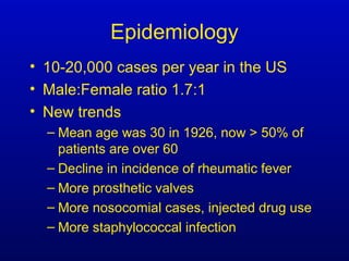
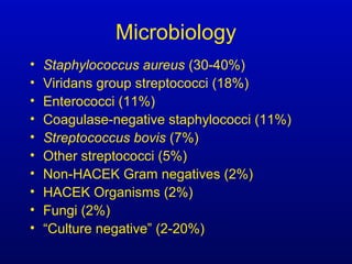
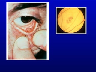
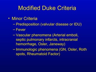
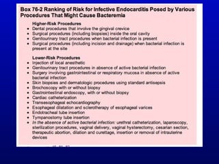
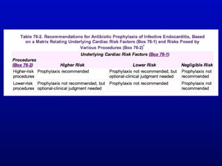
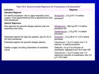
Infective Endocarditis
Epidemiology
• 10-20,000 cases per year in the US
• Male:Female ratio 1.7:1
• New trends
– Mean age was 30 in 1926, now > 50% of
patients are over 60
– Decline in incidence of rheumatic fever
– More prosthetic valves
– More nosocomial cases, injected drug use
– More staphylococcal infection
Epidemiology
• Mitral valve alone 28-45%
• Aortic valve alone 5-36% (bicuspid
valve in 20% of all native valve IE)
• Both mitral and aortic valves 0-36%
• Tricuspid valve 0-6%
• Pulmonic valve <1%
• Right and left sided 0-4%
Classification
• OLD
– Subacute Bacterial Endocarditis
• Death in 3-6 months
– Acute Bacterial Endocarditis
• Death in < 6 weeks
• NEW
– Native Valve Endocarditis
– Prosthetic Valve Endocarditis
Pathogenesis
• Alteration of the valvular endothelial
surface leading to deposition of
platelets and fibrin
• Bacteremia with seeding of non-
bacterial thrombotic vegetation (NBTE)
• Adherence and growth, further platelet
and fibrin deposition
• Extension to adjacent structures
– Papillary muscle, aortic valve ring abscess,
conduction system
Pathogenesis
• Low pressure side of structural lesion
– Atrial side of mitral valve (MR)
– Ventricular side of aortic valve (AR, AS with R)
– Congenital abnormality (MV prolapse, bicuspid AV)
– Scarring from rheumatic heart disease or sclerosis
as a consequence of aging
– Prosthetic valves
• Other turbulence, high-velocity jets
– Ventricular septal defect
– Stenotic valve
• Direct mechanical damage from catheters,
pacemaker leads
Pathogenesis
• Transient bacteremia
– Traumatization of mucosal surface
colonized with bacteria (oral, GI)
– Low grade, cleared in 15-30 minutes
– Susceptibility to complement-mediated
bacterial killing
• Leads to concept of prophylaxis
Microbiology
• Staphylococcus aureus (30-40%)
• Viridans group streptococci (18%)
• Enterococci (11%)
• Coagulase-negative staphylococci (11%)
• Streptococcus bovis (7%)
• Other streptococci (5%)
• Non-HACEK Gram negatives (2%)
• HACEK Organisms (2%)
• Fungi (2%)
• “Culture negative” (2-20%)
Characteristics of Causative Organisms
• Adherence factors critical for growth in the
vegetation
– Can adhere to damaged valves (Staph, Strep and
Enterococci have adhesins that mediate
attachment)
– Staph adhesin binds fibrinogen and fibronectin
– Bacteria trigger tissue-factor production from local
monocytes and induce platelet aggregation so the
organisms become enveloped in the vegetation
– Protection from immune clearance leads to large
numbers of bacteria (109
-1010
per g of tissue)
Risk Factors
• Structural heart disease
– Rheumatic, congenital, aging
– Prosthetic heart valves
• Injected drug use
• Invasive procedures (?)
• Indwelling vascular devices
• Other infection with bacteremia (e.g.
pneumonia, meningitis)
• History of infective endocarditis
Clinical Manifestations
• Symptoms
– Fever, sweats, chills
– Anorexia, malaise, weight loss
• Signs
– Anemia (normochromic, normocytic)
– Splenomegaly
– Microscopic hematuria, proteinuria
– New or changing heart murmur, CHF
– Embolic or immunologic dermatologic signs
– Hypergammaglobulinemia, elevated ESR, CRP,
RF
Cardiac Pathologic Changes
• Vegetations on valve closure lines
• Destruction and perforation of valve
leaflet
• Rupture of chordae tendinae,
intraventricular septum, papillary muscles
• Valve ring abscess
• Myocardial abscess
• Conduction abnormalities
S. Aureus mitral valve vegetation, anterior
leaflet
Pathologic Changes
• Kidney
– Immune complex glomerulonephritis
– Emboli with infarction, abscess
• Aortic mycotic aneurysms
• Cerebral embolism
– Infarction, abscess, mycotic aneurysms
– Purulent meningitis is rare
Pathologic Changes
• Splenic enlargement, infarction
• Septic or bland pulmonary embolism
• Skin
– Petechiae
– Osler nodes: diffuse infiltrate of neutrophils, and
monocytes in the dermal vessels with immune
complex deposition. Tender and erythematous
– Janeway lesions: septic emboli with bacteria,
neutrophils and SQ hemorrhage and necrosis.
Blanching and non-tender. Palms and soles
Case Definition
• 1977 Pelletier and Petersdorf criteria
• 1981 von Reyn criteria
• 1994 Duke criteria
• 2000 Modified Duke criteria
Modified Duke Criteria
• Major Criteria
–Positive blood cultures with typical
organisms
–Persistently positive blood cultures
–Evidence of Endocardial involvement
• Positive Echocardiogram
– Oscillating intracardiac mass
– Abscess
– Dehiscence of prosthetic valve
• New Valvular regurgitation
Modified Duke Criteria
• Minor Criteria
– Predisposition (valvular disease or IDU)
– Fever
– Vascular phenomena (Arterial emboli,
septic pulmonary infarcts, intracranial
hemorrhage, Osler, Janeway)
– Immunologic phenomena (GN, Osler, Roth
spots, Rheumatoid Factor)
Modified Duke Criteria
• Definite IE
– Pathologic criteria
– Clinical criteria
• 2 Major Criteria OR
• 1 Major and 3 minor Criteria OR
• 5 Minor Criteria
• Possible IE
• 1 Major and 1 Minor OR
• 3 Minor
• Rejected IE
Blood Cultures
• MULTIPLE BLOOD CULTURES
BEFORE EMPIRIC THERAPY
• If not critically ill
– 3 blood cultures over 12-24 hour period
– ? Delay therapy until diagnosis confirmed
• If critically ill
– 3 blood cultures over one hour
• No more than 2 from same venipuncture
• Relatively constant bacteremia
“Culture Negative” IE
• Less common with improved blood
culture methods
• Special media required
– Brucella, Mycoplasma, Chlamydia,
Histoplasma, Legionella, Bartonella
• Longer incubation may be required
– HACEK
• Coxiella burnetii (Q Fever), Trophyrema
whipplei will not grow in cell-free media
HACEK
• Haemophilus aphrophilus, H.
paraphrophilus, parainfluenzae
• Actinobacillus actinomycetemcomitans
• Cardiobacterium hominis
• Eikenella corrodens
• Kingella kingae
Other microbiologic methods
• PCR
– Coxiella burnetii
– Tropheryma whipplei
– Bartonella henselae
• Serology
– Coxiella burnetii
– Bartonella
– Brucella
– Legionella
– Chlamydophila psittaci
Echocardiography
• Transthoracic
– Relatively low sensitivity
– Good specificity
• Transesophageal
– Detection of valve ring abscess (87% vs.
28% sensitivity for TTE)
– Detection of prosthetic valve IE
When to go to TEE first?
• Limited thoracic windows = TTE low
sensitivity
• Prosthetic valves
• Prior valvular abnormality
• S. aureus bacteremia and suspected IE
• Bacteremia with organisms likely to
cause IE
= high prior probability of IE
Other tests
• Electrocardiogram
– Conduction delays
– Ischemia or infarction
• Chest X-ray
– Septic emboli in right-sided IE
– Valve calcification
– CHF
Treatment of IE
• Native vs. Prosthetic Valve
• Bactericidal therapy is necessary
• Eradication of bacteria in the vegetation
– May be metabolically inactive (stationary
phase)
– May need higher concentrations of
antimicrobial agents
Antimicrobial Therapy
• Most patients are afebrile in 3-5 days
• Long duration of therapy (4-6 weeks or
more)
• Combination therapy most important for
– Shorter course regimens
– Enterococcal endocarditis
– Prosthetic valve infections
Native Valve IE
• Viridans Streptococci and S. bovis
– Aqueous Penicillin G 12-20 million
units/day continuously or divided q4 or q6
for 4 weeks
– If intermediate susceptibility to penicillin,
aqueous penicillin G 24 million units or
ceftriaxone 2 g q24 PLUS aminoglycoside
for the first 2 weeks
Native Valve IE
• Aminoglycosides for synergy
– Low concentrations are adequate (1-3
mcg/ml)
– Gentamicin 3 mg/kg divided q12 or q8
– Little data for q24 dosing
Native Valve IE
• Enterococci, ampicillin sensitive
– High rates of failure
– β-lactams are bacteriostatic, must combine with
aminoglycoside for optimal therapy
– High-level gentamicin resistance occurs in 35%
• High-dose ampicillin for 8-12 weeks
• Enterococci, ampicillin resistant
– Vancomycin plus gentamicin
• Enterococci, vancomycin resistant
– Linezolid or daptomycin
– Penicillin + vancomycin + gentamicin ?
Native Valve IE
• S. aureus
– Penicillinase-resistant semi-synthetic
penicillin (oxacillin or nafcillin) 1.5-2 g IV q4
or cephalosporin (cefazolin 1-2 g IV q8) for
4-6 weeks
– Aminoglycoside synergistic but does not
affect survival, not recommended
– Short course in right-sided IE
• 2 weeks of semi-synthetic penicillin and
aminoglycoside
Native Valve IE
• Methicillin-resistant S. aureus
– Vancomycin is bacteriostatic
– Vancomycin plus aminoglycoside or
rifampin
– Daptomycin
– Linezolid
Native Valve IE
• HACEK
– Ceftriaxone 2 g IV q 24 x 4-6 weeks
• Fungal
– Amphotericin
– Fluconazole
– Caspofungin, little data
– Surgery usually necessary 1-2 weeks into
treatment
Native Valve IE
• Indications for surgery
– Refractory CHF
– More than one systemic embolic event
– Uncontrolled infection
– Physiologically significant valvular
dysfunction
– Ineffective antimicrobial therapy (e.g.
fungal)
– Local suppurative complications
– Mycotic aneurysm
Prosthetic Valve IE
• Staphylococci most common
– Coagulase negative staphylococci
• Enterococcus
• Nutritonally variant streptococci
• Fungi
Prosthetic Valve IE
• Risk is greatest in the first 3 months
and first year (early PV IE)
– Coagulase-negative staphylococci in early
endocarditis, S. aureus
– Late-onset more similar to native valve
disease in microbiology but more
coagulase-negative staphylococci. Valve is
endothelialized
Prosthetic Valve IE
• TEE should be used first
• Staphylococci
– Vancomycin or oxacillin plus rifampin for at
least six weeks, gentamicin for the first two
weeks (3 mg/kg q24)
– Rifampin started at least 2 days after 2
other agents to avoid resistance
Prophylaxis of IE
• Uncertainty and controversy
• No randomized trials
• Indirect evidence (uncontrolled clinical
series, case-control studies)
• Decision analysis
Endocarditis
Endocarditis
Endocarditis
Endocarditis

Endocarditis

  • 1.
  • 2.
    Epidemiology • 10-20,000 casesper year in the US • Male:Female ratio 1.7:1 • New trends – Mean age was 30 in 1926, now > 50% of patients are over 60 – Decline in incidence of rheumatic fever – More prosthetic valves – More nosocomial cases, injected drug use – More staphylococcal infection
  • 3.
    Epidemiology • Mitral valvealone 28-45% • Aortic valve alone 5-36% (bicuspid valve in 20% of all native valve IE) • Both mitral and aortic valves 0-36% • Tricuspid valve 0-6% • Pulmonic valve <1% • Right and left sided 0-4%
  • 4.
    Classification • OLD – SubacuteBacterial Endocarditis • Death in 3-6 months – Acute Bacterial Endocarditis • Death in < 6 weeks • NEW – Native Valve Endocarditis – Prosthetic Valve Endocarditis
  • 5.
    Pathogenesis • Alteration ofthe valvular endothelial surface leading to deposition of platelets and fibrin • Bacteremia with seeding of non- bacterial thrombotic vegetation (NBTE) • Adherence and growth, further platelet and fibrin deposition • Extension to adjacent structures – Papillary muscle, aortic valve ring abscess, conduction system
  • 6.
    Pathogenesis • Low pressureside of structural lesion – Atrial side of mitral valve (MR) – Ventricular side of aortic valve (AR, AS with R) – Congenital abnormality (MV prolapse, bicuspid AV) – Scarring from rheumatic heart disease or sclerosis as a consequence of aging – Prosthetic valves • Other turbulence, high-velocity jets – Ventricular septal defect – Stenotic valve • Direct mechanical damage from catheters, pacemaker leads
  • 7.
    Pathogenesis • Transient bacteremia –Traumatization of mucosal surface colonized with bacteria (oral, GI) – Low grade, cleared in 15-30 minutes – Susceptibility to complement-mediated bacterial killing • Leads to concept of prophylaxis
  • 9.
    Microbiology • Staphylococcus aureus(30-40%) • Viridans group streptococci (18%) • Enterococci (11%) • Coagulase-negative staphylococci (11%) • Streptococcus bovis (7%) • Other streptococci (5%) • Non-HACEK Gram negatives (2%) • HACEK Organisms (2%) • Fungi (2%) • “Culture negative” (2-20%)
  • 10.
    Characteristics of CausativeOrganisms • Adherence factors critical for growth in the vegetation – Can adhere to damaged valves (Staph, Strep and Enterococci have adhesins that mediate attachment) – Staph adhesin binds fibrinogen and fibronectin – Bacteria trigger tissue-factor production from local monocytes and induce platelet aggregation so the organisms become enveloped in the vegetation – Protection from immune clearance leads to large numbers of bacteria (109 -1010 per g of tissue)
  • 11.
    Risk Factors • Structuralheart disease – Rheumatic, congenital, aging – Prosthetic heart valves • Injected drug use • Invasive procedures (?) • Indwelling vascular devices • Other infection with bacteremia (e.g. pneumonia, meningitis) • History of infective endocarditis
  • 12.
    Clinical Manifestations • Symptoms –Fever, sweats, chills – Anorexia, malaise, weight loss • Signs – Anemia (normochromic, normocytic) – Splenomegaly – Microscopic hematuria, proteinuria – New or changing heart murmur, CHF – Embolic or immunologic dermatologic signs – Hypergammaglobulinemia, elevated ESR, CRP, RF
  • 14.
    Cardiac Pathologic Changes •Vegetations on valve closure lines • Destruction and perforation of valve leaflet • Rupture of chordae tendinae, intraventricular septum, papillary muscles • Valve ring abscess • Myocardial abscess • Conduction abnormalities
  • 15.
    S. Aureus mitralvalve vegetation, anterior leaflet
  • 17.
    Pathologic Changes • Kidney –Immune complex glomerulonephritis – Emboli with infarction, abscess • Aortic mycotic aneurysms • Cerebral embolism – Infarction, abscess, mycotic aneurysms – Purulent meningitis is rare
  • 19.
    Pathologic Changes • Splenicenlargement, infarction • Septic or bland pulmonary embolism • Skin – Petechiae – Osler nodes: diffuse infiltrate of neutrophils, and monocytes in the dermal vessels with immune complex deposition. Tender and erythematous – Janeway lesions: septic emboli with bacteria, neutrophils and SQ hemorrhage and necrosis. Blanching and non-tender. Palms and soles
  • 24.
    Case Definition • 1977Pelletier and Petersdorf criteria • 1981 von Reyn criteria • 1994 Duke criteria • 2000 Modified Duke criteria
  • 25.
    Modified Duke Criteria •Major Criteria –Positive blood cultures with typical organisms –Persistently positive blood cultures –Evidence of Endocardial involvement • Positive Echocardiogram – Oscillating intracardiac mass – Abscess – Dehiscence of prosthetic valve • New Valvular regurgitation
  • 26.
    Modified Duke Criteria •Minor Criteria – Predisposition (valvular disease or IDU) – Fever – Vascular phenomena (Arterial emboli, septic pulmonary infarcts, intracranial hemorrhage, Osler, Janeway) – Immunologic phenomena (GN, Osler, Roth spots, Rheumatoid Factor)
  • 27.
    Modified Duke Criteria •Definite IE – Pathologic criteria – Clinical criteria • 2 Major Criteria OR • 1 Major and 3 minor Criteria OR • 5 Minor Criteria • Possible IE • 1 Major and 1 Minor OR • 3 Minor • Rejected IE
  • 28.
    Blood Cultures • MULTIPLEBLOOD CULTURES BEFORE EMPIRIC THERAPY • If not critically ill – 3 blood cultures over 12-24 hour period – ? Delay therapy until diagnosis confirmed • If critically ill – 3 blood cultures over one hour • No more than 2 from same venipuncture • Relatively constant bacteremia
  • 29.
    “Culture Negative” IE •Less common with improved blood culture methods • Special media required – Brucella, Mycoplasma, Chlamydia, Histoplasma, Legionella, Bartonella • Longer incubation may be required – HACEK • Coxiella burnetii (Q Fever), Trophyrema whipplei will not grow in cell-free media
  • 30.
    HACEK • Haemophilus aphrophilus,H. paraphrophilus, parainfluenzae • Actinobacillus actinomycetemcomitans • Cardiobacterium hominis • Eikenella corrodens • Kingella kingae
  • 31.
    Other microbiologic methods •PCR – Coxiella burnetii – Tropheryma whipplei – Bartonella henselae • Serology – Coxiella burnetii – Bartonella – Brucella – Legionella – Chlamydophila psittaci
  • 32.
    Echocardiography • Transthoracic – Relativelylow sensitivity – Good specificity • Transesophageal – Detection of valve ring abscess (87% vs. 28% sensitivity for TTE) – Detection of prosthetic valve IE
  • 33.
    When to goto TEE first? • Limited thoracic windows = TTE low sensitivity • Prosthetic valves • Prior valvular abnormality • S. aureus bacteremia and suspected IE • Bacteremia with organisms likely to cause IE = high prior probability of IE
  • 34.
    Other tests • Electrocardiogram –Conduction delays – Ischemia or infarction • Chest X-ray – Septic emboli in right-sided IE – Valve calcification – CHF
  • 35.
    Treatment of IE •Native vs. Prosthetic Valve • Bactericidal therapy is necessary • Eradication of bacteria in the vegetation – May be metabolically inactive (stationary phase) – May need higher concentrations of antimicrobial agents
  • 36.
    Antimicrobial Therapy • Mostpatients are afebrile in 3-5 days • Long duration of therapy (4-6 weeks or more) • Combination therapy most important for – Shorter course regimens – Enterococcal endocarditis – Prosthetic valve infections
  • 37.
    Native Valve IE •Viridans Streptococci and S. bovis – Aqueous Penicillin G 12-20 million units/day continuously or divided q4 or q6 for 4 weeks – If intermediate susceptibility to penicillin, aqueous penicillin G 24 million units or ceftriaxone 2 g q24 PLUS aminoglycoside for the first 2 weeks
  • 38.
    Native Valve IE •Aminoglycosides for synergy – Low concentrations are adequate (1-3 mcg/ml) – Gentamicin 3 mg/kg divided q12 or q8 – Little data for q24 dosing
  • 39.
    Native Valve IE •Enterococci, ampicillin sensitive – High rates of failure – β-lactams are bacteriostatic, must combine with aminoglycoside for optimal therapy – High-level gentamicin resistance occurs in 35% • High-dose ampicillin for 8-12 weeks • Enterococci, ampicillin resistant – Vancomycin plus gentamicin • Enterococci, vancomycin resistant – Linezolid or daptomycin – Penicillin + vancomycin + gentamicin ?
  • 40.
    Native Valve IE •S. aureus – Penicillinase-resistant semi-synthetic penicillin (oxacillin or nafcillin) 1.5-2 g IV q4 or cephalosporin (cefazolin 1-2 g IV q8) for 4-6 weeks – Aminoglycoside synergistic but does not affect survival, not recommended – Short course in right-sided IE • 2 weeks of semi-synthetic penicillin and aminoglycoside
  • 41.
    Native Valve IE •Methicillin-resistant S. aureus – Vancomycin is bacteriostatic – Vancomycin plus aminoglycoside or rifampin – Daptomycin – Linezolid
  • 42.
    Native Valve IE •HACEK – Ceftriaxone 2 g IV q 24 x 4-6 weeks • Fungal – Amphotericin – Fluconazole – Caspofungin, little data – Surgery usually necessary 1-2 weeks into treatment
  • 43.
    Native Valve IE •Indications for surgery – Refractory CHF – More than one systemic embolic event – Uncontrolled infection – Physiologically significant valvular dysfunction – Ineffective antimicrobial therapy (e.g. fungal) – Local suppurative complications – Mycotic aneurysm
  • 44.
    Prosthetic Valve IE •Staphylococci most common – Coagulase negative staphylococci • Enterococcus • Nutritonally variant streptococci • Fungi
  • 45.
    Prosthetic Valve IE •Risk is greatest in the first 3 months and first year (early PV IE) – Coagulase-negative staphylococci in early endocarditis, S. aureus – Late-onset more similar to native valve disease in microbiology but more coagulase-negative staphylococci. Valve is endothelialized
  • 46.
    Prosthetic Valve IE •TEE should be used first • Staphylococci – Vancomycin or oxacillin plus rifampin for at least six weeks, gentamicin for the first two weeks (3 mg/kg q24) – Rifampin started at least 2 days after 2 other agents to avoid resistance
  • 49.
    Prophylaxis of IE •Uncertainty and controversy • No randomized trials • Indirect evidence (uncontrolled clinical series, case-control studies) • Decision analysis