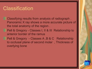
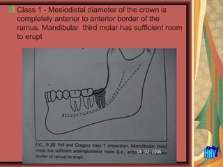
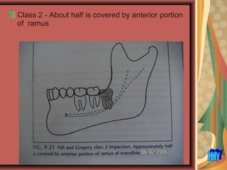
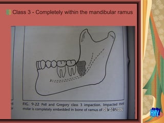
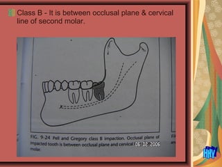
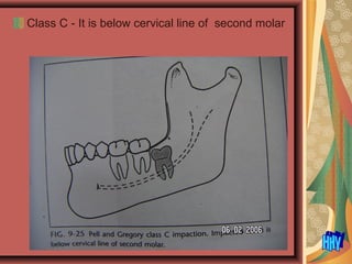
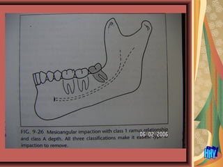
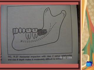
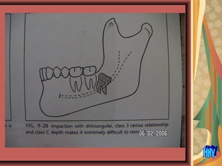
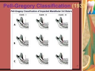
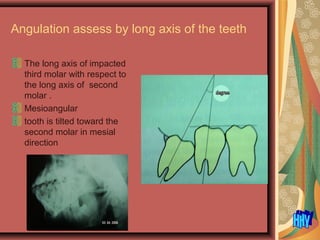
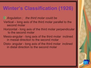
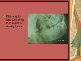
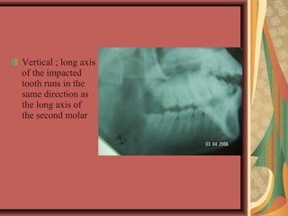
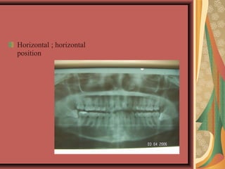
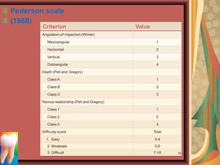
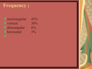
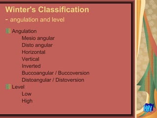
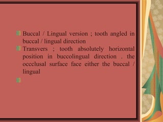
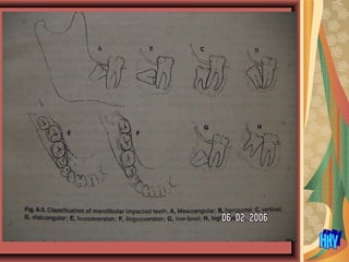
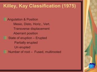
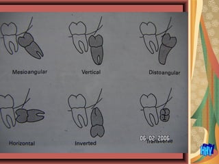
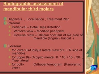
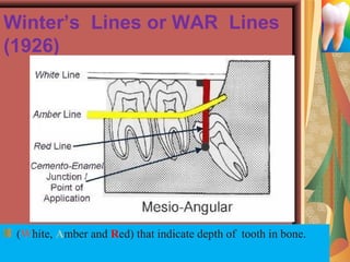
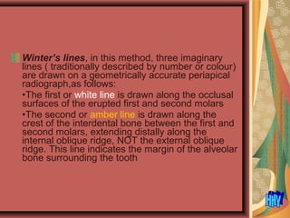
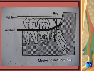
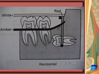
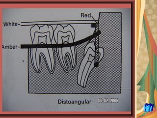
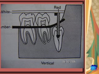
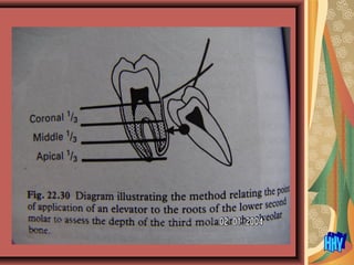
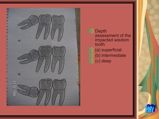
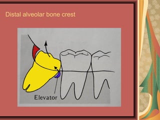
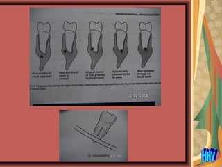
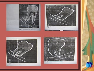
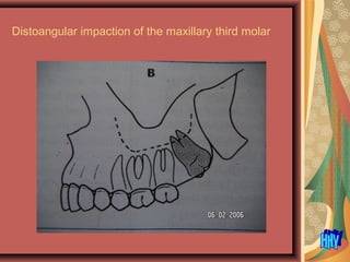
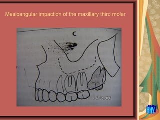
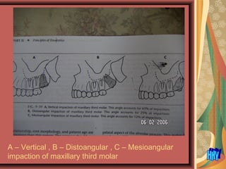
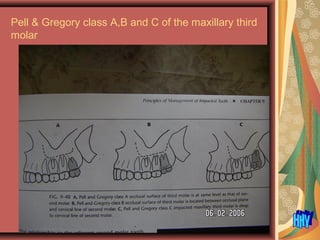
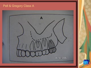
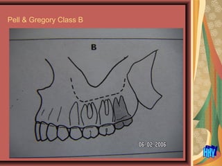
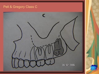
IMPACTION
HTAY HTAY YI
UDM (Mdy.)
Impacted teeth are those prevented from erupting
by some physical barrier in the eruption path
(Shafer,1964)
A tooth that is completely or partially un-erupted
and is positioned against another tooth, bone or
soft tissue, so that its further eruption is unlikely,
described according to its anatomical position.
( Archer, 1975)
One that fails to erupt into the dental arch within
the expected time ( Peterson, 1993)(18-25yrs for
8s)
A tooth that fails to erupt, for whatever reason ,
into the dental arch within the expected time
(Dimitroulis,1997)
Causes
Local causes
-irregularity in the position and pressure of an
adjacent tooth
-density of the overlying and surrounding bone
-long continued chronic inflammation with resultant
increase in density of overlying mucous
membrane
-lack of space due to under developed jaws
-unduly retention of the primary teeth
-acquired diseases such as necrosis due to infection
or abscess
- inflammatory changes in bone due to
exanthomatous diseases in children
Systemic causes
Prenatal causes – heredity
Postnatal causes – Ricket, Anemia , Congenital
Syphilis , Tuberculosis ,
Endocrine dysfunction , Malnutrition
Other rare conditions – Cleidocraiodysostosis ,
Oycephaly , Progeria , Achondroplasia , Cleft
palate
Frequency of impaction
Maxillary and mandibular third molars
Maxillary canine
Mandibular premolars
"Wisdom tooth" is a nickname for Third Molar.
These are the teeth that come into the mouth last,
at age 16-21 (average 18)
Lower last molar
Mesioangular 43 %
Vertical 38 %
Distoangular 6 %
Horizontal 3 %
Indications for removal of impacted teeth
Local ;
- Prevention of pericoronitis (operculitis)-,Ac. pericoronitis
- Prevention of periodontal diseases
- Prevention of infection- cellulitis , osteomyelitis
- Prevention of dental caries
- Prevention of damage to adjacent tooth
- Impacted teeth under dental prosthesis
- Prevention of odontogenic cyst & tumour -
- Prevention of pain of unexplained origin
- Prevention of fracture of jaw
- Facilitation of orthodontic treatment
- Lack of function / occlusion
General ;
- Young – bone elasticity
- Difficult to get treatment - Traveler to the remote area ,
soldier at frontier , seamen
Prevention of pericoronitis
trismus, cellulitis
Prevention of infection , such tooth will be
problematic under the denture
Prevention of caries
Prevention of damage to adjacent tooth
-Removal of embedded supernumerary tooth to facilitate the
orthodontic treatment
- Prevention of fracture of jaw – impacted last molar weaken
the angle of the mandible
Contraindication
Extreme of age - highly calcified & less flexible,
post-op sequelae, greater recovery period
Compromised medical status – systemic diseases
Pregnancy
Probable excessive damage to adjacent structures
– nerves , teeth, prosthesis of precious metal
(consent)
Various Preoperative DifficultyVarious Preoperative Difficulty
Assessment IndicesAssessment Indices
WAR lines or Winter’s lines (1926)
Winter’s classification (1926)
Pell-Gregory classification (1933)
WHARFE’s scale (1985)
Pederson scale (1988)
New index by Yuasa et.al (2002)
New index by Gbotolorun et.al. (2007)
9/6/2012 16
Classification
Classifying results from analysis of radiograph .
Panoramic X ray shows a more accurate picture of
the total anatomy of the region .
Pell & Gregory - Classes I, II & III Relationship to
anterior border of the ramus
Pell & Gregory - Classes A ,B & C Relationship
to occlusal plane of second molar , Thickness of
overlying bone
Class 1 - Mesiodistal diameter of the crown is
completely anterior to anterior border of the
ramus. Mandibular third molar has sufficient room
to erupt
Class 2 - About half is covered by anterior portion
of ramus
Class 3 - Completely within the mandibular ramus
Class A - Occlusal surface of impacted tooth is at
same level as or nearly level with occlusal plane of
second molar
Class B - It is between occlusal plane & cervical
line of second molar.
Class C - It is below cervical line of second molar
Pell-Gregory Classification (1933)
27
Angulation assess by long axis of the teeth
The long axis of impacted
third molar with respect to
the long axis of second
molar .
Mesioangular
tooth is tilted toward the
second molar in mesial
direction
Winter’s Classification (1926)
Angulation ; the third molar could be
Vertical – long axis of the third molar parallel to the
second molar
Horizontal - long axis of the third molar perpendicular
to the second molar
Mesio-angular - long axis of the third molar inclined
in mesial direction to the second molar
Disto- angular - long axis of the third molar inclined
in distal direction to the second molar
Distoangular ;
long axis of the
third molar is
distally inclined
Vertical ; long axis
of the impacted
tooth runs in the
same direction as
the long axis of
the second molar
Horizontal ; horizontal
position
Pederson scale
(1988)
33
Frequency ;
mesioangular 43%
vertical 38%
distoangular 6%
horizontal 3%
 
Shiller 1979
Angle b/t occlusal
surfaces of impacted
8s and 2nd molar.
Vertical <10 degree,
Horizontal > 70 degree
Mesiangular and
Distalangular 10 to 70
degree
Assessed by comparing
the line joining the mesial
and distal images of the
cusps of the wisdom tooth
with the curve of Spee
formed by joining the
cusps of the premolar and
molar teeth
If the wisdom tooth line,
when extended posteriorly
would meet the Spee line
then the tooth is mesio-
obliquely
Conversely , if the wisdom
tooth line never meet the
Spee line , then it is disto-
obliquely
Angulation assess by Curve of Spee
Winter's Classification
- angulation and level
Angulation
Mesio angular
Disto angular
Horizontal
Vertical
Inverted
Buccoangular / Buccoversion
Distoangular / Distoversion
Level
Low
High
Buccal / Lingual version ; tooth angled in 
buccal / lingual direction
Transvers  ;  tooth  absolutely  horizontal 
position  in  buccolingual  direction  .  the 
occclusal  surface  face  either  the  buccal  / 
lingual
Killey, Kay Classification (1975)
Angulation & Position
Mesio, Disto, Horiz., Vert.
Transverse displacement
Aberrant position
State of eruption – Erupted
Partially erupted
Un erupted
Number of root – Fused, multirooted
Radiographic assessment of
mandibular third molars
Diagnosis , Localisation , Treatment Plan
Intraoral
Periapical – Detail, less distortion
Winter's view – Modified periapical
Occlusal view – Oblique occlusal of R/L side of
mandible (lingual / buccal )
Extraoral
for lower 8s-Oblique lateral view of L + R side of
mandible
for upper 8s- Occipito mental 0· / 10· / 15· / 30· ,
True lateral
for both- Orthopantomogram (Panoramic
view)
Radiographic interpretation
The specific features that need to be
identified can be divided into those related
to:
• lower third molar itself
• lower second molar
• surrounding bone
• relationship of the apices with the inferior
dental canal
(a) Lower last molar in bony crypt , crown formation only is completed
(b) Lower last molar tooth bud before calcification
Not a Cyst
(a) (b)
Lower third molar assessment
• Pell& Gregory classification
• Angulation
• buccal or lingual obliquity
• crown
• root
.
The crown
•The size
•The shape
•The presence and extent of caries
•The presence and severity of resorption
The roots
•The number
•The shape
•Curvatures, whether they are favourable or
unfavourable
•The stage of development
The Lower Second molar assessment
The crown
The condition and extent of existing restorations
The presence of caries
The presence and severity of resorption.
The roots
The number
The shape, and if it is conical
The periodontal status
The condition of the apical tissues.
Assessment of the surrounding bone
• Depth of the tooth
• Distal alveolar bone crest
• Bone between lower second and last molar
• Texture and density of the bone
The depth of the tooth in the alveolar bone
Two main methods are used commonly to assess
tooth depth:
•Winter’s lines
•Using the roots of the second molar as a guide.
Winter’s Lines or WAR Lines
(1926)
(White, Amber and Red) that indicate depth of  tooth in bone.
Winter’s lines, in this method, three imaginary
lines ( traditionally described by number or colour)
are drawn on a geometrically accurate periapical
radiograph,as follows:
•The first or white line is drawn along the occlusal
surfaces of the erupted first and second molars
•The second or amber line is drawn along the
crest of the interdental bone between the first and
second molars, extending distally along the
internal oblique ridge, NOT the external oblique
ridge. This line indicates the margin of the alveolar
bone surrounding the tooth
•The third or red line is a perpendicular dropped from
the white line to the point of application for an
elevator, but is measured from the amber line to
this point of application. This line measures the
depth of the third molar within the mandible.
As a general rule, if the red line is 5mm or more in
length the extraction is considered sufficiently
difficult for the tooth to be removed under general
anaesthetic or using local anaesthetic and
sedation
Using the roots of the second molar as a guide
The roots of the adjacent second molar are
divided horizontally into thirds
A horizontal line is then drawn from the point of
application for an elevator to the second molar
If the point of application lies opposite the coronal ,
middle or apical third the extraction is assessed as
being easy , moderate or difficulty , respectively
Depth
assessment of the
impacted wisdom
tooth
(a) superficial
(b) intermediate
(c) deep
Distal alveolar bone crest
* often appear close to the ID canal
* superimposed / intimate relationship
Relationship of the apices to the ID
canal
The normal radiographic appearance of the ID
canal (two thin, parallel radioopaque lines - the so-
called Tram lines)and the variations that indicate a
possible intimate relationship.These variations
include:
•Loss of the tramlines
•Narrowing of the tramlines
•A sudden change in direction of the tramlines
•A radiolucent band evident across the root if the
tooth is grooved or tunnelled through by the ID
bundle.
a - interruption of canal
b - diversion of the canal
(a) (b)
Assessment of the patient –
Indication/ contraindication , Choice of
anesthesia , Treatment plan - one visit, two
visit
Age – Health condition , surgical
stress, inconvenience ½ visit , High calcified, thick
overlying bone , ankylosed , atrophied periodontal
lig. , Recovery / healing
Sex - Male / Female
Occupation – Traveler, soldier, sailor – all
8s clearance
Type – Nervous, apprehensive, Co-
operative, Handicapped
Others – Small mouth , fat cheek ,
TMJ problem , angular stomatitis
General conditions – Host defense (medically
compromised , immunocompromised ) ,
Pregnancy (reproductive age) , R/T – ORN ,
unable to lie (cervical spondilosis)
Clinical assessment - Treatment plan ,
Operative procedure , Timing
History – First attack of pericoronitis , Repeated
attack , Attempted removal , Failure of opeculectomy , Pain
of unknown etiology
Examination –
- General – febrile/ ill
- Local - intraoral examination
No S/S
With S/S – Pain, Tenderness, Trismus, Swelling ,
Lymphadenitis, Dysphagia , Consequences of
untreated infection
8 – condition, portion visible intra-oral , position (tally with x
ray ), function
adjacent tooth (7) condition, caries , periodontal problem
Opposing 8. Impinging, Buccally erupt
Site of Injection – Pus - L.A become ionized , cannot diffuse
- Spread of infection
Oral hygiene status – pre-op prophylaxis
Condition of the 8 - caries
Cl I , Cl A vertical impaction , soft tissue impaction only , half of the
crown can be seen intraorally
Condition of adjacent tooth – cervical caries with pulp exposure
Mesioangular last molar can be easily removed after removal of
the the poor quality adjacent tooth
Extracted badly carious 7 and horizontal 8
Buccally erupted upper 8 traumatize the buccal soft tissue
Treatment Plan
- Treat Ac. condition – ? Hospitalization
- antibiotics –Emperical , mixed gm (+)& (-) and
anaerobic organisms
- analgesic
- anti inflammatory
- mouth wash antiseptic(chlorohexidine) , bland
(hypertonic hot saline)
- Abscess – I & D, pus for C+ S
- Trismus - mouth opening exercise
- Removal of opposing 8 impinging
- Plan for removal of lower 8 ( focal of infection )only
when Ac. condition are subsided
Miscellaneous assessment
Armamentarium – Light, suction , proper
instruments – chisel , mallet , elevator etc.
Operator – Skill, condition, time
Assistant
Choice of anaesthesia
L.A ( Bilateral Block – safe,anesthetize only sensory not
motor)
L.A + Sedation D/Z , Medazolam ( twice as potent as D/Z)
- Sedation; provided that standard safe guards are
applied
- Close monitoring – BP , ECG, Pulse oximeter
- Air way protection – sedated case has reduced gag
reflex
- Flumerzenil – reversal agent for D/Z
L.A + relative analgesia ( O2 + N2O )
G.A. (Emotional status, competence of patient ,convenience
of surgeon) , Anesthetia assessment
Flap
Objective;
for adequate exposure
reflected soft t/s (retract of mucoperiosteal flap)
provides accessible surgery
to access the need for bone removal, create
fulcrum by window , guttering make into hollow) ,
ditching( trench cut). NB; stop cut is mandatory
when chiseling
to divide tooth either by bur or chisel without
excessive bone removal
appropriate elevation
wound cleansed and irrigated under vision
Flap design for removal of wisdom tooth
Tooth removal
Elective surgery – wound prophylaxis
Simple elevation Cl. I, A, vertical , ¾ crown
Surgical removal - open
Sectioning - bone ( widening of exit)/
tooth(reducing of object) , bur and/ or chisel
Germectomy – removal of developmental buds
before anchoring of the roots in the jaw ( 12-19yrs
for lower 8s )
Tooth sectioning
Chisel
Advantages – less damages to adjacent
structures(buccal approach)
Disadvantages – tooth with shallow groove
- sectioning of tooth before elevation
- disturbing , only perform under GA,
premadicated
- cannot perform on elderly , only in young due to
high elasticity
- chisel in line with long axis of tooth, no
transverse section
- force not control(bone/ tooth – need experience)
- necessary of jaw support during tapping
Bur
Advantages- familial with bur
- controlled bone removal
- transverse section possible
- fewer assistant
- no sedation
- no physical blow
- continous wash surgical field
- less swelling & pain ( Hall)
Disadvantages-emphysema
- continuous water syringing is necessary
- damage to adjacent tissue( friction)
- reassembling of tooth impossible
- bur slip
Bone removal by bur
Tooth sectioning
Bone removal and distal aspect of the crown
sectioning
Curvature of the root
Point of elevation on the convex surface
Wound Toilet
saline , chlorohexdine
- inside socket , under flap
soft tissue ( residual tooth sac , granulation tissue)
inside socket , ? biopsy –if in doubt
tooth fragment
bone fragment
dislodged filling
sharp bony edge – squeeze , file especially on
lingual side
Closure (reposition of flap)
reassemble all tooth pieces
before closure
absorbable suture ( 3/0 for
oral ) is more convenient for
patient, no STO
watertight suture is un-
necessary
socket kept open
no tension
no medicated cone inside
socket
Flap closure
Maxillary Third Molar
Vertical impaction of the maxillary third molar
Distoangular impaction of the maxillary third molar
Mesioangular impaction of the maxillary third molar
A – Vertical , B – Distoangular , C – Mesioangular
impaction of maxillary third molar
Pell & Gregory class A,B and C of the maxillary third
molar
Pell & Gregory Class A
Pell & Gregory Class B
Pell & Gregory Class C
Envelop flap is most commonly used flap
When three cornered flap is reflected the bones
more apical portions become more visible
Canine Impaction
Supernumerary impaction
Supernumerary tooth - mesiodens
Embedded mesiodens + high labial frenum attachment – treatment plan
includes of removal of the mesiodens and frenectomy
Impacted lower 7 s
Complications (Mobidity)
addition to all local complications of simple exodontia
Intraoperative;
Haemorrhage – local inflammation
- cutting of vessel- severing of facial vessel along
vertical
incision at lower first molar region upto the lower
border of
mandible
Displaced tooth – lingual pouch/periosteum, lateral
pharyngeal space, air way , GI( antrum, infratemporal
pouch in upper)
Adjacent second molar –subluxation, dislodgement of filling ,
prosthesis
Subluxation / dislocation of TMJ
Jaw fracture
Displaced into the lingual pouch
Postoperative
Haemorrhage – Inferior alveolar , facial, buccal ,
usually primary bleeding not controlled , FB
(tooth fragment, filling)
Pain – excessive trauma , scald, abrasion ,
hematoma under tight suture
Dry socket(alveolar osteitis)
Swelling – directly proportionate to operation time ,
hematoma , infection
Neuroprexia, Axontemesis, Neurotemesis –
lingual , chorda tympani , inferior alveolar , due to
hematoma, odema
Trimus – infection , trauma
Dysphagia
Airway obstruction
Pyrexia- infection
Factors that make the impaction surgery
Less difficult More difficult
1. Mesioangular position Distoangular
2. Class 1 ramus Class 3 ramus
3. Class A Depth Class C Depth
4. Roots 1/3 to 2/3 formed Long, thin roots
5. Fused conical roots Divergent curved root
6. Wide periodontal ligament Narrow periodontal
7. Large Follicle Thin Follicle
8. Elastic Bone Dense, inelastic bone
9. Separated from second molar Contact with
10. Separated from IAN Close to
11. Soft tissue impaction Complete bone impaction
Benefits of Difficulty Assessment
 Treatment
 Suitable anaesthesia
 Surgical instruments
 Surgical technique
 Morbidity
 Operation time
 Referral
122
New Indices
Yuasa et al (2002) proposed a new index
 a combination of Pederson index &
measurement of width of root
Gbotolorun et al (2007) also proposed a new
index with variables of
 age
 body mass index
 depth from point of elevation
 curvature of roots.
123
Variables
Previous assessments - based exclusively on
radiographic variables (MacGregor,1976).
root number and morphology
tooth position
periodontal space
depth from point of elevation
second molar relation
124
Variables in 21st
Century
nonradiographic variables with difficulty assessment of
impacted third molar surgery (Renton et.al, 2001,
Susurla and Dodson, 2004, Akadiri and Obiechina,
2009, Cavalho and Vasconcelos, 2011).
age
gender
body mass index
mouth opening
cheek flexibility
experience of surgeon
procedure type
number of teeth extracted 125
Width of root
Thin - width of middle root →
thinner than width of neck
Thick - (multiple roots), width
of middle root is thicker than
width of neck & roots
separately
Bulbous - width of middle root
is thicker than width of neck
& roots do not separate
Bulbous root. A<B: the width of middle
root (B) is thicker than width of neck (A)
and roots do not separate.
WHARFE's scale
(1985)
Winter’s classification →
expanded by Macgregor to
WHARFE's scale .
Based on six dental factors
Winter's classification
height of mandible
angulation of second molar
root shape and development
follicle morphology
exit path
.
127
Category Score
1. Winter's classification Vertical 0
Mesial 1
Horizontal 2
Distal 2
2. Height of mandible (mm) 01-30 mm 0
31-34 mm 1
35-39 mm 2
3. Angle of second molar (degrees) 1-59• 0
60-69•
1
70-79•
2
80-89• 3
>90•
4
4. Root Shape and development
a)Less than 1/3 complete 2
b)1/3 to 2/3 complete 1
c)More than 2/3 complete 3
Complex 3
Unfavourable curve 2
Favourable curve 1
Normal 0
5. Follicle Normal 0
Possibly enlarged --1
Enlarged -2
Impaction relieved -3
6. Exit path Space 0
Distal cusp Covered 1
Mesial cusp Covered 2
Both covered 3
Total score
During operation
Operation time
Parant scale
Intraoperative findings related to radiographic
variables
Surgeon’s comment
128
Intra-operative Assessment
Operation time
- from bone removal to tooth out or if without bone
removal from use of forceps/elevator to tooth out
-
129
Operative procedure (Parant scale)
130
1. Easy - I
2. Moderate - II, III
3. Difficult - IV
(Sulieman et. al, 2006)
Consent for the surgical
removal of lower last molar
Discussion
Written consent
Documentation in the chart
Seven areas ;
Specific problem
Proposed treatment
Anticipated or common side effects
Possible complications and frequency of
occurence
Anaesthesia
Treatment alternatives
Uncertainities of the outcome

Impaction

  • 1.
  • 2.
    Impacted teeth arethose prevented from erupting by some physical barrier in the eruption path (Shafer,1964) A tooth that is completely or partially un-erupted and is positioned against another tooth, bone or soft tissue, so that its further eruption is unlikely, described according to its anatomical position. ( Archer, 1975) One that fails to erupt into the dental arch within the expected time ( Peterson, 1993)(18-25yrs for 8s) A tooth that fails to erupt, for whatever reason , into the dental arch within the expected time (Dimitroulis,1997)
  • 3.
    Causes Local causes -irregularity inthe position and pressure of an adjacent tooth -density of the overlying and surrounding bone -long continued chronic inflammation with resultant increase in density of overlying mucous membrane -lack of space due to under developed jaws -unduly retention of the primary teeth -acquired diseases such as necrosis due to infection or abscess - inflammatory changes in bone due to exanthomatous diseases in children
  • 4.
    Systemic causes Prenatal causes– heredity Postnatal causes – Ricket, Anemia , Congenital Syphilis , Tuberculosis , Endocrine dysfunction , Malnutrition Other rare conditions – Cleidocraiodysostosis , Oycephaly , Progeria , Achondroplasia , Cleft palate
  • 5.
    Frequency of impaction Maxillaryand mandibular third molars Maxillary canine Mandibular premolars
  • 6.
    "Wisdom tooth" isa nickname for Third Molar. These are the teeth that come into the mouth last, at age 16-21 (average 18)
  • 7.
    Lower last molar Mesioangular43 % Vertical 38 % Distoangular 6 % Horizontal 3 %
  • 8.
    Indications for removalof impacted teeth Local ; - Prevention of pericoronitis (operculitis)-,Ac. pericoronitis - Prevention of periodontal diseases - Prevention of infection- cellulitis , osteomyelitis - Prevention of dental caries - Prevention of damage to adjacent tooth - Impacted teeth under dental prosthesis - Prevention of odontogenic cyst & tumour - - Prevention of pain of unexplained origin - Prevention of fracture of jaw - Facilitation of orthodontic treatment - Lack of function / occlusion General ; - Young – bone elasticity - Difficult to get treatment - Traveler to the remote area , soldier at frontier , seamen
  • 9.
  • 10.
    Prevention of infection, such tooth will be problematic under the denture
  • 11.
  • 12.
    Prevention of damageto adjacent tooth
  • 13.
    -Removal of embeddedsupernumerary tooth to facilitate the orthodontic treatment
  • 14.
    - Prevention offracture of jaw – impacted last molar weaken the angle of the mandible
  • 15.
    Contraindication Extreme of age- highly calcified & less flexible, post-op sequelae, greater recovery period Compromised medical status – systemic diseases Pregnancy Probable excessive damage to adjacent structures – nerves , teeth, prosthesis of precious metal (consent)
  • 16.
    Various Preoperative DifficultyVariousPreoperative Difficulty Assessment IndicesAssessment Indices WAR lines or Winter’s lines (1926) Winter’s classification (1926) Pell-Gregory classification (1933) WHARFE’s scale (1985) Pederson scale (1988) New index by Yuasa et.al (2002) New index by Gbotolorun et.al. (2007) 9/6/2012 16
  • 17.
    Classification Classifying results fromanalysis of radiograph . Panoramic X ray shows a more accurate picture of the total anatomy of the region . Pell & Gregory - Classes I, II & III Relationship to anterior border of the ramus Pell & Gregory - Classes A ,B & C Relationship to occlusal plane of second molar , Thickness of overlying bone
  • 18.
    Class 1 -Mesiodistal diameter of the crown is completely anterior to anterior border of the ramus. Mandibular third molar has sufficient room to erupt
  • 19.
    Class 2 -About half is covered by anterior portion of ramus
  • 20.
    Class 3 -Completely within the mandibular ramus
  • 21.
    Class A -Occlusal surface of impacted tooth is at same level as or nearly level with occlusal plane of second molar
  • 22.
    Class B -It is between occlusal plane & cervical line of second molar.
  • 23.
    Class C -It is below cervical line of second molar
  • 27.
  • 28.
    Angulation assess bylong axis of the teeth The long axis of impacted third molar with respect to the long axis of second molar . Mesioangular tooth is tilted toward the second molar in mesial direction
  • 29.
    Winter’s Classification (1926) Angulation; the third molar could be Vertical – long axis of the third molar parallel to the second molar Horizontal - long axis of the third molar perpendicular to the second molar Mesio-angular - long axis of the third molar inclined in mesial direction to the second molar Disto- angular - long axis of the third molar inclined in distal direction to the second molar
  • 30.
    Distoangular ; long axisof the third molar is distally inclined
  • 31.
    Vertical ; longaxis of the impacted tooth runs in the same direction as the long axis of the second molar
  • 32.
  • 33.
  • 34.
    Frequency ; mesioangular 43% vertical38% distoangular 6% horizontal 3%  
  • 35.
    Shiller 1979 Angle b/tocclusal surfaces of impacted 8s and 2nd molar. Vertical <10 degree, Horizontal > 70 degree Mesiangular and Distalangular 10 to 70 degree
  • 36.
    Assessed by comparing theline joining the mesial and distal images of the cusps of the wisdom tooth with the curve of Spee formed by joining the cusps of the premolar and molar teeth If the wisdom tooth line, when extended posteriorly would meet the Spee line then the tooth is mesio- obliquely Conversely , if the wisdom tooth line never meet the Spee line , then it is disto- obliquely Angulation assess by Curve of Spee
  • 37.
    Winter's Classification - angulationand level Angulation Mesio angular Disto angular Horizontal Vertical Inverted Buccoangular / Buccoversion Distoangular / Distoversion Level Low High
  • 38.
    Buccal / Lingual version ; tooth angled in  buccal / lingual direction Transvers  ;  tooth absolutely  horizontal  position  in  buccolingual  direction  .  the  occclusal  surface  face  either  the  buccal  /  lingual
  • 40.
    Killey, Kay Classification(1975) Angulation & Position Mesio, Disto, Horiz., Vert. Transverse displacement Aberrant position State of eruption – Erupted Partially erupted Un erupted Number of root – Fused, multirooted
  • 42.
    Radiographic assessment of mandibularthird molars Diagnosis , Localisation , Treatment Plan Intraoral Periapical – Detail, less distortion Winter's view – Modified periapical Occlusal view – Oblique occlusal of R/L side of mandible (lingual / buccal ) Extraoral for lower 8s-Oblique lateral view of L + R side of mandible for upper 8s- Occipito mental 0· / 10· / 15· / 30· , True lateral for both- Orthopantomogram (Panoramic view)
  • 43.
    Radiographic interpretation The specificfeatures that need to be identified can be divided into those related to: • lower third molar itself • lower second molar • surrounding bone • relationship of the apices with the inferior dental canal
  • 44.
    (a) Lower lastmolar in bony crypt , crown formation only is completed (b) Lower last molar tooth bud before calcification Not a Cyst (a) (b)
  • 45.
    Lower third molarassessment • Pell& Gregory classification • Angulation • buccal or lingual obliquity • crown • root .
  • 46.
    The crown •The size •Theshape •The presence and extent of caries •The presence and severity of resorption The roots •The number •The shape •Curvatures, whether they are favourable or unfavourable •The stage of development
  • 47.
    The Lower Secondmolar assessment The crown The condition and extent of existing restorations The presence of caries The presence and severity of resorption. The roots The number The shape, and if it is conical The periodontal status The condition of the apical tissues.
  • 48.
    Assessment of thesurrounding bone • Depth of the tooth • Distal alveolar bone crest • Bone between lower second and last molar • Texture and density of the bone
  • 49.
    The depth ofthe tooth in the alveolar bone Two main methods are used commonly to assess tooth depth: •Winter’s lines •Using the roots of the second molar as a guide.
  • 50.
    Winter’s Lines orWAR Lines (1926) (White, Amber and Red) that indicate depth of  tooth in bone.
  • 51.
    Winter’s lines, inthis method, three imaginary lines ( traditionally described by number or colour) are drawn on a geometrically accurate periapical radiograph,as follows: •The first or white line is drawn along the occlusal surfaces of the erupted first and second molars •The second or amber line is drawn along the crest of the interdental bone between the first and second molars, extending distally along the internal oblique ridge, NOT the external oblique ridge. This line indicates the margin of the alveolar bone surrounding the tooth
  • 52.
    •The third orred line is a perpendicular dropped from the white line to the point of application for an elevator, but is measured from the amber line to this point of application. This line measures the depth of the third molar within the mandible. As a general rule, if the red line is 5mm or more in length the extraction is considered sufficiently difficult for the tooth to be removed under general anaesthetic or using local anaesthetic and sedation
  • 57.
    Using the rootsof the second molar as a guide The roots of the adjacent second molar are divided horizontally into thirds A horizontal line is then drawn from the point of application for an elevator to the second molar If the point of application lies opposite the coronal , middle or apical third the extraction is assessed as being easy , moderate or difficulty , respectively
  • 59.
    Depth assessment of the impactedwisdom tooth (a) superficial (b) intermediate (c) deep
  • 60.
  • 61.
    * often appearclose to the ID canal * superimposed / intimate relationship Relationship of the apices to the ID canal
  • 63.
    The normal radiographicappearance of the ID canal (two thin, parallel radioopaque lines - the so- called Tram lines)and the variations that indicate a possible intimate relationship.These variations include: •Loss of the tramlines •Narrowing of the tramlines •A sudden change in direction of the tramlines •A radiolucent band evident across the root if the tooth is grooved or tunnelled through by the ID bundle.
  • 65.
    a - interruptionof canal b - diversion of the canal (a) (b)
  • 67.
    Assessment of thepatient – Indication/ contraindication , Choice of anesthesia , Treatment plan - one visit, two visit Age – Health condition , surgical stress, inconvenience ½ visit , High calcified, thick overlying bone , ankylosed , atrophied periodontal lig. , Recovery / healing Sex - Male / Female Occupation – Traveler, soldier, sailor – all 8s clearance Type – Nervous, apprehensive, Co- operative, Handicapped Others – Small mouth , fat cheek , TMJ problem , angular stomatitis General conditions – Host defense (medically compromised , immunocompromised ) , Pregnancy (reproductive age) , R/T – ORN , unable to lie (cervical spondilosis)
  • 68.
    Clinical assessment -Treatment plan , Operative procedure , Timing History – First attack of pericoronitis , Repeated attack , Attempted removal , Failure of opeculectomy , Pain of unknown etiology Examination – - General – febrile/ ill - Local - intraoral examination No S/S With S/S – Pain, Tenderness, Trismus, Swelling , Lymphadenitis, Dysphagia , Consequences of untreated infection 8 – condition, portion visible intra-oral , position (tally with x ray ), function adjacent tooth (7) condition, caries , periodontal problem Opposing 8. Impinging, Buccally erupt Site of Injection – Pus - L.A become ionized , cannot diffuse - Spread of infection Oral hygiene status – pre-op prophylaxis
  • 69.
    Condition of the8 - caries
  • 70.
    Cl I ,Cl A vertical impaction , soft tissue impaction only , half of the crown can be seen intraorally
  • 71.
    Condition of adjacenttooth – cervical caries with pulp exposure Mesioangular last molar can be easily removed after removal of the the poor quality adjacent tooth
  • 72.
    Extracted badly carious7 and horizontal 8
  • 73.
    Buccally erupted upper8 traumatize the buccal soft tissue
  • 74.
    Treatment Plan - TreatAc. condition – ? Hospitalization - antibiotics –Emperical , mixed gm (+)& (-) and anaerobic organisms - analgesic - anti inflammatory - mouth wash antiseptic(chlorohexidine) , bland (hypertonic hot saline) - Abscess – I & D, pus for C+ S - Trismus - mouth opening exercise - Removal of opposing 8 impinging - Plan for removal of lower 8 ( focal of infection )only when Ac. condition are subsided
  • 76.
    Miscellaneous assessment Armamentarium –Light, suction , proper instruments – chisel , mallet , elevator etc. Operator – Skill, condition, time Assistant
  • 77.
    Choice of anaesthesia L.A( Bilateral Block – safe,anesthetize only sensory not motor) L.A + Sedation D/Z , Medazolam ( twice as potent as D/Z) - Sedation; provided that standard safe guards are applied - Close monitoring – BP , ECG, Pulse oximeter - Air way protection – sedated case has reduced gag reflex - Flumerzenil – reversal agent for D/Z L.A + relative analgesia ( O2 + N2O ) G.A. (Emotional status, competence of patient ,convenience of surgeon) , Anesthetia assessment
  • 78.
    Flap Objective; for adequate exposure reflectedsoft t/s (retract of mucoperiosteal flap) provides accessible surgery to access the need for bone removal, create fulcrum by window , guttering make into hollow) , ditching( trench cut). NB; stop cut is mandatory when chiseling to divide tooth either by bur or chisel without excessive bone removal appropriate elevation wound cleansed and irrigated under vision
  • 79.
    Flap design forremoval of wisdom tooth
  • 82.
    Tooth removal Elective surgery– wound prophylaxis Simple elevation Cl. I, A, vertical , ¾ crown Surgical removal - open Sectioning - bone ( widening of exit)/ tooth(reducing of object) , bur and/ or chisel Germectomy – removal of developmental buds before anchoring of the roots in the jaw ( 12-19yrs for lower 8s )
  • 83.
  • 84.
    Chisel Advantages – lessdamages to adjacent structures(buccal approach) Disadvantages – tooth with shallow groove - sectioning of tooth before elevation - disturbing , only perform under GA, premadicated - cannot perform on elderly , only in young due to high elasticity - chisel in line with long axis of tooth, no transverse section - force not control(bone/ tooth – need experience) - necessary of jaw support during tapping
  • 85.
    Bur Advantages- familial withbur - controlled bone removal - transverse section possible - fewer assistant - no sedation - no physical blow - continous wash surgical field - less swelling & pain ( Hall) Disadvantages-emphysema - continuous water syringing is necessary - damage to adjacent tissue( friction) - reassembling of tooth impossible - bur slip
  • 87.
  • 89.
  • 90.
    Bone removal anddistal aspect of the crown sectioning
  • 92.
    Curvature of theroot Point of elevation on the convex surface
  • 94.
    Wound Toilet saline ,chlorohexdine - inside socket , under flap soft tissue ( residual tooth sac , granulation tissue) inside socket , ? biopsy –if in doubt tooth fragment bone fragment dislodged filling sharp bony edge – squeeze , file especially on lingual side
  • 95.
    Closure (reposition offlap) reassemble all tooth pieces before closure absorbable suture ( 3/0 for oral ) is more convenient for patient, no STO watertight suture is un- necessary socket kept open no tension no medicated cone inside socket
  • 96.
  • 99.
  • 100.
    Vertical impaction ofthe maxillary third molar
  • 101.
    Distoangular impaction ofthe maxillary third molar
  • 102.
    Mesioangular impaction ofthe maxillary third molar
  • 103.
    A – Vertical, B – Distoangular , C – Mesioangular impaction of maxillary third molar
  • 104.
    Pell & Gregoryclass A,B and C of the maxillary third molar
  • 105.
  • 106.
  • 107.
  • 108.
    Envelop flap ismost commonly used flap When three cornered flap is reflected the bones more apical portions become more visible
  • 110.
  • 112.
  • 113.
  • 114.
    Embedded mesiodens +high labial frenum attachment – treatment plan includes of removal of the mesiodens and frenectomy
  • 117.
  • 118.
    Complications (Mobidity) addition toall local complications of simple exodontia Intraoperative; Haemorrhage – local inflammation - cutting of vessel- severing of facial vessel along vertical incision at lower first molar region upto the lower border of mandible Displaced tooth – lingual pouch/periosteum, lateral pharyngeal space, air way , GI( antrum, infratemporal pouch in upper) Adjacent second molar –subluxation, dislodgement of filling , prosthesis Subluxation / dislocation of TMJ Jaw fracture
  • 119.
    Displaced into thelingual pouch
  • 120.
    Postoperative Haemorrhage – Inferioralveolar , facial, buccal , usually primary bleeding not controlled , FB (tooth fragment, filling) Pain – excessive trauma , scald, abrasion , hematoma under tight suture Dry socket(alveolar osteitis) Swelling – directly proportionate to operation time , hematoma , infection Neuroprexia, Axontemesis, Neurotemesis – lingual , chorda tympani , inferior alveolar , due to hematoma, odema Trimus – infection , trauma Dysphagia Airway obstruction Pyrexia- infection
  • 121.
    Factors that makethe impaction surgery Less difficult More difficult 1. Mesioangular position Distoangular 2. Class 1 ramus Class 3 ramus 3. Class A Depth Class C Depth 4. Roots 1/3 to 2/3 formed Long, thin roots 5. Fused conical roots Divergent curved root 6. Wide periodontal ligament Narrow periodontal 7. Large Follicle Thin Follicle 8. Elastic Bone Dense, inelastic bone 9. Separated from second molar Contact with 10. Separated from IAN Close to 11. Soft tissue impaction Complete bone impaction
  • 122.
    Benefits of DifficultyAssessment  Treatment  Suitable anaesthesia  Surgical instruments  Surgical technique  Morbidity  Operation time  Referral 122
  • 123.
    New Indices Yuasa etal (2002) proposed a new index  a combination of Pederson index & measurement of width of root Gbotolorun et al (2007) also proposed a new index with variables of  age  body mass index  depth from point of elevation  curvature of roots. 123
  • 124.
    Variables Previous assessments -based exclusively on radiographic variables (MacGregor,1976). root number and morphology tooth position periodontal space depth from point of elevation second molar relation 124
  • 125.
    Variables in 21st Century nonradiographicvariables with difficulty assessment of impacted third molar surgery (Renton et.al, 2001, Susurla and Dodson, 2004, Akadiri and Obiechina, 2009, Cavalho and Vasconcelos, 2011). age gender body mass index mouth opening cheek flexibility experience of surgeon procedure type number of teeth extracted 125
  • 126.
    Width of root Thin- width of middle root → thinner than width of neck Thick - (multiple roots), width of middle root is thicker than width of neck & roots separately Bulbous - width of middle root is thicker than width of neck & roots do not separate Bulbous root. A<B: the width of middle root (B) is thicker than width of neck (A) and roots do not separate.
  • 127.
    WHARFE's scale (1985) Winter’s classification→ expanded by Macgregor to WHARFE's scale . Based on six dental factors Winter's classification height of mandible angulation of second molar root shape and development follicle morphology exit path . 127 Category Score 1. Winter's classification Vertical 0 Mesial 1 Horizontal 2 Distal 2 2. Height of mandible (mm) 01-30 mm 0 31-34 mm 1 35-39 mm 2 3. Angle of second molar (degrees) 1-59• 0 60-69• 1 70-79• 2 80-89• 3 >90• 4 4. Root Shape and development a)Less than 1/3 complete 2 b)1/3 to 2/3 complete 1 c)More than 2/3 complete 3 Complex 3 Unfavourable curve 2 Favourable curve 1 Normal 0 5. Follicle Normal 0 Possibly enlarged --1 Enlarged -2 Impaction relieved -3 6. Exit path Space 0 Distal cusp Covered 1 Mesial cusp Covered 2 Both covered 3 Total score
  • 128.
    During operation Operation time Parantscale Intraoperative findings related to radiographic variables Surgeon’s comment 128
  • 129.
    Intra-operative Assessment Operation time -from bone removal to tooth out or if without bone removal from use of forceps/elevator to tooth out - 129
  • 130.
    Operative procedure (Parantscale) 130 1. Easy - I 2. Moderate - II, III 3. Difficult - IV (Sulieman et. al, 2006)
  • 131.
    Consent for thesurgical removal of lower last molar Discussion Written consent Documentation in the chart
  • 132.
    Seven areas ; Specificproblem Proposed treatment Anticipated or common side effects Possible complications and frequency of occurence Anaesthesia Treatment alternatives Uncertainities of the outcome