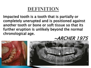
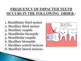
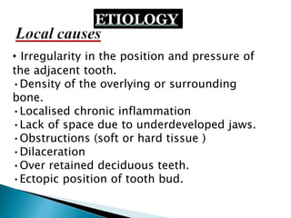
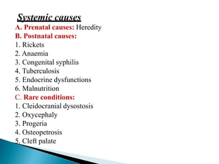
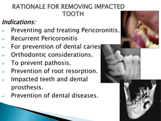
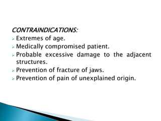
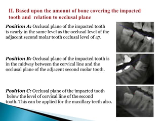
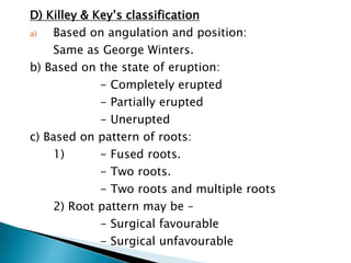
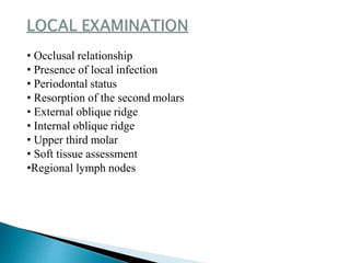
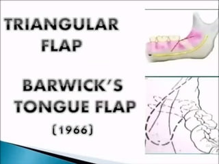
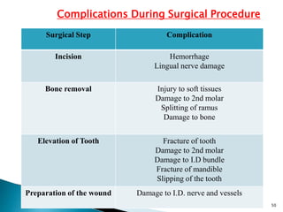
PRESENTED BY –
Dr.ANKITA RAJ (MDS Reader)
Oral & Maxillofacial Surgery
Department
Rama Dental College, Kanpur
Impacted tooth is a tooth that is partially or
completely unerupted and is positioned against
another tooth or bone or soft tissue so that its
further eruption is unlikely beyond the normal
chronological age.
~ARCHER 1975
1. Mandibular third molars
2. Maxillary third molars
3. Maxillary cuspids
4. Mandibular bicuspids
5. Mandibular cuspids
6. Maxillary bicuspids
7. Maxillary central incisors
8. Maxillary lateral incisors
•Orthodontic theory (small jaw-decreased
space):
Growth of the jaw and movement of teeth occurs in forward
direction, anything that interferes with such moment will
cause an impaction (small jaw-decreased space).
A dense bone decreases the movement of the teeth:
(1)acute infection
(2)Local inflammation of PDL
(3)Malocclusion,
(4)Trauma
(5)Early loss of primary teeth and arrested growth of the jaw
•Mendelian theory:
Heredity is the most common cause. An individual may
inherit small jaws from one parent and a complement of
large teeth from the other, i.e. hereditary transmission of
small jaws and large teeth from parents to children.
• Pathological theory:
Osteosclerosis in the third molar area, caused by the early disease of
adjacent molars, cause chronic infections affecting an individual and may
bring the condensation of osseous tissue further preventing the growth
and development of the jaws.
• Endocrinal theory:
Increase or decrease in the growth hormone secretion may
affect the size of the jaws.
• Nature and nurture theory:
A. J. MacGregor explains that impaction can occur due to a
mismatch in size and shape of teeth and jaws.
ETIOLOGY
• Irregularity in the position and pressure of
the adjacent tooth.
•Density of the overlying or surrounding
bone.
•Localised chronic inflammation
•Lack of space due to underdeveloped jaws.
•Obstructions (soft or hard tissue )
•Dilaceration
•Over retained deciduous teeth.
•Ectopic position of tooth bud.
A. Prenatal causes: Heredity
B. Postnatal causes:
1. Rickets
2. Anaemia
3. Congenital syphilis
4. Tuberculosis
5. Endocrine dysfunctions
6. Malnutrition
C. Rare conditions:
1. Cleidocranial dysostosis
2. Oxycephaly
3. Progeria
4. Osteopetrosis
5. Cleft palate
RATIONALE FOR REMOVING IMPACTED
TOOTH
Indications:
 Preventing and treating Pericoronitis.
 Recurrent Pericoronitis
 For prevention of dental caries.
 Orthodontic considerations.
 To prevent pathosis.
 Prevention of root resorption.
 Impacted teeth and dental
prosthesis.
 Prevention of dental diseases.
CONTRAINDICATIONS:
 Extremes of age.
 Medically compromised patient.
 Probable excessive damage to the adjacent
structures.
 Prevention of fracture of jaws.
 Prevention of pain of unexplained origin.
Based on the nature of the overlying
tissue
Winter’s classification
Pell and Gregory’s classification
Killey & Key’s classification
A. BASED ON THE NATURE OF
THE OVERLYING TISSUE
i. Soft tissue impaction
ii. Hard tissue impaction
I. Mesioangular: Long axis of 3rd molar bisects the long axis
2nd molar at or above occlusal plane
Mesioangular 38—(A) long axis of 38 bisects the long axis 37
above the occlusal plane. (B) Interradicular bone width
between 37 and 38 is more than interradicular bone width
between 36 and 37.
Distoangular—(A) long axis of 48 is away from long axis of 47 at the
level of occlusal plane. (B) The interradicular bone between 47 and 48
is almost obliterated and less than that between 46 and 47.
Horizontal—long axis of 38 bisects long axis of 37
at right angle.
(A)Vertical—the long axis of the impacted 48 runs parallel to the long
axis of the 47.
(B)Vertical—interradicular bone width between 47 and 48 equal to
interradicular bone width between 46 and 47.
WINTERS
CLASSIFICATION
Buccoangul
ar
I. Based on their relationship with the anterior
border of the mandible :
Class I: The anteroposterior diameter of the tooth is equal to the
space between the anterior border of ramus of the mandible
and distal surface of the second molar tooth
Class II: A small amount of bone covers the distal surface of the
tooth and The space is inadequate for eruption of the tooth,
i.e. mesiodistal diameter of the tooth is greater than the
space available.
Class III: Tooth is located completely within the ramus of the
mandible– least accessible.
Position A: Occlusal plane of the impacted tooth
is nearly in the same level as the occlusal level of the
adjacent second molar tooth occlusal level of 47.
Position B: Occlusal plane of the impacted tooth is
in the midway between the cervical line and the
occlusal plane of the adjacent second molar tooth.
Position C: Occlusal plane of the impacted tooth
below the level of cervical line of the second
tooth. This can be applied for the maxillary teeth also.
III. Based on long axis of the impacted tooth :
It is similar to the one as proposed in the Winter’s classification
D) Killey & Key’s classification
a) Based on angulation and position:
Same as George Winters.
b) Based on the state of eruption:
- Completely erupted
- Partially erupted
- Unerupted
c) Based on pattern of roots:
1) - Fused roots.
- Two roots.
- Two roots and multiple roots
2) Root pattern may be –
- Surgical favourable
- Surgical unfavourable
• Occlusal relationship
• Presence of local infection
• Periodontal status
• Resorption of the second molars
• External oblique ridge
• Internal oblique ridge
• Upper third molar
• Soft tissue assessment
•Regional lymph nodes
Types of radiographs used
• Intraoral periapical (IOPA) radiograph
• Bitewing radiograph
• Occlusal radiograph
• Lateral oblique radiograph
• Orthopantomograph (OPG)
• CBCT (in indicated cases)
WAR lines
White line
White line is drawn along the occlusal surfaces of the erupted
mandibular molars and extended over the third molar region
posteriorly.
Indicates
▪The depth of the tooth within the mandible.
▪Relationship of occlusal surface of impacted
tooth with the erupted molars.
 Amber line
Amber line is drawn from the surface of the bone on the distal aspect of the
third molar to the crest of the interdental septum between the first and
second mandibular molars.
This line represents the margin
of the alveolar bone covering the third molar.
 Red line
It is the perpendicular line drawn from the amber line to the imaginary
point of application for the elevator (all types-mesial, distoangular-
distal)
The length of the red line indicates depth of the impacted tooth.
With each increase in length of the red line by 1 mm, the impacted tooth
becomes three times more difficult to remove.
A) Spatial Relationship Value
- Mesioangular 1
- Horizontal / transverse 2
- Vertical 3
- Distoangular 4
B) Depth
- Level A 1
- Level B 2
- Level C 3
C) Ramus relationship
- Class I 1
- Class II 2
- Class III 3
PEDERSON DIFFICULTY INDEX
Classification:
Difficulty scores:
Very difficult 7-10
Moderately 5-7
Minimally 3-4
Example: Mesioangular tooth 1 difficulty score is
Level B 2 5-7
Class III 3 Moderately difficult
 Relationship of 3rd molar to the
INFERIOR DENTAL CANAL.
Darkening of
roots
Deflection of
roots
Narrowing of
roots
Dark & Bifid apex Interruption of
white line of canal
Diversion of canal Narrowing of canal
34
• Cheek retraction and visualization - Mouth mirror,
prop,
of the surgical area cheek retractor
• Incision - BP Handle & No. 15 blade
• Flap development & reflection - Moon’s probe, Howarth’s
periosteal elevator
• Flap retraction - Austin’s retractor
• Bone removal - Handpiece, bur, chisel
• Luxation - Elevators
• Tooth removal - Forceps
• Suturing - Sutures & needle holder
35
STEPS IN REMOVAL OF IMPACTED TEETH
1. Isolation
2. Anaesthesia
3. Incision and flap design (flap elevation and retraction)
4. Bone removal
5. Sectioning/division of tooth (if required)
6. Elevation and extraction of tooth
7. Debridement and smoothening of bone
8. Control of bleeding
9. Flap repositioning and Suturing
10. Follow up
36
Step 4: Incisions placed
Step 5: Buccal Mucoperiosteal flap raised
Step 6: Lingual Mucoperiosteal flap raised and complete exposure of the tooth done.
Step 7: Guttering of the mesial and distal bone.
Step 8: Odontectomy performed.
Step 10: Removal of mesial segment
Step 9: Removal of distal segment
Step 11: Extraoral reorientation of the extraoral fragments.
Step 12: Wound debridement and primary closure.
A. LATERAL TREPHINATION TECHNIQUE
The external oblique ridge is palpated and an S-shaped incision is made.
Incision line starts from the retromolar fossa and extends across the
external oblique ridge curving down along the reflection of the mucous
membrane above the vestibule and ends anterior to the first permanent molar.
Using a round bur, the buccal cortical plate over the third molar crypt is
trephined and is fractured to expose the third molar crypt using a chisel
Using an elevator, the impacted tooth is delivered out of the crypt.
Takes advantage of the thinness of the lingual cortical plate, avoids and preserves
plate and hence preserves the buccal plate and external oblique ridge.
ENVELOP FLAP
Surgical Step Complication
Incision Hemorrhage
Lingual nerve damage
Bone removal Injury to soft tissues
Damage to 2nd molar
Splitting of ramus
Damage to bone
Elevation of Tooth Fracture of tooth
Damage to 2nd molar
Damage to I.D bundle
Fracture of mandible
Slipping of the tooth
Preparation of the wound Damage to I.D. nerve and vessels
Complications During Surgical Procedure
50
◦ Dry socket
◦ Pain
◦ Swelling
◦ Trismus
◦ Paraesthesia
◦ Sensitivity
◦ Loss of vitality
◦ Pocket formation
51
Thank you for
listening

MANDIBULAR 3RD MOLAR IMPACTION

  • 1.
    PRESENTED BY – Dr.ANKITARAJ (MDS Reader) Oral & Maxillofacial Surgery Department Rama Dental College, Kanpur
  • 3.
    Impacted tooth isa tooth that is partially or completely unerupted and is positioned against another tooth or bone or soft tissue so that its further eruption is unlikely beyond the normal chronological age. ~ARCHER 1975
  • 4.
    1. Mandibular thirdmolars 2. Maxillary third molars 3. Maxillary cuspids 4. Mandibular bicuspids 5. Mandibular cuspids 6. Maxillary bicuspids 7. Maxillary central incisors 8. Maxillary lateral incisors
  • 5.
    •Orthodontic theory (smalljaw-decreased space): Growth of the jaw and movement of teeth occurs in forward direction, anything that interferes with such moment will cause an impaction (small jaw-decreased space). A dense bone decreases the movement of the teeth: (1)acute infection (2)Local inflammation of PDL (3)Malocclusion, (4)Trauma (5)Early loss of primary teeth and arrested growth of the jaw
  • 6.
    •Mendelian theory: Heredity isthe most common cause. An individual may inherit small jaws from one parent and a complement of large teeth from the other, i.e. hereditary transmission of small jaws and large teeth from parents to children.
  • 7.
    • Pathological theory: Osteosclerosisin the third molar area, caused by the early disease of adjacent molars, cause chronic infections affecting an individual and may bring the condensation of osseous tissue further preventing the growth and development of the jaws. • Endocrinal theory: Increase or decrease in the growth hormone secretion may affect the size of the jaws. • Nature and nurture theory: A. J. MacGregor explains that impaction can occur due to a mismatch in size and shape of teeth and jaws.
  • 8.
    ETIOLOGY • Irregularity inthe position and pressure of the adjacent tooth. •Density of the overlying or surrounding bone. •Localised chronic inflammation •Lack of space due to underdeveloped jaws. •Obstructions (soft or hard tissue ) •Dilaceration •Over retained deciduous teeth. •Ectopic position of tooth bud.
  • 9.
    A. Prenatal causes:Heredity B. Postnatal causes: 1. Rickets 2. Anaemia 3. Congenital syphilis 4. Tuberculosis 5. Endocrine dysfunctions 6. Malnutrition C. Rare conditions: 1. Cleidocranial dysostosis 2. Oxycephaly 3. Progeria 4. Osteopetrosis 5. Cleft palate
  • 10.
    RATIONALE FOR REMOVINGIMPACTED TOOTH Indications:  Preventing and treating Pericoronitis.  Recurrent Pericoronitis  For prevention of dental caries.  Orthodontic considerations.  To prevent pathosis.  Prevention of root resorption.  Impacted teeth and dental prosthesis.  Prevention of dental diseases.
  • 11.
    CONTRAINDICATIONS:  Extremes ofage.  Medically compromised patient.  Probable excessive damage to the adjacent structures.  Prevention of fracture of jaws.  Prevention of pain of unexplained origin.
  • 12.
    Based on thenature of the overlying tissue Winter’s classification Pell and Gregory’s classification Killey & Key’s classification
  • 13.
    A. BASED ONTHE NATURE OF THE OVERLYING TISSUE i. Soft tissue impaction ii. Hard tissue impaction
  • 14.
    I. Mesioangular: Longaxis of 3rd molar bisects the long axis 2nd molar at or above occlusal plane Mesioangular 38—(A) long axis of 38 bisects the long axis 37 above the occlusal plane. (B) Interradicular bone width between 37 and 38 is more than interradicular bone width between 36 and 37.
  • 15.
    Distoangular—(A) long axisof 48 is away from long axis of 47 at the level of occlusal plane. (B) The interradicular bone between 47 and 48 is almost obliterated and less than that between 46 and 47.
  • 16.
    Horizontal—long axis of38 bisects long axis of 37 at right angle.
  • 17.
    (A)Vertical—the long axisof the impacted 48 runs parallel to the long axis of the 47. (B)Vertical—interradicular bone width between 47 and 48 equal to interradicular bone width between 46 and 47.
  • 18.
  • 19.
    I. Based ontheir relationship with the anterior border of the mandible : Class I: The anteroposterior diameter of the tooth is equal to the space between the anterior border of ramus of the mandible and distal surface of the second molar tooth Class II: A small amount of bone covers the distal surface of the tooth and The space is inadequate for eruption of the tooth, i.e. mesiodistal diameter of the tooth is greater than the space available. Class III: Tooth is located completely within the ramus of the mandible– least accessible.
  • 20.
    Position A: Occlusalplane of the impacted tooth is nearly in the same level as the occlusal level of the adjacent second molar tooth occlusal level of 47. Position B: Occlusal plane of the impacted tooth is in the midway between the cervical line and the occlusal plane of the adjacent second molar tooth. Position C: Occlusal plane of the impacted tooth below the level of cervical line of the second tooth. This can be applied for the maxillary teeth also.
  • 21.
    III. Based onlong axis of the impacted tooth : It is similar to the one as proposed in the Winter’s classification
  • 22.
    D) Killey &Key’s classification a) Based on angulation and position: Same as George Winters. b) Based on the state of eruption: - Completely erupted - Partially erupted - Unerupted c) Based on pattern of roots: 1) - Fused roots. - Two roots. - Two roots and multiple roots 2) Root pattern may be – - Surgical favourable - Surgical unfavourable
  • 24.
    • Occlusal relationship •Presence of local infection • Periodontal status • Resorption of the second molars • External oblique ridge • Internal oblique ridge • Upper third molar • Soft tissue assessment •Regional lymph nodes
  • 25.
    Types of radiographsused • Intraoral periapical (IOPA) radiograph • Bitewing radiograph • Occlusal radiograph • Lateral oblique radiograph • Orthopantomograph (OPG) • CBCT (in indicated cases)
  • 26.
    WAR lines White line Whiteline is drawn along the occlusal surfaces of the erupted mandibular molars and extended over the third molar region posteriorly. Indicates ▪The depth of the tooth within the mandible. ▪Relationship of occlusal surface of impacted tooth with the erupted molars.
  • 27.
     Amber line Amberline is drawn from the surface of the bone on the distal aspect of the third molar to the crest of the interdental septum between the first and second mandibular molars. This line represents the margin of the alveolar bone covering the third molar.
  • 28.
     Red line Itis the perpendicular line drawn from the amber line to the imaginary point of application for the elevator (all types-mesial, distoangular- distal) The length of the red line indicates depth of the impacted tooth. With each increase in length of the red line by 1 mm, the impacted tooth becomes three times more difficult to remove.
  • 31.
    A) Spatial RelationshipValue - Mesioangular 1 - Horizontal / transverse 2 - Vertical 3 - Distoangular 4 B) Depth - Level A 1 - Level B 2 - Level C 3 C) Ramus relationship - Class I 1 - Class II 2 - Class III 3 PEDERSON DIFFICULTY INDEX Classification: Difficulty scores: Very difficult 7-10 Moderately 5-7 Minimally 3-4 Example: Mesioangular tooth 1 difficulty score is Level B 2 5-7 Class III 3 Moderately difficult
  • 32.
     Relationship of3rd molar to the INFERIOR DENTAL CANAL. Darkening of roots Deflection of roots Narrowing of roots Dark & Bifid apex Interruption of white line of canal Diversion of canal Narrowing of canal
  • 34.
    34 • Cheek retractionand visualization - Mouth mirror, prop, of the surgical area cheek retractor • Incision - BP Handle & No. 15 blade • Flap development & reflection - Moon’s probe, Howarth’s periosteal elevator • Flap retraction - Austin’s retractor • Bone removal - Handpiece, bur, chisel • Luxation - Elevators • Tooth removal - Forceps • Suturing - Sutures & needle holder
  • 35.
  • 36.
    STEPS IN REMOVALOF IMPACTED TEETH 1. Isolation 2. Anaesthesia 3. Incision and flap design (flap elevation and retraction) 4. Bone removal 5. Sectioning/division of tooth (if required) 6. Elevation and extraction of tooth 7. Debridement and smoothening of bone 8. Control of bleeding 9. Flap repositioning and Suturing 10. Follow up 36
  • 38.
  • 39.
    Step 5: BuccalMucoperiosteal flap raised Step 6: Lingual Mucoperiosteal flap raised and complete exposure of the tooth done.
  • 40.
    Step 7: Gutteringof the mesial and distal bone. Step 8: Odontectomy performed.
  • 41.
    Step 10: Removalof mesial segment Step 9: Removal of distal segment
  • 42.
    Step 11: Extraoralreorientation of the extraoral fragments. Step 12: Wound debridement and primary closure.
  • 43.
    A. LATERAL TREPHINATIONTECHNIQUE The external oblique ridge is palpated and an S-shaped incision is made. Incision line starts from the retromolar fossa and extends across the external oblique ridge curving down along the reflection of the mucous membrane above the vestibule and ends anterior to the first permanent molar.
  • 44.
    Using a roundbur, the buccal cortical plate over the third molar crypt is trephined and is fractured to expose the third molar crypt using a chisel Using an elevator, the impacted tooth is delivered out of the crypt.
  • 45.
    Takes advantage ofthe thinness of the lingual cortical plate, avoids and preserves plate and hence preserves the buccal plate and external oblique ridge.
  • 48.
  • 50.
    Surgical Step Complication IncisionHemorrhage Lingual nerve damage Bone removal Injury to soft tissues Damage to 2nd molar Splitting of ramus Damage to bone Elevation of Tooth Fracture of tooth Damage to 2nd molar Damage to I.D bundle Fracture of mandible Slipping of the tooth Preparation of the wound Damage to I.D. nerve and vessels Complications During Surgical Procedure 50
  • 51.
    ◦ Dry socket ◦Pain ◦ Swelling ◦ Trismus ◦ Paraesthesia ◦ Sensitivity ◦ Loss of vitality ◦ Pocket formation 51
  • 52.