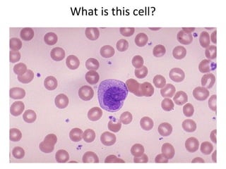
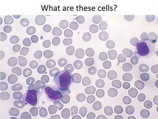
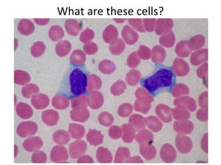
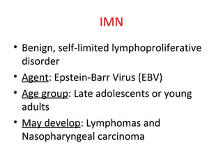
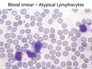
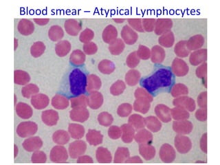
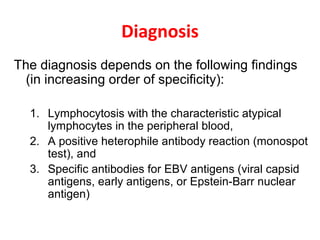
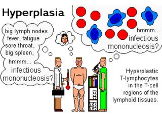
Infectious mononucleosis
IMN -IMN - The Kissing Disease
Dr.CSBR.Prasad, M.D.,
What are these cells?
What is this cell?
What are these cells?
IMN
• Benign, self-limited lymphoproliferative
disorder
• Agent: Epstein-Barr Virus (EBV)
• Age group: Late adolescents or young
adults
• May develop: Lymphomas and
Nasopharyngeal carcinoma
Clinical Features
• Fever
• Sore throat
• Generalized lymphadenopathy
• Splenomegaly
• In some: Hepatitis, Meningoencephalitis,
and Pneumonitis
• EBV occurs in childhood and is usually
asymptomatic
Exudate
covered tonsils
Palatal
petechiae
Pathogenesis
• Transmitted by close human contact, frequently
with the saliva during kissing
• EBV envelope glycoprotein binds to CD21 (CR2),
the receptor for the C3d component of
complement, present on B cells
• The viral infection begins in nasopharyngeal and
oropharyngeal lymphoid tissues, particularly the
tonsils
• infection of B cells may take one of two forms
– In a minority of B cells there is productive infection
– In most B cells, EBV establishes latent infection
• Production of Heterophil antibodies
Outcome of
EBV
infection
Morphology
Peripheral blood:
• Lymphocytosis (Diff count: 60%)
• Atypical lymphocytes constitute 5-80%
• Atypical lymphocytes express CD8, are
sufficiently distinctive to strongly
suggest the diagnosis
Blood smear – Atypical Lymphocytes
Blood smear – Atypical Lymphocytes - SEM
Morphology
Lymph nodes:
• Generalized lymphadenopathy
(Mainly: Posterior cervical, Axillary & Inguinal)
• On histologic examination:
• Expansion of paracortical areas by activated T cells
(immunoblasts), may resemble Malignant lymphoma
• Reed-Sternberg (RS-like) cells
• EBNA2, LMP1, can be demonstrated by IHC
• B-cell areas may also be hyperplastic, but this is
usually mild
• Similar changes commonly occur in the tonsils and
lymphoid tissue of the oropharynx
IHC for EBV
proteins
EBV infection may raise a specter of
leukemia or lymphoma
• Lymphocytosis
• Organomegaly
• Atypical lymphocytes mimicking leukemic blasts
Morphology
The spleen:
• Enlarged in most cases 300 - 500 gm.
• Very soft and fleshy, with a hyperemic cut surface.
• Histology:
• Expansion of white pulp follicles and red pulp
sinusoids
The liver:
• Hepatomegaly is moderate
• Histology:
– Atypical lymphocytes in the portal areas & sinusoids
– Foci of parenchymal necrosis may be present
Splenomegaly in IMN
Spleen is vulnerable to rupture
possibly in part because the rapid increase
in size produces a tense, fragile splenic
capsule
“Acute splenic tumor”
Exercise caution when you are
palpating the abdomen for
spleen in IMN
“you may rupture the spleen!”
Diagnosis
The diagnosis depends on the following findings
(in increasing order of specificity):
1. Lymphocytosis with the characteristic atypical
lymphocytes in the peripheral blood,
2. A positive heterophile antibody reaction (monospot
test), and
3. Specific antibodies for EBV antigens (viral capsid
antigens, early antigens, or Epstein-Barr nuclear
antigen)
Mono spot test
In most patients, infectious
mononucleosis resolves within 4 to 6
weeks,
But the fatigue lasts longer in some
EBV is implicated in Chronic Fatigue
Syndrome
Complications
• Chronic fatigue
• Most common: marked hepatic dysfunction with
jaundice, elevated hepatic enzyme levels,
disturbed appetite, and rarely even liver failure
• Other complications: involve the nervous system,
kidneys, bone marrow, lungs, eyes, heart
• Splenic rupture
• In immunodeficient patients: B-cell lymphomas
• Burkitt’s lymphoma in Africa
E N D

Imn csbrp

  • 1.
    Infectious mononucleosis IMN -IMN- The Kissing Disease Dr.CSBR.Prasad, M.D.,
  • 2.
  • 3.
  • 4.
  • 5.
  • 6.
    IMN • Benign, self-limitedlymphoproliferative disorder • Agent: Epstein-Barr Virus (EBV) • Age group: Late adolescents or young adults • May develop: Lymphomas and Nasopharyngeal carcinoma
  • 7.
    Clinical Features • Fever •Sore throat • Generalized lymphadenopathy • Splenomegaly • In some: Hepatitis, Meningoencephalitis, and Pneumonitis • EBV occurs in childhood and is usually asymptomatic
  • 8.
  • 9.
    Pathogenesis • Transmitted byclose human contact, frequently with the saliva during kissing • EBV envelope glycoprotein binds to CD21 (CR2), the receptor for the C3d component of complement, present on B cells • The viral infection begins in nasopharyngeal and oropharyngeal lymphoid tissues, particularly the tonsils • infection of B cells may take one of two forms – In a minority of B cells there is productive infection – In most B cells, EBV establishes latent infection • Production of Heterophil antibodies
  • 10.
  • 11.
    Morphology Peripheral blood: • Lymphocytosis(Diff count: 60%) • Atypical lymphocytes constitute 5-80% • Atypical lymphocytes express CD8, are sufficiently distinctive to strongly suggest the diagnosis
  • 12.
    Blood smear –Atypical Lymphocytes
  • 13.
    Blood smear –Atypical Lymphocytes
  • 14.
    Blood smear –Atypical Lymphocytes - SEM
  • 15.
    Morphology Lymph nodes: • Generalizedlymphadenopathy (Mainly: Posterior cervical, Axillary & Inguinal) • On histologic examination: • Expansion of paracortical areas by activated T cells (immunoblasts), may resemble Malignant lymphoma • Reed-Sternberg (RS-like) cells • EBNA2, LMP1, can be demonstrated by IHC • B-cell areas may also be hyperplastic, but this is usually mild • Similar changes commonly occur in the tonsils and lymphoid tissue of the oropharynx
  • 17.
  • 18.
    EBV infection mayraise a specter of leukemia or lymphoma • Lymphocytosis • Organomegaly • Atypical lymphocytes mimicking leukemic blasts
  • 19.
    Morphology The spleen: • Enlargedin most cases 300 - 500 gm. • Very soft and fleshy, with a hyperemic cut surface. • Histology: • Expansion of white pulp follicles and red pulp sinusoids The liver: • Hepatomegaly is moderate • Histology: – Atypical lymphocytes in the portal areas & sinusoids – Foci of parenchymal necrosis may be present
  • 20.
    Splenomegaly in IMN Spleenis vulnerable to rupture possibly in part because the rapid increase in size produces a tense, fragile splenic capsule “Acute splenic tumor” Exercise caution when you are palpating the abdomen for spleen in IMN “you may rupture the spleen!”
  • 21.
    Diagnosis The diagnosis dependson the following findings (in increasing order of specificity): 1. Lymphocytosis with the characteristic atypical lymphocytes in the peripheral blood, 2. A positive heterophile antibody reaction (monospot test), and 3. Specific antibodies for EBV antigens (viral capsid antigens, early antigens, or Epstein-Barr nuclear antigen)
  • 22.
  • 23.
    In most patients,infectious mononucleosis resolves within 4 to 6 weeks, But the fatigue lasts longer in some EBV is implicated in Chronic Fatigue Syndrome
  • 24.
    Complications • Chronic fatigue •Most common: marked hepatic dysfunction with jaundice, elevated hepatic enzyme levels, disturbed appetite, and rarely even liver failure • Other complications: involve the nervous system, kidneys, bone marrow, lungs, eyes, heart • Splenic rupture • In immunodeficient patients: B-cell lymphomas • Burkitt’s lymphoma in Africa
  • 26.

Editor's Notes

  • #7 Benign, self-limited lymphoproliferative disorder Agent: Epstein-Barr Virus (EBV) Age group: Late adolescents or young adults associated with the development of a number of neoplasms, most notably certain lymphomas and nasopharyngeal carcinoma
  • #9 Fig-1 Within the mouth, the most typical manifestation of infectious mononucleosis is the merging creamy ooze in the passageway of the mouth, a spot of ruptured blood vessels at the junction of the soft and hard palate and inflammation. Ulceration in the mouth or gums may be seen, but the oral changes are non-specific. Fig-2 Palatal petechiae (A minute red or purple spot on the surface of the palate)
  • #10 During latent infection, a small number of EBV genes are expressed and they are involved in the establishment of latency. The gene products include EBNA1, which binds the EBV genome to chromosomes, mediating episomal persistence and maintenance, and EBNA2 and latent membrane protein 1 (LMP1), which drive B-cell activation and proliferation.[59] LMP1 appears to act by binding to TNF receptor–associated factors, and activates signaling pathways that mimic B-cell activation by CD40, which is involved in normal B-cell responses ( Chapter 6 ). EBNA2 stimulates transcription of many host cell genes, including genes that drive cell cycle entry. The activated B cells then disseminate in the circulation and secrete antibodies with several specificities, including the heterophile anti–sheep red blood cell antibodies used for the diagnosis of infectious mononucleosis. Heterophile antibodies bind to antigens that differ from the antigens that induced them. Thus, people with mononucleosis make antibodies that agglutinate sheep or horse red blood cells in the laboratory, but these antibodies do not react with EBV.
  • #11 FIGURE 8-16  Outcome of Epstein-Barr virus (EBV) infection. In an individual with normal immune function, infection is usually either asymptomatic or leads to mononucleosis. In the setting of cellular immunodeficiency, the proliferation of infected B cells may be uncontrolled, leading to the development of B-cell neoplasms. In other instances, persons without overt evidence of immunodeficiency develop EBV-positive tumors, which are usually (but not always) also derived from B cells. One secondary genetic event that collaborates with EBV to cause B-cell transformation is a balanced 8;14 chromosomal translocation, which is seen in Burkitt lymphoma. EBV is also implicated in the pathogenesis of nasopharyngeal carcinoma, Hodgkin lymphoma, and certain other rare non-Hodgkin lymphomas.
  • #13 Peripheral blood:shows a bsolute lymphocytosis; more than 60% of white blood cells are lymphocytes. Between 5% and 80% of these are large, atypical lymphocytes, 12 to 16 μm in diameter, characterized by an abundant cytoplasm containing multiple clear vacuolations, an oval, indented, or folded nucleus, and scattered cytoplasmic azurophilic granules These atypical lymphocytes, most of which express CD8, are sufficiently distinctive to strongly suggest the diagnosis.
  • #17 T-cell proliferation is sometimes so exuberant that it is difficult to distinguish the nodal morphology from that seen in malignant lymphomas. Similar changes commonly occur in the tonsils and lymphoid tissue of the oropharynx.
  • #21 The spleen is enlarged in most cases, weighing between 300 and 500 gm. It is usually soft and fleshy, with a hyperemic cut surface. The histologic changes are analogous to those of the lymph nodes, showing an expansion of white pulp follicles and red pulp sinusoids due to the presence of numerous activated T cells. These spleens are especially vulnerable to rupture, possibly in part because the rapid increase in size produces a tense, fragile splenic capsule. The liver is usually involved to some degree, although hepatomegaly is at most moderate. On histologic examination, atypical lymphocytes are seen in the portal areas and sinusoids, and scattered, isolated cells or foci of parenchymal necrosis may be present. This histologic picture is similar to that of other forms of viral hepatitis.
  • #23 The diagnosis depends on the following findings (in increasing order of specificity): (1) lymphocytosis with the characteristic atypical lymphocytes in the peripheral blood, (2) a positive heterophile antibody reaction (monospot test), and (3) specific antibodies for EBV antigens (viral capsid antigens, early antigens, or Epstein-Barr nuclear antigen). In most patients, infectious mononucleosis resolves within 4 to 6 weeks, but sometimes the fatigue lasts longer
  • #26 In most patients, infectious mononucleosis resolves within 4 to 6 weeks, but sometimes the fatigue lasts longer. One or more complications occasionally supervene. Perhaps most common is marked hepatic dysfunction with jaundice, elevated hepatic enzyme levels, disturbed appetite, and rarely even liver failure. Other complications involve the nervous system, kidneys, bone marrow, lungs, eyes, heart, and spleen. Splenic rupture can occur even with minor trauma, leading to hemorrhage that may be fatal. A more serious complication in those suffering from some form of immunodeficiency, such as AIDS, or receiving immunosuppressive therapy (e.g., bone marrow or solid-organ transplant recipients) is B-cell lymphomas. As detailed in Chapter 13 , EBV also causes another distinctive form of lymphoma, called Burkitt lymphoma, particularly in certain geographic locales.
  • #27 Unfortunate consequences also occur in individuals suffering from the X-linked lymphoproliferation syndrome (also known as Duncan disease), a disorder caused by a defect in a gene, SH2D1A, that is expressed primarily in cytotoxic T cells and NK cells.[60] SH2D1A (also called SAP) participates in a signaling pathway critical for an effective cellular response to EBV-infected B cells. Patients are often normal until they are acutely infected with EBV, often during adolescence. The failure to control EBV infection variously leads to chronic infectious mononucleosis, agammaglobulinemia, and B-cell lymphoma, each of which proves fatal in about a third of patients.