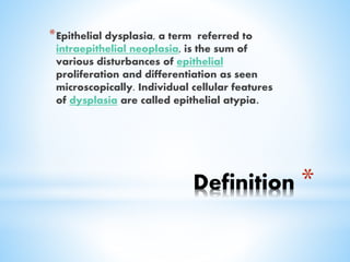
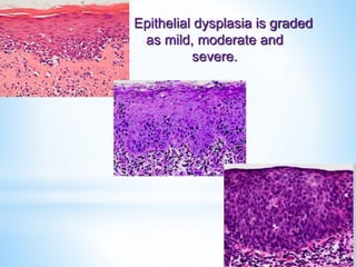
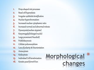
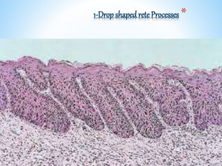
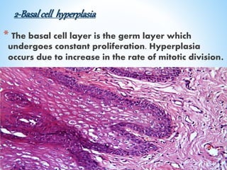
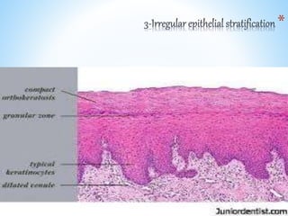
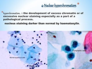
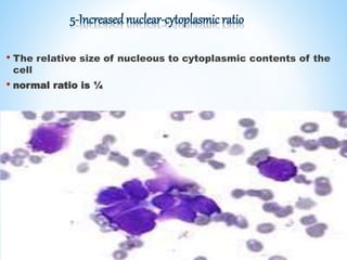
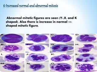
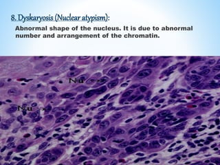
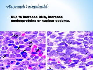
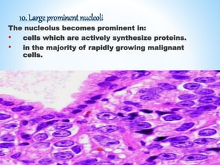
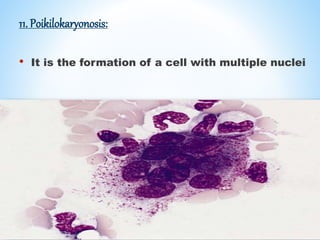
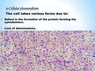
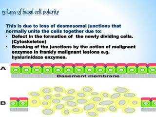
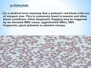
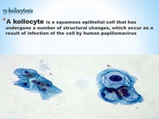
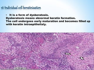
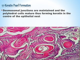
Presented By : AsmaaA.Rahman
*Definition
*Epithelial dysplasia, a term referred to
intraepithelial neoplasia, is the sum of
various disturbances of epithelial
proliferation and differentiation as seen
microscopically. Individual cellular features
of dysplasia are called epithelial atypia.
Epithelial dysplasia is graded
as mild, moderate and
severe.
*
1. Drop-shapedreteprocesses
2. Basalcellhyperplasia
3. Irregularepithelialstratification
4. Nuclearhyperchromatism
5. Increasednuclear-cytoplasmicratio
6. Increasednormalandabnormalmitosis
7. Dyscaryosis(nuclearatypism)
8. Karyomegaly(Enlargednuclei)
9. LargeprominentNucelioli)
10. Poikilokarynosis
11. Cellularpoleomorphism
12. Lossof polarity& Disorientation
13. Anisocytosis
14. Koilocytosis
15. IndividualCellkeratinization
16. KeratinpearlformATion
*1-Drop shaped reteProcesses
2-Basalcell hyperplasia
* The basal cell layer is the germ layer which
undergoes constant proliferation. Hyperplasia
occurs due to increase in the rate of mitotic division.
*
*4-Nuclear hyperchromatism
*hyperchromatism. : the development of excess chromatin or of
excessive nuclear staining especially as a part of a
pathological process.
nucleus staining darker than normal by haematoxylin.
5-Increased nuclear-cytoplasmic ratio
• The relative size of nucleous to cytoplasmic contents of the
cell
• normal ratio is ¼
6-Increased normal and abnormal mitosis
Abnormal mitotic figures are seen (Y-,X, and K
shaped). Also there is increase in normal 11-
shaped mitotic figure.
8. Dyskaryosis (Nuclear atypism):
Abnormal shape of the nucleus. It is due to abnormal
number and arrangement of the chromatin.
9-Karyomegaly( enlarged nuclei )
• Due to increase DNA, increase
nucleoproteins or nuclear oedema.
10. Large prominent nucleoli
The nucleolus becomes prominent in:
• cells which are actively synthesize proteins.
• in the majority of rapidly growing malignant
cells.
11. Poikilokaryonosis:
• It is the formation of a cell with multiple nuclei
12-Cellular pleomorphism
The cell takes various forms due to:
• Defect in the formation of the protein forming the
cytoskeleton.
• Loss of desmosomes.
13-Loss of basal cell polarity
This is due to loss of desmosomal junctions that
normally unite the cells together due to:
• Defect in the formation of the newly dividing cells.
(Cytoskeleton)
• Breaking of the junctions by the action of malignant
enzymes in frankly malignant lesions e.g.
hyalurinidaze enzymes.
14-Anisocytosis
is a medical term meaning that a patient's red blood cells are
of unequal size. This is commonly found in anemia and other
blood conditions. False diagnostic flagging may be triggered
by an elevated WBC count, agglutinated RBCs, RBC
fragments, giant platelets or platelet clumps.
15-koilocytosis
*A koilocyte is a squamous epithelial cell that has
undergone a number of structural changes, which occur as a
result of infection of the cell by human papillomavirus
16-Individual cell keratinization
• It is a form of dyskeratosis.
Dyskeratosis means abnormal keratin formation.
The cell undergoes early maturation and becomes filled up
with keratin intraepithelialy.
17-Keratin Pearl Formation
• Desmosomal junctions are maintained and the
polyhedral cells mature thus forming keratin in the
centre of the epithelial nest
Epithelial dysplasia

Epithelial dysplasia

  • 1.
    Presented By :AsmaaA.Rahman
  • 2.
    *Definition *Epithelial dysplasia, aterm referred to intraepithelial neoplasia, is the sum of various disturbances of epithelial proliferation and differentiation as seen microscopically. Individual cellular features of dysplasia are called epithelial atypia.
  • 3.
    Epithelial dysplasia isgraded as mild, moderate and severe.
  • 4.
    * 1. Drop-shapedreteprocesses 2. Basalcellhyperplasia 3.Irregularepithelialstratification 4. Nuclearhyperchromatism 5. Increasednuclear-cytoplasmicratio 6. Increasednormalandabnormalmitosis 7. Dyscaryosis(nuclearatypism) 8. Karyomegaly(Enlargednuclei) 9. LargeprominentNucelioli) 10. Poikilokarynosis 11. Cellularpoleomorphism 12. Lossof polarity& Disorientation 13. Anisocytosis 14. Koilocytosis 15. IndividualCellkeratinization 16. KeratinpearlformATion
  • 5.
  • 6.
    2-Basalcell hyperplasia * Thebasal cell layer is the germ layer which undergoes constant proliferation. Hyperplasia occurs due to increase in the rate of mitotic division.
  • 7.
  • 8.
    *4-Nuclear hyperchromatism *hyperchromatism. :the development of excess chromatin or of excessive nuclear staining especially as a part of a pathological process. nucleus staining darker than normal by haematoxylin.
  • 9.
    5-Increased nuclear-cytoplasmic ratio •The relative size of nucleous to cytoplasmic contents of the cell • normal ratio is ¼
  • 10.
    6-Increased normal andabnormal mitosis Abnormal mitotic figures are seen (Y-,X, and K shaped). Also there is increase in normal 11- shaped mitotic figure.
  • 11.
    8. Dyskaryosis (Nuclearatypism): Abnormal shape of the nucleus. It is due to abnormal number and arrangement of the chromatin.
  • 12.
    9-Karyomegaly( enlarged nuclei) • Due to increase DNA, increase nucleoproteins or nuclear oedema.
  • 13.
    10. Large prominentnucleoli The nucleolus becomes prominent in: • cells which are actively synthesize proteins. • in the majority of rapidly growing malignant cells.
  • 14.
    11. Poikilokaryonosis: • Itis the formation of a cell with multiple nuclei
  • 15.
    12-Cellular pleomorphism The celltakes various forms due to: • Defect in the formation of the protein forming the cytoskeleton. • Loss of desmosomes.
  • 16.
    13-Loss of basalcell polarity This is due to loss of desmosomal junctions that normally unite the cells together due to: • Defect in the formation of the newly dividing cells. (Cytoskeleton) • Breaking of the junctions by the action of malignant enzymes in frankly malignant lesions e.g. hyalurinidaze enzymes.
  • 17.
    14-Anisocytosis is a medicalterm meaning that a patient's red blood cells are of unequal size. This is commonly found in anemia and other blood conditions. False diagnostic flagging may be triggered by an elevated WBC count, agglutinated RBCs, RBC fragments, giant platelets or platelet clumps.
  • 18.
    15-koilocytosis *A koilocyte isa squamous epithelial cell that has undergone a number of structural changes, which occur as a result of infection of the cell by human papillomavirus
  • 19.
    16-Individual cell keratinization •It is a form of dyskeratosis. Dyskeratosis means abnormal keratin formation. The cell undergoes early maturation and becomes filled up with keratin intraepithelialy.
  • 20.
    17-Keratin Pearl Formation •Desmosomal junctions are maintained and the polyhedral cells mature thus forming keratin in the centre of the epithelial nest