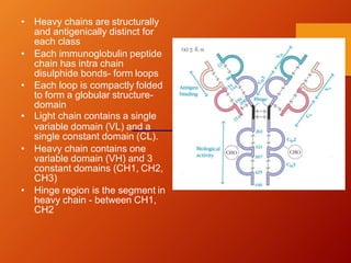
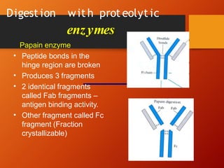
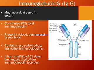
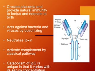
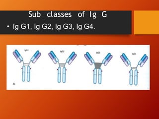
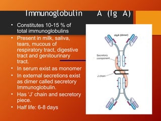
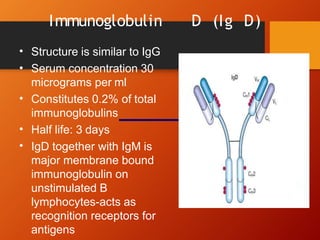
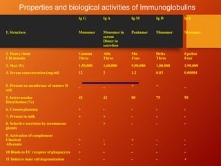
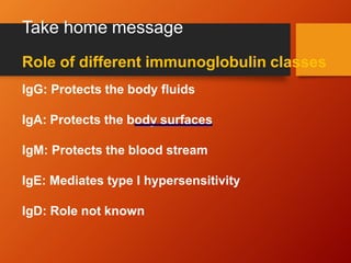
Immunoglobulins
Mrs.ShubhadaA
Kulkarni
AssistantProfessor
SYIPER
Immunoglobulin
Immunoglobulin is a glycoprotein that is
made in response to an antigen and can
recognize and bind to the antigen that
caused its production.
• Are gamma globulins
• Synthesized by plasma cells
• Constitute 25-30 % of total serum proteins
• Antibodies are present in serum, tissue
fluids and mucosal surfaces.
• All antibodies are immunoglobulins, but all
immunoglobulins may not be antibodies
IMMUNOGLOBULIN
Basic structure
• Composed of 4 polypeptide
chains.
• 2 identical light and 2 identical
heavy chains
Linked by disulphide bonds
•
• Light chains similar in all
immunoglobulins
• Light chains occur in 2
varieties kappa and lambda
• Light and Heavy chains are
subdivided into variable and
constant region.
• Each heavy and light chain
contains amino terminal in
variable region carboxy
terminal in constant region
• Heavy chains are structurally
and antigenically distinct for
each class
• Each immunoglobulin peptide
chain has intra chain
disulphide bonds- form loops
• Each loop is compactly folded
to form a globular structure-
domain
• Light chain contains a single
variable domain (VL) and a
single constant domain (CL).
• Heavy chain contains one
variable domain (VH) and 3
constant domains (CH1, CH2,
CH3)
• Hinge region is the segment in
heavy chain - between CH1,
CH2
Digest ion wit h prot eolyt ic
enzymes
Papain enzyme
• Peptide bonds in the
hinge region are broken
Produces 3 fragments
•
• 2 identical fragments
called Fab fragments –
antigen binding activity.
• Other fragment called Fc
fragment (Fraction
crystallizable)
Pepsin digestion
• Produce a single fragment composed of two Fab
like subunits F(ab)2 binds antigen
• Fc fragment is not recovered- digested to small
numerous peptides.
Classification
• Based on structure and antigenic nature of H
chain the immunoglobulins are classified into 5
classes.
• Ig G- (gamma)
• Ig A- (alpha)
• Ig M- (mu)
• Ig D- (delta)
• Ig E - (epsilon)
Immunoglobulin G (Ig G)
• Most abundant class in
serum
• Constitutes 80% total
immunoglobulin
• Present in blood, plasma and
tissue fluids
• Contains less carbohydrate
than other immunoglobulins
• It has a half life of 23 days:
the longest of all of the
immunoglobulin isotypes
• Crosses placenta and
provide natural immunity
to foetus and neonate at
birth
• Acts against bacteria and
viruses by opsonizing
• Neutralize toxin
• Activate complement by
classical pathway
• Catabolism of IgG is
unique in that it varies with
Sub classes of Ig G
• Ig G1, Ig G2, Ig G3, Ig G4.
Biological funct ion of subclasses
• IgG1, IgG3, IgG4 – cross placenta and
protect foetus
• IgG3 activates complement
• IgG1 and IgG3 binds to Fc receptor on
phagocytic cells, monocytes and
macrophages and mediate opsinization.
Immunoglobulin A (Ig A)
• Constitutes 10-15 % of
total immunoglobulins
• Present in milk, saliva,
tears, mucous of
respiratory tract, digestive
tract and genitourinary
tract.
In serum exist as monomer
•
• In external secretions exist
as dimer called secretory
Immunoglobulin.
• Has ‘J’ chain and secretory
piece.
• Half life: 6-8 days
Format ion of secret ory Ig A
• Dimeric Ig A binds to the
receptor on the surface of the
epithelial cells -endocytosed
and transported across the
cell to the luminal surface
• After reaching the surface, the
poly-Ig receptor is cleaved
• The portion of the receptor
that remains attached to the
Ig A dimer – secretory
component
• Secretory piece protects Ig A
from digestive enzymes and
denaturation by bacterial
proteases
Functions
Provides local immunity.
•
• Secretory Ig A binds to surface antigens of microorganism
and prevent its attachment and invasion of the mucosal
surfaces of respiratory and digestive tract- immune
elimination.
• Secretory IgA provides important line of defense against
salmonella, Vibrio cholerae, N. gonorrhoeae, influenza virus
and poliovirus.
• Secretory IgA present in breast milk protects newborn
during first months of life.
Activates complement by the alternative pathway
•
• Promotes phagocytosis and intracellular killing of
microorganisms
Immunoglobulin M (Ig M)
• Accounts for 5-10% of
total serum proteins
• Polymer of five monomeric
units (pentamer)
• Held together by disulfide
bonds and ‘J’ chain
• Mol. Wt. of 900,000-
10,00,000 (millionaire
molecule)
• Half life: 5 days
• Most of IgM (80%) present intravascularly
• Present in low concentration in intercellular tissue
fluids
• Cannot cross placenta
• Presence of IgM antibody in serum of newborn
indicate congenital infection.
• Earliest immunoglobulin to be synthesized by foetus
(20 weeks)
• First immunoglobulin to be produced in primary
response to antigen
• Relatively short-lived hence it’s demonstration in the
serum indicates recent infection
• Monomeric IgM appears on the surface of
unstimulated B lymphocytes and act as receptors
for antigens
Functions
• It agglutinates bacteria
• Activates complement by
classical pathway
• Causes opsonization and
immune heamolysis
• Believed to be responsible for
protection against blood invasion
by microorganisms
Immunoglobulin E (Ig E)
Structure is similar to Ig G
•
• Has 4 constant region
domains.
Mol. Wt. 1,90,000
Half life: 2 days
•
•
• Heat labile (inactivated at
560C in 1 hour)
• Normal serum
concentration 0.3 ug/ml
• Mostly present extra
cellularly
• Does not cross placenta
• Produced in the lining of respiratory and
intestinal tract
• Known as reagin antibody
• Does not activate complement nor agglutinate
antigens
• Binds to the Fc receptors on the membranes of
blood basophils and tissue mast cells
• Mediates immediate hypersensitivity reaction
and P.K. reaction
• Responsible for symptoms of anaphylactic
shock, hay fever and asthma.
• Play a role in immunity against helminthic
parasites
• IgE binds to Fc receptors on
the membrane of blood
basophils and tissue mast
cells.
• When two IgE molecules on
the surface of these cells are
cross linked by binding of the
same antigen- cells
degranulates.
• Release histamine and
pharmacological mediators
of anaphylaxis from cell.
• The physiological role of IgE
appears to be protection
against pathogens by mast
cell degranulation and
release of inflammatory
mediators
• Mediates P.K. reaction (PRAUSNITZ & KUSTNER)
• The presence of a serum component responsible for
allergic reaction was first demonstrated by Prausnitz
and Kustner in 1921.
• Kustner was suffering from atopic hypersensitivity to
certain species of fish
• Kustner’s serum was injected intracutaneously in
Prausnitz
• After 24 hrs small quantity of cooked fish antigen
was injected at the same site
• A wheal and flare reaction occurred within minutes.
Immunoglobulin D (Ig D)
Structure is similar to IgG
•
• Serum concentration 30
micrograms per ml
• Constitutes 0.2% of total
immunoglobulins
Half life: 3 days
•
• IgD together with IgM is
major membrane bound
immunoglobulin on
unstimulated B
lymphocytes-acts as
recognition receptors for
antigens
Properties and biological activities of Immunoglobulins
Ig G Ig A Ig M Ig D Ig E
1. Structure Monomer Monomer in
serum
Dimer in
secretion
Pentamer Monomer Monomer
2. Heavy chain Gamma
Three
Alfa
Three
Mu
Four
Delta
Three
Epsilon
Four
CH domain
3. Mol. Wt. 1,50,000 1,60,000 9,00,000 1,80,000 1,90,000
4. Serum concentration (mg/ml) 12 2 1.2 0.03 0.00004
5. Present on membrane of mature B
cell
_ _ + + _
5. Intravascular
Distribution (%)
45 42 80 75 50
6. Crosses placenta + - - -
7. Present in milk + + - -
8. Selective secretion by seromucous - + - -
-
-
-
glands
9. Activation of complement
Classical
Alternate
+
-
-
+
+
-
-
-
-
-
10 Binds to FC receptor of phagocytes + - - - -
11 Induces mast cell degranulation - - - - +
Take home message
Role of different immunoglobulin classes
IgG: Protects the body fluids
IgA: Protects the body surfaces
IgM: Protects the blood stream
IgE: Mediates type I hypersensitivity
IgD: Role not known
immunoglobulins.pptx

immunoglobulins.pptx

  • 1.
  • 2.
    Immunoglobulin Immunoglobulin is aglycoprotein that is made in response to an antigen and can recognize and bind to the antigen that caused its production.
  • 3.
    • Are gammaglobulins • Synthesized by plasma cells • Constitute 25-30 % of total serum proteins • Antibodies are present in serum, tissue fluids and mucosal surfaces. • All antibodies are immunoglobulins, but all immunoglobulins may not be antibodies IMMUNOGLOBULIN
  • 4.
    Basic structure • Composedof 4 polypeptide chains. • 2 identical light and 2 identical heavy chains Linked by disulphide bonds • • Light chains similar in all immunoglobulins • Light chains occur in 2 varieties kappa and lambda • Light and Heavy chains are subdivided into variable and constant region. • Each heavy and light chain contains amino terminal in variable region carboxy terminal in constant region
  • 5.
    • Heavy chainsare structurally and antigenically distinct for each class • Each immunoglobulin peptide chain has intra chain disulphide bonds- form loops • Each loop is compactly folded to form a globular structure- domain • Light chain contains a single variable domain (VL) and a single constant domain (CL). • Heavy chain contains one variable domain (VH) and 3 constant domains (CH1, CH2, CH3) • Hinge region is the segment in heavy chain - between CH1, CH2
  • 6.
    Digest ion with prot eolyt ic enzymes Papain enzyme • Peptide bonds in the hinge region are broken Produces 3 fragments • • 2 identical fragments called Fab fragments – antigen binding activity. • Other fragment called Fc fragment (Fraction crystallizable)
  • 7.
    Pepsin digestion • Producea single fragment composed of two Fab like subunits F(ab)2 binds antigen • Fc fragment is not recovered- digested to small numerous peptides.
  • 8.
    Classification • Based onstructure and antigenic nature of H chain the immunoglobulins are classified into 5 classes. • Ig G- (gamma) • Ig A- (alpha) • Ig M- (mu) • Ig D- (delta) • Ig E - (epsilon)
  • 9.
    Immunoglobulin G (IgG) • Most abundant class in serum • Constitutes 80% total immunoglobulin • Present in blood, plasma and tissue fluids • Contains less carbohydrate than other immunoglobulins • It has a half life of 23 days: the longest of all of the immunoglobulin isotypes
  • 10.
    • Crosses placentaand provide natural immunity to foetus and neonate at birth • Acts against bacteria and viruses by opsonizing • Neutralize toxin • Activate complement by classical pathway • Catabolism of IgG is unique in that it varies with
  • 11.
    Sub classes ofIg G • Ig G1, Ig G2, Ig G3, Ig G4.
  • 12.
    Biological funct ionof subclasses • IgG1, IgG3, IgG4 – cross placenta and protect foetus • IgG3 activates complement • IgG1 and IgG3 binds to Fc receptor on phagocytic cells, monocytes and macrophages and mediate opsinization.
  • 13.
    Immunoglobulin A (IgA) • Constitutes 10-15 % of total immunoglobulins • Present in milk, saliva, tears, mucous of respiratory tract, digestive tract and genitourinary tract. In serum exist as monomer • • In external secretions exist as dimer called secretory Immunoglobulin. • Has ‘J’ chain and secretory piece. • Half life: 6-8 days
  • 14.
    Format ion ofsecret ory Ig A • Dimeric Ig A binds to the receptor on the surface of the epithelial cells -endocytosed and transported across the cell to the luminal surface • After reaching the surface, the poly-Ig receptor is cleaved • The portion of the receptor that remains attached to the Ig A dimer – secretory component • Secretory piece protects Ig A from digestive enzymes and denaturation by bacterial proteases
  • 15.
    Functions Provides local immunity. • •Secretory Ig A binds to surface antigens of microorganism and prevent its attachment and invasion of the mucosal surfaces of respiratory and digestive tract- immune elimination. • Secretory IgA provides important line of defense against salmonella, Vibrio cholerae, N. gonorrhoeae, influenza virus and poliovirus. • Secretory IgA present in breast milk protects newborn during first months of life. Activates complement by the alternative pathway • • Promotes phagocytosis and intracellular killing of microorganisms
  • 16.
    Immunoglobulin M (IgM) • Accounts for 5-10% of total serum proteins • Polymer of five monomeric units (pentamer) • Held together by disulfide bonds and ‘J’ chain • Mol. Wt. of 900,000- 10,00,000 (millionaire molecule) • Half life: 5 days
  • 17.
    • Most ofIgM (80%) present intravascularly • Present in low concentration in intercellular tissue fluids • Cannot cross placenta • Presence of IgM antibody in serum of newborn indicate congenital infection. • Earliest immunoglobulin to be synthesized by foetus (20 weeks) • First immunoglobulin to be produced in primary response to antigen • Relatively short-lived hence it’s demonstration in the serum indicates recent infection • Monomeric IgM appears on the surface of unstimulated B lymphocytes and act as receptors for antigens
  • 18.
    Functions • It agglutinatesbacteria • Activates complement by classical pathway • Causes opsonization and immune heamolysis • Believed to be responsible for protection against blood invasion by microorganisms
  • 19.
    Immunoglobulin E (IgE) Structure is similar to Ig G • • Has 4 constant region domains. Mol. Wt. 1,90,000 Half life: 2 days • • • Heat labile (inactivated at 560C in 1 hour) • Normal serum concentration 0.3 ug/ml • Mostly present extra cellularly • Does not cross placenta
  • 20.
    • Produced inthe lining of respiratory and intestinal tract • Known as reagin antibody • Does not activate complement nor agglutinate antigens • Binds to the Fc receptors on the membranes of blood basophils and tissue mast cells • Mediates immediate hypersensitivity reaction and P.K. reaction • Responsible for symptoms of anaphylactic shock, hay fever and asthma. • Play a role in immunity against helminthic parasites
  • 21.
    • IgE bindsto Fc receptors on the membrane of blood basophils and tissue mast cells. • When two IgE molecules on the surface of these cells are cross linked by binding of the same antigen- cells degranulates. • Release histamine and pharmacological mediators of anaphylaxis from cell. • The physiological role of IgE appears to be protection against pathogens by mast cell degranulation and release of inflammatory mediators
  • 22.
    • Mediates P.K.reaction (PRAUSNITZ & KUSTNER) • The presence of a serum component responsible for allergic reaction was first demonstrated by Prausnitz and Kustner in 1921. • Kustner was suffering from atopic hypersensitivity to certain species of fish • Kustner’s serum was injected intracutaneously in Prausnitz • After 24 hrs small quantity of cooked fish antigen was injected at the same site • A wheal and flare reaction occurred within minutes.
  • 23.
    Immunoglobulin D (IgD) Structure is similar to IgG • • Serum concentration 30 micrograms per ml • Constitutes 0.2% of total immunoglobulins Half life: 3 days • • IgD together with IgM is major membrane bound immunoglobulin on unstimulated B lymphocytes-acts as recognition receptors for antigens
  • 24.
    Properties and biologicalactivities of Immunoglobulins Ig G Ig A Ig M Ig D Ig E 1. Structure Monomer Monomer in serum Dimer in secretion Pentamer Monomer Monomer 2. Heavy chain Gamma Three Alfa Three Mu Four Delta Three Epsilon Four CH domain 3. Mol. Wt. 1,50,000 1,60,000 9,00,000 1,80,000 1,90,000 4. Serum concentration (mg/ml) 12 2 1.2 0.03 0.00004 5. Present on membrane of mature B cell _ _ + + _ 5. Intravascular Distribution (%) 45 42 80 75 50 6. Crosses placenta + - - - 7. Present in milk + + - - 8. Selective secretion by seromucous - + - - - - - glands 9. Activation of complement Classical Alternate + - - + + - - - - - 10 Binds to FC receptor of phagocytes + - - - - 11 Induces mast cell degranulation - - - - +
  • 25.
    Take home message Roleof different immunoglobulin classes IgG: Protects the body fluids IgA: Protects the body surfaces IgM: Protects the blood stream IgE: Mediates type I hypersensitivity IgD: Role not known