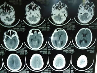
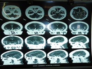
1. A 41-year-old man presented with vomiting, altered mental status, and difficulty walking. On examination, he showed signs of brainstem involvement including nystagmus and facial weakness.
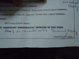
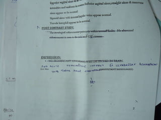
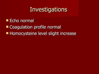
2. Imaging and lab tests did not reveal any coagulation abnormalities or structural causes like tumors. A slight elevation in homocysteine levels was found.
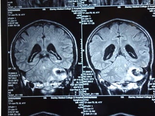
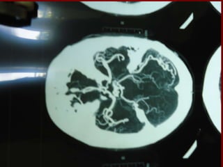
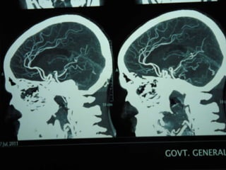
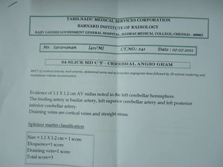
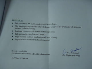
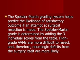
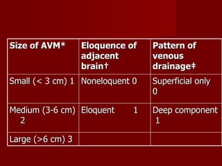
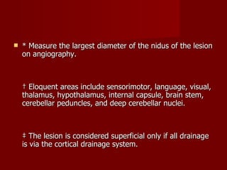
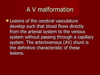
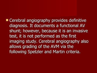
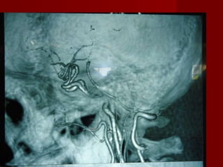
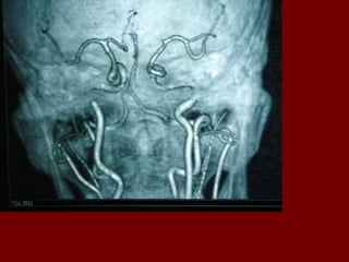
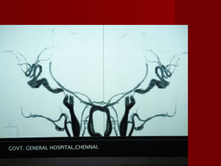
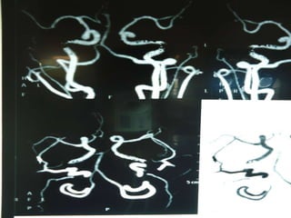
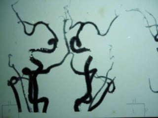
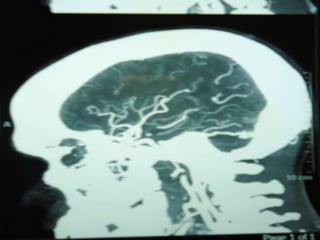
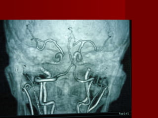
3. Cerebral angiography confirmed an arteriovenous malformation (AVM), which are congenital lesions caused by a failure of the embryonic vasculature to fully develop. The Spetzler-Martin grading system was then used to assess the AVM based on its size, location, and venous drainage patterns.