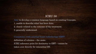
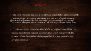
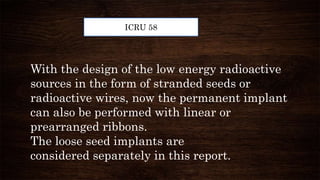
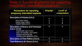
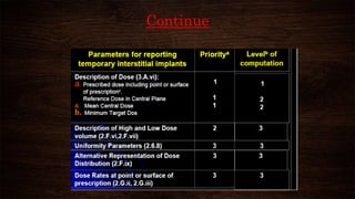
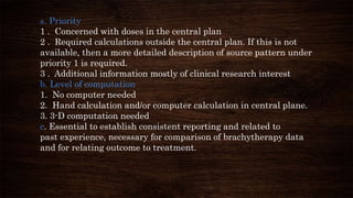
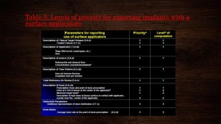
ANJU.K.V.
MSC RADIATION PHYSICS
CALICUT UNIVERSITY
INTRODUCTION
With the introduction of miniaturized brachytherapy sources, the
sophisticated 3-D source localization methods have been
introduced, which can be linked to computerized methods for dose
calculation.
Therefore, a common language is valuable to provide a method of
dose specification and reporting which can be used for implants of
all types of interstitial brachytherapy procedures.
1985: ICRU Report 38,
Was written regarding the Dose and volume specification as
well as reporting intracavitary Brachytherapy in GYN.
1997: ICRU Report 58,
Was written to generate a guideline about the dose and
volumes specification for reporting interstitial Brachytherapy.
ICRU 58
Aim: to develop a common language based on existing Concepts.
1. usable to describe what has been done.
2. closely related to the outcome of the treatment.
3. generally understood.
Consistency with external beam radiotherapy (EBT)
definition of volumes – the same.
ICRU reference point for dosimetry in EBT – cannot be
taken over directly for interstitial BT.
ICRU 58
There are several historic systems of prescribing brachytherapy
• –Manchester system
• –Paris system
• Quimby system
• The term “system” denotes a set of rules which takes into account the
source types , strengths, geometry and method of application to
obtain suitable dose distributions over the volume(s) to be treated.
The system also provides a means of calculating and specifying dose.
• It is important to remember that while an implant may follow the
source distribution rules of a system, it does not comply with the
system unless the method of dose specification and prescription
are also followed.
ICRU 58
ICRU
Two important type of implants in ICRU 58 are,
1. Temporary implant
2. Permanent implant
Temporary Implant
• The radioactive sources are removed from the tissue after the treatment is
completed.
• Radionuclide used have typically longer half life.
Permanent Implant
• The brachytherapy sources remain in the patient, and they will not be removed.
• Radionuclide used have typically shorter half life.
ICRU 58
ICRU 58
Traditionally, the temporary implants were performed
with the linear (like wires) radioactive sources or seeds
arranged in a linear fashion (i.e. ribbons, etc.).
However, in a permanent implants, multiple loose
sources were distributed in random orientations.
With the design of the low energy radioactive
sources in the form of stranded seeds or
radioactive wires, now the permanent implant
can also be performed with linear or
prearranged ribbons.
The loose seed implants are
considered separately in this report.
ICRU 58
For Planning
permanent implants
The number of sources
depends on,
a. Their initial strength.
b. Type of the radioisotope.
For Planning temporary
implants
The total time of implantation
depends on,
a. Number of sources
b. Strengths and source
c. Pattern of distribution of sources
Planning boost permanent and temporary implants,
the number of seeds and the initial activity of the seeds are
adjusted upon the external beam radiation dose.
KERMA (Kinetic Energy
Released in Matter)
The sum of the initial kinetic energie of
all the ionizing charged particles
liberated by uncharged ionizing particles.
K = dEtr/ dm
ABSORBED DOSE
The mean energy imparted by radiation
to a unit
mass of medium
D=dE/dm
1Gy = 1J/Kg
Reference Air Kerma rate
Reference Air Kerma Rate = Air Kerma Rate at a reference point of 1 cm
or 1 m.
OR
It is defined as the air kerma rate measured at one meter
along the perpendicular bisector of the long axis of the
source,corrected for air attenuation and scattering.
Unit : 1 U = μGy/h
The term m^2 is not included inthe unit,since it is inherent
in the definition itself.
Total Reference Air Kerma (TRAK)
TRAK= Σti . Si
Where;
Si = Reference air kerma rate for
each source.
ti = Irradiation time for each source.
Description of Source Patterns
single plane: implant is defined as an implant containing two or more
sources, which lie in the same plane.
two-plane (double-plane): implant contains two planes, which are
generally parallel to each other.
Multi-plane: implant, contains three or more planes.
non-plane: sphere or cylinder.
VOLUMES
(Gross Tumor
volume)
(Clinical
Target Volume)
(Planning
Target Volume)
(Treatment
Volume)
(Irradiated
Volume)
Gross Tumor Volume (GTV)
The palpable or visible/demonstrable extent
and location of the malignant growth.
Clinical Target Volume (CTV)
The volume of the tissue that contains a
gross tumor volume and/or subclinical
microscopic malignant disease which has to
be eliminated.
Planning Target Volume (PTV)
The volume of tissue receiving the
prescribed irradiation.
For an interstitial brachytherapy, the PTV is,
in general, identical to the CTV.
Treatment Volume (TV)
The volume of tissue, which is encompassed
by an isodose surface that has been
specified by the radiation oncologist.
The dose value at this isodose surface is the
minimum target dose
CTV
The maximum diameters measured in three orthogonal directions must be
reported.
CENTRAL PLANE
The plane that is perpendicular to the main
direction of the linear sources and passing
through the estimated center of the implant.
Central Plane (continue)
The plane that is perpendicular to the main
direction of the linear sources and passing
through the estimated center of the implant.
Description of Dose
distribution
Interstitial Brachytherapy
Implants:
1. Non Homogeneous dose
Distribution .
2. High Regions around
Sources.
3. Regions of plateau dose
or local minimum doses.
The dose distribution in a plane perpendicular to linear
and parallel sources, shows a plateau dose region of low
dose gradient. In this example of three sources of 6 cm
long and with 1.5 cm spacing, the dose varies by less than
2% between the sources.
Dose Distribution in One or More Planes
through the Implant
1. The minimum information needed for the dose distribution of
an implant is,
The isodose curves in at least one plane either in tabular form or
by graphical Presentation.
2. The central plane of the implant should be used, If only one
plane is chose.
Prescription Dose
The prescribed dose is defined as the dose, which the physician
intends to give, and enters in the patient’s treatment chart.
Minimum Target Dose (MTD): The minimum dose at the
periphery of the CTV = Minimum dose decided upon by the clinician as
adequate to treat the CTV (minimum peripheral dose).
MTD≅ 90% of the prescribed dose in the Manchsteer system for
interstitial therapy.
Prescription Dose (continue)
Mean Central Dose (MCD) : Dm
The minimum dose at the periphery of the CTV
= Arithmetic mean of the local minimum doses between sources, in the central
planes.
Prescription Dose (continue)
High Dose Volume
The volume of tissue that is encompassed that will receive more than
150% of the mean central dose be reported.
Low Dose Volume
A low dose volume should be defined as a volume within the clinical
target volume, encompassed by an isodose corresponding to 90% of the
prescribed dose. The maximum dimension of the low dose volume in
any plane calculated should be reported.
Prescription Dose (continue)
Dose Uniformity Parameters
ICRU defines the following two methods for the dose uniformity
1. The spread of the individual minimum doses used to calculate the mean
central dose in the central plane (expressed as a percentage of the
mean central dose).
2. The dose homogeneity index; defined as the ratio of minimum
target dose to the mean central dose.
Time Dose Factors
Time and Dose Rates for Temporary Implants
Time = Prescribed dose/initial dose rate
Time Dose Pattern for Temporary Implants
a. Continuous irradiation
b. Non-continuous irradiation
c. Pulsed irradiation
Recommendations for Recording and Reporting
Parameters Required for Recording and Reporting
. Description of Volumes : GTV, PTV,…….
. Description of Sources :
1. Radioisotope and filtration
2. Pattern of source distribution
• Description of Technique and Source pattern
Table 1: Levels of priority for reporting
temporary interstitial implants
Continue
a. Priority
1 . Concerned with doses in the central plan
2 . Required calculations outside the central plan. If this is not
available, then a more detailed description of source pattern under
priority 1 is required.
3 . Additional information mostly of clinical research interest
b. Level of computation
1. No computer needed
2. Hand calculation and/or computer calculation in central plane.
3. 3-D computation needed
c. Essential to establish consistent reporting and related to
past experience, necessary for comparison of brachytherapy data
and for relating outcome to treatment.
Table 3: Levels of priority for reporting implants
with a single stationary source line.
Table 4: Levels of priority for reporting
implants with a moving source.
Table 5: Levels of priority for reporting implants with a
surface applicators
CONCLUSION
 It is very important to follow these
recommendations to achieve the aim of
brachytherapy.
 In this presentation, the ICRU-58
recommended concepts and procedures for dose
specifications and reporting of the interstitial
brachytherapy implants has reviewed.
Selected reference
1. ICRU Report 38: Dose and Volume Specification for Reporting
Intracavitary Gynecology (1985)
2. ICRU Report 58: Dose and Volume Specification for Reporting
Interstitial Therapy (1997)
3. The GEC ESTRO Handbook of Brachytherapy, Editors: Alain
Gerbaulet, Richard Pötter, Jean-Jacques Mazeron, Erik van
Limbergen (2002;107;3225-332
4. ESMP 2013 Reporting in Brachytherapy,Lecture 5 Brachytherapy
Implantation.s
Icru 58.
Icru 58.

Icru 58.

  • 1.
  • 2.
    INTRODUCTION With the introductionof miniaturized brachytherapy sources, the sophisticated 3-D source localization methods have been introduced, which can be linked to computerized methods for dose calculation. Therefore, a common language is valuable to provide a method of dose specification and reporting which can be used for implants of all types of interstitial brachytherapy procedures.
  • 3.
    1985: ICRU Report38, Was written regarding the Dose and volume specification as well as reporting intracavitary Brachytherapy in GYN. 1997: ICRU Report 58, Was written to generate a guideline about the dose and volumes specification for reporting interstitial Brachytherapy.
  • 4.
    ICRU 58 Aim: todevelop a common language based on existing Concepts. 1. usable to describe what has been done. 2. closely related to the outcome of the treatment. 3. generally understood. Consistency with external beam radiotherapy (EBT) definition of volumes – the same. ICRU reference point for dosimetry in EBT – cannot be taken over directly for interstitial BT.
  • 5.
    ICRU 58 There areseveral historic systems of prescribing brachytherapy • –Manchester system • –Paris system • Quimby system
  • 6.
    • The term“system” denotes a set of rules which takes into account the source types , strengths, geometry and method of application to obtain suitable dose distributions over the volume(s) to be treated. The system also provides a means of calculating and specifying dose. • It is important to remember that while an implant may follow the source distribution rules of a system, it does not comply with the system unless the method of dose specification and prescription are also followed. ICRU 58
  • 7.
    ICRU Two important typeof implants in ICRU 58 are, 1. Temporary implant 2. Permanent implant
  • 8.
    Temporary Implant • Theradioactive sources are removed from the tissue after the treatment is completed. • Radionuclide used have typically longer half life. Permanent Implant • The brachytherapy sources remain in the patient, and they will not be removed. • Radionuclide used have typically shorter half life. ICRU 58
  • 9.
    ICRU 58 Traditionally, thetemporary implants were performed with the linear (like wires) radioactive sources or seeds arranged in a linear fashion (i.e. ribbons, etc.). However, in a permanent implants, multiple loose sources were distributed in random orientations.
  • 10.
    With the designof the low energy radioactive sources in the form of stranded seeds or radioactive wires, now the permanent implant can also be performed with linear or prearranged ribbons. The loose seed implants are considered separately in this report. ICRU 58
  • 11.
    For Planning permanent implants Thenumber of sources depends on, a. Their initial strength. b. Type of the radioisotope. For Planning temporary implants The total time of implantation depends on, a. Number of sources b. Strengths and source c. Pattern of distribution of sources
  • 12.
    Planning boost permanentand temporary implants, the number of seeds and the initial activity of the seeds are adjusted upon the external beam radiation dose.
  • 13.
    KERMA (Kinetic Energy Releasedin Matter) The sum of the initial kinetic energie of all the ionizing charged particles liberated by uncharged ionizing particles. K = dEtr/ dm ABSORBED DOSE The mean energy imparted by radiation to a unit mass of medium D=dE/dm 1Gy = 1J/Kg
  • 14.
    Reference Air Kermarate Reference Air Kerma Rate = Air Kerma Rate at a reference point of 1 cm or 1 m. OR It is defined as the air kerma rate measured at one meter along the perpendicular bisector of the long axis of the source,corrected for air attenuation and scattering. Unit : 1 U = μGy/h The term m^2 is not included inthe unit,since it is inherent in the definition itself.
  • 15.
    Total Reference AirKerma (TRAK) TRAK= Σti . Si Where; Si = Reference air kerma rate for each source. ti = Irradiation time for each source.
  • 16.
    Description of SourcePatterns single plane: implant is defined as an implant containing two or more sources, which lie in the same plane. two-plane (double-plane): implant contains two planes, which are generally parallel to each other. Multi-plane: implant, contains three or more planes. non-plane: sphere or cylinder.
  • 17.
    VOLUMES (Gross Tumor volume) (Clinical Target Volume) (Planning TargetVolume) (Treatment Volume) (Irradiated Volume)
  • 18.
    Gross Tumor Volume(GTV) The palpable or visible/demonstrable extent and location of the malignant growth. Clinical Target Volume (CTV) The volume of the tissue that contains a gross tumor volume and/or subclinical microscopic malignant disease which has to be eliminated.
  • 19.
    Planning Target Volume(PTV) The volume of tissue receiving the prescribed irradiation. For an interstitial brachytherapy, the PTV is, in general, identical to the CTV. Treatment Volume (TV) The volume of tissue, which is encompassed by an isodose surface that has been specified by the radiation oncologist. The dose value at this isodose surface is the minimum target dose
  • 20.
    CTV The maximum diametersmeasured in three orthogonal directions must be reported.
  • 21.
    CENTRAL PLANE The planethat is perpendicular to the main direction of the linear sources and passing through the estimated center of the implant.
  • 22.
    Central Plane (continue) Theplane that is perpendicular to the main direction of the linear sources and passing through the estimated center of the implant.
  • 23.
    Description of Dose distribution InterstitialBrachytherapy Implants: 1. Non Homogeneous dose Distribution . 2. High Regions around Sources. 3. Regions of plateau dose or local minimum doses. The dose distribution in a plane perpendicular to linear and parallel sources, shows a plateau dose region of low dose gradient. In this example of three sources of 6 cm long and with 1.5 cm spacing, the dose varies by less than 2% between the sources.
  • 24.
    Dose Distribution inOne or More Planes through the Implant 1. The minimum information needed for the dose distribution of an implant is, The isodose curves in at least one plane either in tabular form or by graphical Presentation. 2. The central plane of the implant should be used, If only one plane is chose.
  • 25.
    Prescription Dose The prescribeddose is defined as the dose, which the physician intends to give, and enters in the patient’s treatment chart. Minimum Target Dose (MTD): The minimum dose at the periphery of the CTV = Minimum dose decided upon by the clinician as adequate to treat the CTV (minimum peripheral dose). MTD≅ 90% of the prescribed dose in the Manchsteer system for interstitial therapy.
  • 26.
    Prescription Dose (continue) MeanCentral Dose (MCD) : Dm The minimum dose at the periphery of the CTV = Arithmetic mean of the local minimum doses between sources, in the central planes.
  • 27.
    Prescription Dose (continue) HighDose Volume The volume of tissue that is encompassed that will receive more than 150% of the mean central dose be reported. Low Dose Volume A low dose volume should be defined as a volume within the clinical target volume, encompassed by an isodose corresponding to 90% of the prescribed dose. The maximum dimension of the low dose volume in any plane calculated should be reported.
  • 28.
    Prescription Dose (continue) DoseUniformity Parameters ICRU defines the following two methods for the dose uniformity 1. The spread of the individual minimum doses used to calculate the mean central dose in the central plane (expressed as a percentage of the mean central dose). 2. The dose homogeneity index; defined as the ratio of minimum target dose to the mean central dose.
  • 29.
    Time Dose Factors Timeand Dose Rates for Temporary Implants Time = Prescribed dose/initial dose rate Time Dose Pattern for Temporary Implants a. Continuous irradiation b. Non-continuous irradiation c. Pulsed irradiation
  • 30.
    Recommendations for Recordingand Reporting Parameters Required for Recording and Reporting . Description of Volumes : GTV, PTV,……. . Description of Sources : 1. Radioisotope and filtration 2. Pattern of source distribution • Description of Technique and Source pattern
  • 31.
    Table 1: Levelsof priority for reporting temporary interstitial implants
  • 32.
  • 33.
    a. Priority 1 .Concerned with doses in the central plan 2 . Required calculations outside the central plan. If this is not available, then a more detailed description of source pattern under priority 1 is required. 3 . Additional information mostly of clinical research interest b. Level of computation 1. No computer needed 2. Hand calculation and/or computer calculation in central plane. 3. 3-D computation needed c. Essential to establish consistent reporting and related to past experience, necessary for comparison of brachytherapy data and for relating outcome to treatment.
  • 34.
    Table 3: Levelsof priority for reporting implants with a single stationary source line.
  • 35.
    Table 4: Levelsof priority for reporting implants with a moving source.
  • 36.
    Table 5: Levelsof priority for reporting implants with a surface applicators
  • 37.
    CONCLUSION  It isvery important to follow these recommendations to achieve the aim of brachytherapy.  In this presentation, the ICRU-58 recommended concepts and procedures for dose specifications and reporting of the interstitial brachytherapy implants has reviewed.
  • 38.
    Selected reference 1. ICRUReport 38: Dose and Volume Specification for Reporting Intracavitary Gynecology (1985) 2. ICRU Report 58: Dose and Volume Specification for Reporting Interstitial Therapy (1997) 3. The GEC ESTRO Handbook of Brachytherapy, Editors: Alain Gerbaulet, Richard Pötter, Jean-Jacques Mazeron, Erik van Limbergen (2002;107;3225-332 4. ESMP 2013 Reporting in Brachytherapy,Lecture 5 Brachytherapy Implantation.s