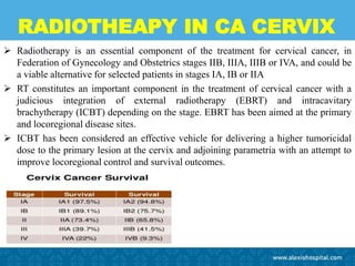
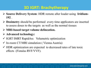
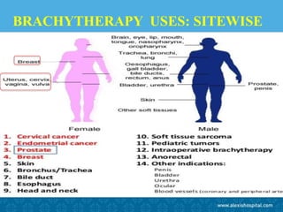
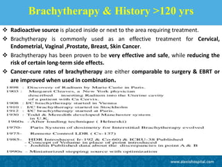
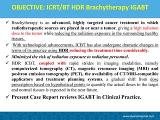
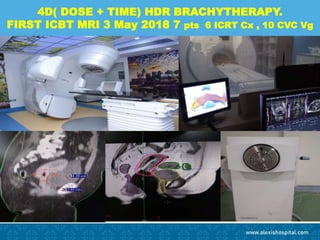
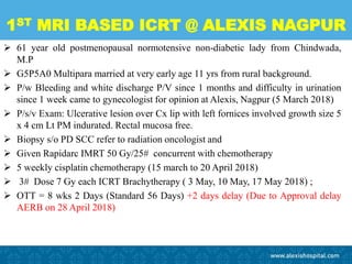
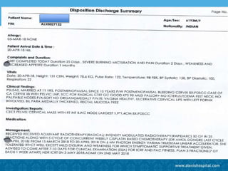
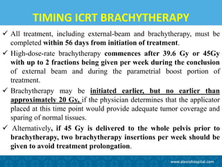
6. 3D IGRT: Brachytherapy
Source Delivery System: HDR remote after loader using Iridium-
192 .
Dosimetry should be performed every time applicators are inserted
to assess doses to the targets as well as the normal tissues
MRI-based target volume delineation.
Advanced technology:
IGRT IMRT RapidArc Volumetric optimization
In-room CT/MRI simulation ( Vienna Austria)
HDR optimization are expected to decreased rates of late toxic
effects (Fistulas RVF/VVF).
8. Brachytherapy & History >120 yrs
Radioactive source is placed inside or next to the area requiring treatment.
Brachytherapy is commonly used as an effective treatment for Cervical,
Endometrial, Vaginal ,Prostate, Breast, Skin Cancer.
Brachytherapy has been proven to be very effective and safe, while reducing the
risk of certain long-term side effects.
Cancer-cure rates of brachytherapy are either comparable to surgery & EBRT or
are improved when used in combination.
9. Introduction: History & Market BT
‘BRACHY’ term by Forsell in 1931. Precise placement of short-range radiation-
sources (radioisotopes Iridium 192 or Cobalt 60) enclosed in a protective
capsule or wire.
Affects only a very localized area around the radiation sources. Exposure to
radiation of healthy tissues farther away from the sources is therefore reduced.
BT OPD 1hrs / IPD 1-3 DAYS fewer visits to the radiotherapy clinic compared
with EBRT 3-6 WEEKS as outpatients. This makes treatment accessible and
convenient for many patients
Global market US$680 million in 2013, high-dose rate (HDR) and Low dose rate
(LDR) accounted for 70%. Microspheres and electronic brachytherapy comprised
the remaining 30%. May reach over US$2.4 billion in 2030, growing by 8%
annually.
15. OBJECTIVE: ICRT/BT HDR Brachytherapy IGABT
Brachytherapy is an advanced, highly targeted cancer treatment in which
radiotherapeutic sources are placed in or near a tumor, giving a high radiation
dose to the tumor while reducing the radiation exposure in the surrounding healthy
tissues.
With technological advancements, ICBT has also undergone dramatic changes in
terms of its practice using HDR reducing the treatment time considerably.
Minimized the risk of radiation exposure to radiation personnel.
HDR ICBT, coupled with rapid strides in imaging modalities, namely
computerized tomography (CT), magnetic resonance imaging (MRI) and
positron emission tomography (PET), the availability of CT/MRI-compatible
applicators and treatment planning systems, a gradual shift from dose
prescription based on hypothetical points to quantify the actual doses to the target
and normal tissues is expected in the near future.
Present Case Report reviews IGABT in Clinical Practice.
16. Learning Objective IGABT LA CA Cx
Standard treatment of locally advanced cervical ca is External Beam
Radiotherapy (EBRT) concomitant Chemotherapy & Brachytherapy
(BT). Often significant tumour regression observed during the first
few weeks of radio-chemotherapy (30-45 Gy), making cervical cancer
an obvious candidate for an adaptive radiotherapeutic strategy.
17.
18. WHATS IGABT ?
IGABT is based on an adaptive 4D target concept and requires the use of 3D
imaging (preferably MRI) with the BT applicator in situ at the time of BT.
Since the publication of the GEC ESTRO recommendations in 2005, the concept
of IGABT has gained significant momentum
IGABT is now considered the gold standard as their strong clinical indications
that MR image guidance improves local control.
The major advantage is possibility to conform the dose given by BT with regard to
both volume (3D) and time (4D).
20. IGABT : ADAPT DOSE WITH IMAGING
Repetitive imaging performed before each BT implant it is possible adapt the
dose given by BT to the anatomy for the tumour regression.
Major impact on the clinical outcome with a concomitant decrease in the rates of
both local failure and morbidity.
A multi-centric collaborative study
(intErnational study on MRI guided BRAchytherapy in locally advanced
Cervical cancer) was launched in 2008 to validate study attempts to use MRI
based IGABT in locally advanced cervical cancers in a multi-centric setting to
establish benchmark for local control, overall survival, morbidity and quality of
life.
With an expected sample size of more than 600 patients and a proposed long term
follow up of 3-6 years, the outcomes from this study are keenly awaited to
reconfirm the utility and efficacy of MRI based IGABT.
21. WHY IGABT ?
The practice of image guided adaptive brachytherapy (IGABT) is gradually gaining
momentum and the number of centres opting for IGABT is increasing.
In United Kingdom, 71% of the centres in 2011 had on IGABT compared to just
26% in 2008. In Canada, although point A is still the most common dose
prescription point, but 73% of the centers have expressed their desire to change to
3D IGABT
The ICRU Report 38 bladder reference point underestimated the 2 cm3 bladder dose
by 75%-300%, while it overestimated the rectal dose in 75% patients. 3D MRI
based planning, improved the optimization to the various dose-volume parameters
Pötter et al have reported excellent local control rates of 95%-100% at 3 years in
limited/favourable tumours (stage IB1/IIB proximal, less that 4-5 cm) and 85%-90%
in larger tumours (stage IIB-IV) with acceptable treatment related morbidity (< 5%).
Pelvic recurrences in this series had decreased by 65%-70%, as compared to
historical series
27. PELVIC FAILURE & CONTROL
Value of ICBT has been adequately demonstrated from various studies that
have shown a better locoregional control when ICBT and EXRT are used in
combination compared with EXRT alone
Incidence of pelvic failures with EBRT alone 86% in patients in stage III
compared with 50% when both EBRT and ICBT were used (P < 0.0002) .
Longsdon and Eifel reported the pelvic failure rate to be 45% with EBRT
alone versus 24% in stage IIIB patients receiving both EBRT and ICBT (P <
0.0001).
Coia et al.observed a significant increase in the pelvic failure rate by 31%
when ICBT was not included in treatment protocols for stages I, II and III
(53% versus 22%, P < 0.01).
33. 1ST MRI BASED ICRT @ ALEXIS NAGPUR
61 year old postmenopausal normotensive non-diabetic lady from Chindwada,
M.P
G5P5A0 Multipara married at very early age 11 yrs from rural background.
P/w Bleeding and white discharge P/V since 1 months and difficulty in urination
since 1 week came to gynecologist for opinion at Alexis, Nagpur (5 March 2018)
P/s/v Exam: Ulcerative lesion over Cx lip with left fornices involved growth size 5
x 4 cm Lt PM indurated. Rectal mucosa free.
Biopsy s/o PD SCC refer to radiation oncologist and
Given Rapidarc IMRT 50 Gy/25# concurrent with chemotherapy
5 weekly cisplatin chemotherapy (15 march to 20 April 2018)
3# Dose 7 Gy each ICRT Brachytherapy ( 3 May, 10 May, 17 May 2018) ;
OTT = 8 wks 2 Days (Standard 56 Days) +2 days delay (Due to Approval delay
AERB on 28 April 2018)
34.
35. TIMING ICRT BRACHYTHERAPY
All treatment, including external-beam and brachytherapy, must be
completed within 56 days from initiation of treatment.
High-dose-rate brachytherapy commences after 39.6 Gy or 45Gy
with up to 2 fractions being given per week during the conclusion
of external beam and during the parametrial boost portion of
treatment.
Brachytherapy may be initiated earlier, but no earlier than
approximately 20 Gy, if the physician determines that the applicator
placed at this time point would provide adequate tumor coverage and
sparing of normal tissues.
Alternatively, if 45 Gy is delivered to the whole pelvis prior to
brachytherapy, two brachytherapy insertions per week should be
given to avoid treatment prolongation.
36. TREATMENT PREPARATION BT
• Bowel preparation(Laxative/Enema) should always be used to ensure an empty
rectum and sigmoid colon.
• Supportive treatment such as LMW heparin, antibiotics and analgesics are given
according to individual patient needs.
• EUA Before placement of the BT applicator a clinical assessment of the tumour
extension is performed describing tumour dimensions (width, height and thickness)
as well as the possible involvement of parametria, vagina, bladder and rectum.
• A Foley catheter is placed in the bladder and 7 ml of diluted contrast medium is
injected into the balloon suitable for MRI
39. ICRT BT APPLICATOR INSERTION
Dilatation of the uterine canal can be guided by ultrasound and the depth of the
uterine cavity is measured. MRI compatible applicator
Vaginal packing can then be performed with gauze filled with diluted betadine to
push away the rectum and bladder and to fix the applicator against the cervix or
other customized procedures may be used for fixation of the applicator
Patient is transferred to the MRI
in the supine treatment position.
40. 3 TESLA MRI VARIAN BRACHYVISION TPS
BASED PLANNING/CONTOURING
46. DOSE CONSTRAINTS OAR
Hot spots in the bladder (D0.1cc and D2cc) as these small volumes may have an
impact on clinical outcome.
For the HR-CTV D90 doses 75-96 Gy
For the IR CTV D90 doses 60-75 Gy
Total cumulative dose of EBRT and BT
EQD2
• Rectum D2cc < 70-75 Gy
• Sigmoid colon D2cc < 75 Gy
• Bladder D2cc < 90 Gy
49. Post-Treatment Evaluation: 4P’s
• Pap smear follow-ups every 3 months for 2 years,
every 6 months for 3 years then yearly.
• PET evaluation 3 months (10-12 weeks)
prognostic.
• Physical examination for recurrence or complications.
Biopsy if suspicious recurrence.
• Patient satisfaction & Quality of life QOL.
50. OUTCOME MEASURES & EVALUTION
Primary outcome measures
Local control within the specific BT targets (HR-CTV and IR-CTV)
Morbidity related to OAR in the pelvis
Secondary endpoints
Regional control
Disease free survival DFS and
Overall survival OS.
Remission status evaluated 3 month after treatment by pelvic MRI
and gynaecological examination.
Complete CR, Partial PR,
Stable SD Progressive disease PD
51. IGABT CONCLUSION
IGABT requires volumetric imaging, preferably MRI Availability
of advanced ICBT applicators.
3D cross sectional imaging prior to brachytherapy to correctly
estimate the residual disease either with CT based or preferably MRI
based planning.
Lack of imaging scanners and personnel are barriers to
implementation
IGRT and IGABT requires multidisciplinary knowledge,
experience and skills involving radiation oncology, medical
physics, radiology, anaesthesiology and nursing.
Teaching and training for high-quality IGABT needed.
ESTRO(http://www.estro.org/school) workshops organised by Medical
University of Vienna (Elekta, Stockholm, Sweden) and Aarhus University
Hospital (Varian Medical Systems, Palo Alto, CA, USA)
52. RADIOTHEAPY IN CA CERVIX
Radiotherapy is an essential component of the treatment for cervical cancer, in
Federation of Gynecology and Obstetrics stages IIB, IIIA, IIIB or IVA, and could be
a viable alternative for selected patients in stages IA, IB or IIA
RT constitutes an important component in the treatment of cervical cancer with a
judicious integration of external radiotherapy (EBRT) and intracavitary
brachytherapy (ICBT) depending on the stage. EBRT has been aimed at the primary
and locoregional disease sites.
ICBT has been considered an effective vehicle for delivering a higher tumoricidal
dose to the primary lesion at the cervix and adjoining parametria with an attempt to
improve locoregional control and survival outcomes.