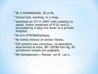
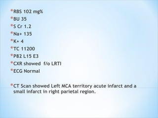
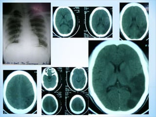
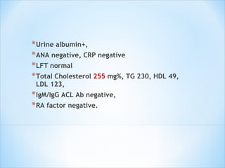
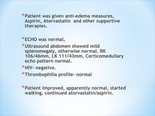
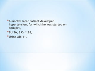
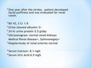
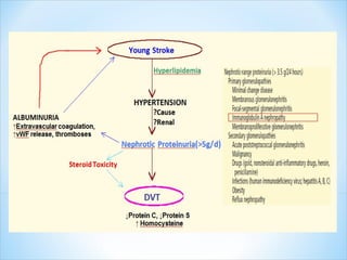
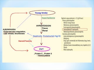
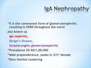
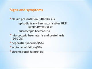
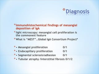
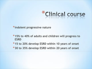
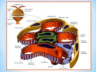
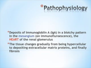
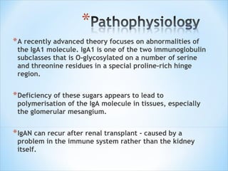
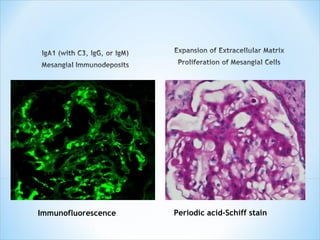
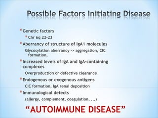
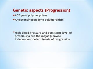
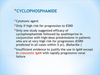
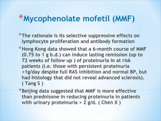
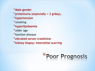
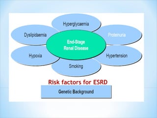
Dr Jishanth M Prof Dr A Gowrisankar’s Unit, M4
Mr A SHANMUGAM, 28 yr/M,  Unmarried, working  in a shop,  admitted on 27-11-2007 with inability to speak, motor weakness of R UL and LL, preceded by 2 days h/o fever to a private hospital.  No h/o HTN/DM/Epilepsy No family history of similar illness.  O/E patient was conscious, co-operative, disoriented at time, BP-120/80 mm Hg, all peripheral vessels are palpable, (R) Hemiparesis+, Plantar ↑on R, ↓on L.
RBS 102 mg%  BU 35  S Cr 1.2  Na+ 135  K+ 4  TC 11200 P82 L15 E3  CXR showed  f/o LRTI  ECG Normal  CT Scan showed Left MCA territory acute infarct and a  small infarct in right parietal region.
 
Urine albumin+,  ANA negative, CRP negative LFT normal Total Cholesterol  255  mg%, TG 230, HDL 49, LDL 123,  IgM/IgG ACL Ab negative,  RA factor negative.
Patient was given anti-edema measures, Aspirin, Atorvastatin  and other supportive therapies.  ECHO was normal,  Ultrasound abdomen showed mild splenomegaly, otherwise normal. RK 106/46mm, LK 111/43mm, Corticomedullary echo pattern normal.  HIV- negative. Thrombophilia profile- normal Patient improved, apparently normal, started walking, continued atorvastatin/aspirin.
 
6 months later patient developed hypertension, for which he was started on Ramipril,  BU 36, S Cr 1.28,  Urine Alb 1+.
One year after the stroke,  patient developed facial puffiness and was evaluated for renal cause.  BU 42, S Cr 1.8  Urine showed albumin 3+  24 hr urine protein 5.5 g/day  Ultrasonogram- normal sized kidneys Medical Renal disease+, Splenomegaly+ Dopplerstudy of renal arteries normal Serum Calcium- 8.1 mg% Serum Uric Acid 6.9 mg%
FBS 90/ PPBS 118, Urine sugar-negative ANA neg, CRP neg  HIV neg  HBsAg neg, Anti HCV negative  PT 11.7s(12s), aPTT 20s(30s)  IgG/IgM ACL Ab negative  CXR normal, ECG normal  Peripheral smear study -normal.  Patient underwent renal biopsy which showed segmental sclerosis with mesangial proliferation, focal tubular atrophy, and Immunofluroscence  showed mesangial and peripheral deposits in IgA, C3c and IgM with fibrinogen.
Patient was given diuretics and steroid. Elevated renal parameters came down, BU 28, S Cr 1.1.  Patient continued Ramipril, Atorvastatin, Aspirin.  Patient was put on prednisolone.
 
After 3 months RBS 238, BU 32, S Cr 1.3,  BP 110/90 mmHg,  Serum total protein 5.8 g%, Alb 3.8, Globulin 2g Urine albmin 3+.  Steroids were slowly tapered off.  Sugar levels came down.  BP was fluctuating.
2 years after the initial stroke, patient developed swelling of  left lower limb associated with pain,  Doppler study showed left popliteal vein thrombosis extending upto adductor canal,  Blood Sugar 99, BU 28, S Cr 1.2  Na+ 138, K+ 3.9 CX- normal  Urine albumin 2+  Cholesterol 182  S Total protein 5.9 g, Albumin 3.7, Glob 2.2.  Patient again readmitted and continued Atorvastatin, Prednisolone, Ramipril, +Acitrom 3 mg OD.
At  discharge 24 hr urine protein was 1.7g,  BP 140/90 mmHg,  Protein C 42.3(70-140),  Protein S 54.9(60-150),  Serum Homocysteine 16.50(5.9-16),  Anti thrombin III level-normal.  He was discharged with advice to continue aspirin, atorvastatin, ramipril, acitrom, and prednisolone.  After 3 months 24 hr urine protein was 360 mg and steroid was slowly tapered.  BU 27, S Cr 0.7, RBS 92 mg%.  Ultrasonogram-no splenomegaly.
 
 
Scanty glomeruli with sclerosing proliferative glomerulonephritis, focal tubular atrophy and abundant IgA deposits, consistent with  “ IgA Nephropathy”
To rule out remote possibilities of pauci-immune glomerulonephritis, ANCA antibodies were done- both were negative.


It is the commonest form of glomerulonephritis resulting in ESRD throughout the world also known as  IgA nephritis, Berger's disease ,  Synpharyngitic glomerulonephritis Prevalence 25-50/1,00,000 Male preponderence, peaks in 2/3 rd  decade Rare familial clustering
Signs and symptoms classic presentation ( 40-50% ) is  episodic frank haematuria after URTI  (synpharyngitic) or microscopic haematuria microscopic haematuria and proteinuria (20-30%) nephrotic syndrome(5%) acute renal failure(5%) chronic renal failure(5%)
Immunohistochemical findings of mesangial deposition of IgA light microscopy: mesangial cell proliferation is the commonest feature What is “MEST”…Global IgA Consortium Project* •  Mesangial proliferation  0/1 •  Endocapillary proliferation 0/1 •  Segmental sclerosis/adhesion 0/1 •  Tubular atrophy /interstitial fibrosis 0/1/2
indolent progressive nature 15% to 40% of adults and children will progress to ESRD 15 to 20% develop ESRD within 10 years of onset 30 to 35% develop ESRD within 20 years of onset
 
Deposits of Immunogloblin A (IgA) in a blotchy pattern in the  mesangium  (on immunofluroescence), the  HEART  of the renal glomerulus The tissue changes gradually from being hypercellular to depositing extracellular matrix proteins, and finally fibrosis
A recently advanced theory focuses on abnormalities of the IgA1 molecule. IgA1 is one of the two immunoglobulin subclasses that is O-glycosylated on a number of serine and threonine residues in a special proline-rich hinge region. Deficiency of these sugars appears to lead to polymerisation of the IgA molecule in tissues, especially the glomerular mesangium. IgAN can recur after renal transplant - caused by a problem in the immune system rather than the kidney itself.
Immunofluorescence Periodic acid-Schiff stain
Genetic factors Chr 6q 22-23 Aberrancy of structure of IgA1 molecules Glycosylation aberrancy -> aggregation, CIC formation, Increased levels of IgA and IgA-containing complexes Overproduction or defective clearance Endogenous or exogenous antigens CIC formation, IgA renal deposition Immunological defects (allergy, complement, coagulation, ...) “ AUTOIMMUNE DISEASE”
Genetic aspects (Progression) ACE gene polymorphism Angiotensinogen gene polymorphism High Blood Pressure and persistant level of proteinuria are the major (known) independent determinants of progression
Conservative  Blockers of the renin-angiotensin system Corticosteroids Fish oil (n-3 Polyunsaturated Fatty Acids) supplementation Cyclophosphamide Mycophenolate mofetil (MMF)
CONSERVATIVE TREATMENT normal renal function, normotension and  only minor urinary abnormalities  isolated microscopic haematuria, and/or mild proteinuria  keep such patients under review Up to 23% of patients will have a spontaneous complete remission .
BLOCKERS OF THE RENIN-ANGIOTENSIN SYSTEM Reduction in proteinuria is considered to be the hallmark of effective treatment in preserving renal function in nondiabetic renal diseases. ACEi and more recently angiotensin II type 1 receptor blockers (ARB) control blood pressure and proteinuria Controls both proteinuria and hypertension(modifiable risk factors for progression of disease) Superior to other antihypertensive drugs in lowering proteinuria by virtue of their capacity to reduce intraglomerular pressure.
CORTICOSTEROIDS Steroid does not appear to consistently offer any beneficial effect other than modest amelioration of proteinuria. The only exception is in children with IgA deposition in the setting of minimal change nephrotic syndrome. Such patients respond to corticosteroid promptly. In adults, in view of the toxicities associated with steroid therapy, it should be considered only when  proteinuria persists >1 g/24 h despite optimal BP  control and maximum RAS blockade.
Fish oil (n-3 Polyunsaturated Fatty Acids) supplementation The rationale of using fish oil in IgAN is based on the premise that n-3 PUFAs alter the production or action of cytokines and eicosanoids evoked by the initial or repeated immunological renal injury, alleviating ongoing renal inflammation and glomerulosclerosis (hallmarks of progressive renal disease) Mayo investigators published their long-term data in 1999 to conclude that early and prolonged treatment with fish oil slows renal progression for high-risk patients with IgAN. Still under debate.
CYCLOPHOSPHAMIDE Cytotoxic agent Only if high risk for progression to ESRD Only one study suggested efficacy of cyclophosphamide followed by azathioprine in conjunction with high-dose prednisolone in patients who are at very high risk for progression (ESRD predicted in all cases within 5 yr). (Ballardie ) Insufficient evidence to justify the use in IgAN except in  crescentic IgAN  with rapidly progressive renal failure
Mycophenolate mofetil (MMF) The rationale is its selective suppressive effects on lymphocyte proliferation and antibody formation Hong Kong data showed that a 6-month course of MMF (0.75 to 1 g b.d.) can induce lasting remission (up to 72 weeks of follow up ) of proteinuria in at risk patients (i.e. those with persistent proteinuria >1g/day despite full RAS inhibition and normal BP, but had histology that did not reveal advanced sclerosis). ( Tang S ) Beijing data suggested that MMF is more effective than prednisone in reducing proteinuria in patients with urinary proteinuria > 2 g/d. ( Chen X )
Male gender proteinuria (especially > 2 g/day), hypertension smoking hyperlipidaemia older age familial disease elevated serum creatinine kidney biopsy: interstitial scarring
Risk factors for ESRD
 
Harrison’s Principles of Internal Medicine: 17ed Proteinuria:How to evaluate an important finding:  WAQAR   KASHIF, MD Department of Medicine, Medical College of Wisconsin,Milwaukee(Review ) IgA nephropathy:The Value of Proteinuria,   Daniel Cattran,CNC-ASN,Philadelphia, PA,2008 UpToDate 17.1

IgA Nephropathy

  • 1.
    Dr Jishanth MProf Dr A Gowrisankar’s Unit, M4
  • 2.
    Mr A SHANMUGAM,28 yr/M, Unmarried, working in a shop, admitted on 27-11-2007 with inability to speak, motor weakness of R UL and LL, preceded by 2 days h/o fever to a private hospital. No h/o HTN/DM/Epilepsy No family history of similar illness. O/E patient was conscious, co-operative, disoriented at time, BP-120/80 mm Hg, all peripheral vessels are palpable, (R) Hemiparesis+, Plantar ↑on R, ↓on L.
  • 3.
    RBS 102 mg% BU 35 S Cr 1.2 Na+ 135 K+ 4 TC 11200 P82 L15 E3 CXR showed f/o LRTI ECG Normal CT Scan showed Left MCA territory acute infarct and a small infarct in right parietal region.
  • 4.
  • 5.
    Urine albumin+, ANA negative, CRP negative LFT normal Total Cholesterol 255 mg%, TG 230, HDL 49, LDL 123, IgM/IgG ACL Ab negative, RA factor negative.
  • 6.
    Patient was givenanti-edema measures, Aspirin, Atorvastatin and other supportive therapies. ECHO was normal, Ultrasound abdomen showed mild splenomegaly, otherwise normal. RK 106/46mm, LK 111/43mm, Corticomedullary echo pattern normal. HIV- negative. Thrombophilia profile- normal Patient improved, apparently normal, started walking, continued atorvastatin/aspirin.
  • 7.
  • 8.
    6 months laterpatient developed hypertension, for which he was started on Ramipril, BU 36, S Cr 1.28, Urine Alb 1+.
  • 9.
    One year afterthe stroke, patient developed facial puffiness and was evaluated for renal cause. BU 42, S Cr 1.8 Urine showed albumin 3+ 24 hr urine protein 5.5 g/day Ultrasonogram- normal sized kidneys Medical Renal disease+, Splenomegaly+ Dopplerstudy of renal arteries normal Serum Calcium- 8.1 mg% Serum Uric Acid 6.9 mg%
  • 10.
    FBS 90/ PPBS118, Urine sugar-negative ANA neg, CRP neg HIV neg HBsAg neg, Anti HCV negative PT 11.7s(12s), aPTT 20s(30s) IgG/IgM ACL Ab negative CXR normal, ECG normal Peripheral smear study -normal. Patient underwent renal biopsy which showed segmental sclerosis with mesangial proliferation, focal tubular atrophy, and Immunofluroscence showed mesangial and peripheral deposits in IgA, C3c and IgM with fibrinogen.
  • 11.
    Patient was givendiuretics and steroid. Elevated renal parameters came down, BU 28, S Cr 1.1. Patient continued Ramipril, Atorvastatin, Aspirin. Patient was put on prednisolone.
  • 12.
  • 13.
    After 3 monthsRBS 238, BU 32, S Cr 1.3, BP 110/90 mmHg, Serum total protein 5.8 g%, Alb 3.8, Globulin 2g Urine albmin 3+. Steroids were slowly tapered off. Sugar levels came down. BP was fluctuating.
  • 14.
    2 years afterthe initial stroke, patient developed swelling of left lower limb associated with pain, Doppler study showed left popliteal vein thrombosis extending upto adductor canal, Blood Sugar 99, BU 28, S Cr 1.2 Na+ 138, K+ 3.9 CX- normal Urine albumin 2+ Cholesterol 182 S Total protein 5.9 g, Albumin 3.7, Glob 2.2. Patient again readmitted and continued Atorvastatin, Prednisolone, Ramipril, +Acitrom 3 mg OD.
  • 15.
    At discharge24 hr urine protein was 1.7g, BP 140/90 mmHg, Protein C 42.3(70-140), Protein S 54.9(60-150), Serum Homocysteine 16.50(5.9-16), Anti thrombin III level-normal. He was discharged with advice to continue aspirin, atorvastatin, ramipril, acitrom, and prednisolone. After 3 months 24 hr urine protein was 360 mg and steroid was slowly tapered. BU 27, S Cr 0.7, RBS 92 mg%. Ultrasonogram-no splenomegaly.
  • 16.
  • 17.
  • 18.
    Scanty glomeruli withsclerosing proliferative glomerulonephritis, focal tubular atrophy and abundant IgA deposits, consistent with “ IgA Nephropathy”
  • 19.
    To rule outremote possibilities of pauci-immune glomerulonephritis, ANCA antibodies were done- both were negative.
  • 20.
  • 21.
  • 22.
  • 23.
    It is thecommonest form of glomerulonephritis resulting in ESRD throughout the world also known as IgA nephritis, Berger's disease , Synpharyngitic glomerulonephritis Prevalence 25-50/1,00,000 Male preponderence, peaks in 2/3 rd decade Rare familial clustering
  • 24.
    Signs and symptomsclassic presentation ( 40-50% ) is episodic frank haematuria after URTI (synpharyngitic) or microscopic haematuria microscopic haematuria and proteinuria (20-30%) nephrotic syndrome(5%) acute renal failure(5%) chronic renal failure(5%)
  • 25.
    Immunohistochemical findings ofmesangial deposition of IgA light microscopy: mesangial cell proliferation is the commonest feature What is “MEST”…Global IgA Consortium Project* • Mesangial proliferation 0/1 • Endocapillary proliferation 0/1 • Segmental sclerosis/adhesion 0/1 • Tubular atrophy /interstitial fibrosis 0/1/2
  • 26.
    indolent progressive nature15% to 40% of adults and children will progress to ESRD 15 to 20% develop ESRD within 10 years of onset 30 to 35% develop ESRD within 20 years of onset
  • 27.
  • 28.
    Deposits of ImmunogloblinA (IgA) in a blotchy pattern in the mesangium (on immunofluroescence), the HEART of the renal glomerulus The tissue changes gradually from being hypercellular to depositing extracellular matrix proteins, and finally fibrosis
  • 29.
    A recently advancedtheory focuses on abnormalities of the IgA1 molecule. IgA1 is one of the two immunoglobulin subclasses that is O-glycosylated on a number of serine and threonine residues in a special proline-rich hinge region. Deficiency of these sugars appears to lead to polymerisation of the IgA molecule in tissues, especially the glomerular mesangium. IgAN can recur after renal transplant - caused by a problem in the immune system rather than the kidney itself.
  • 30.
  • 31.
    Genetic factors Chr6q 22-23 Aberrancy of structure of IgA1 molecules Glycosylation aberrancy -> aggregation, CIC formation, Increased levels of IgA and IgA-containing complexes Overproduction or defective clearance Endogenous or exogenous antigens CIC formation, IgA renal deposition Immunological defects (allergy, complement, coagulation, ...) “ AUTOIMMUNE DISEASE”
  • 32.
    Genetic aspects (Progression)ACE gene polymorphism Angiotensinogen gene polymorphism High Blood Pressure and persistant level of proteinuria are the major (known) independent determinants of progression
  • 33.
    Conservative Blockersof the renin-angiotensin system Corticosteroids Fish oil (n-3 Polyunsaturated Fatty Acids) supplementation Cyclophosphamide Mycophenolate mofetil (MMF)
  • 34.
    CONSERVATIVE TREATMENT normalrenal function, normotension and only minor urinary abnormalities isolated microscopic haematuria, and/or mild proteinuria keep such patients under review Up to 23% of patients will have a spontaneous complete remission .
  • 35.
    BLOCKERS OF THERENIN-ANGIOTENSIN SYSTEM Reduction in proteinuria is considered to be the hallmark of effective treatment in preserving renal function in nondiabetic renal diseases. ACEi and more recently angiotensin II type 1 receptor blockers (ARB) control blood pressure and proteinuria Controls both proteinuria and hypertension(modifiable risk factors for progression of disease) Superior to other antihypertensive drugs in lowering proteinuria by virtue of their capacity to reduce intraglomerular pressure.
  • 36.
    CORTICOSTEROIDS Steroid doesnot appear to consistently offer any beneficial effect other than modest amelioration of proteinuria. The only exception is in children with IgA deposition in the setting of minimal change nephrotic syndrome. Such patients respond to corticosteroid promptly. In adults, in view of the toxicities associated with steroid therapy, it should be considered only when proteinuria persists >1 g/24 h despite optimal BP control and maximum RAS blockade.
  • 37.
    Fish oil (n-3Polyunsaturated Fatty Acids) supplementation The rationale of using fish oil in IgAN is based on the premise that n-3 PUFAs alter the production or action of cytokines and eicosanoids evoked by the initial or repeated immunological renal injury, alleviating ongoing renal inflammation and glomerulosclerosis (hallmarks of progressive renal disease) Mayo investigators published their long-term data in 1999 to conclude that early and prolonged treatment with fish oil slows renal progression for high-risk patients with IgAN. Still under debate.
  • 38.
    CYCLOPHOSPHAMIDE Cytotoxic agentOnly if high risk for progression to ESRD Only one study suggested efficacy of cyclophosphamide followed by azathioprine in conjunction with high-dose prednisolone in patients who are at very high risk for progression (ESRD predicted in all cases within 5 yr). (Ballardie ) Insufficient evidence to justify the use in IgAN except in crescentic IgAN with rapidly progressive renal failure
  • 39.
    Mycophenolate mofetil (MMF)The rationale is its selective suppressive effects on lymphocyte proliferation and antibody formation Hong Kong data showed that a 6-month course of MMF (0.75 to 1 g b.d.) can induce lasting remission (up to 72 weeks of follow up ) of proteinuria in at risk patients (i.e. those with persistent proteinuria >1g/day despite full RAS inhibition and normal BP, but had histology that did not reveal advanced sclerosis). ( Tang S ) Beijing data suggested that MMF is more effective than prednisone in reducing proteinuria in patients with urinary proteinuria > 2 g/d. ( Chen X )
  • 40.
    Male gender proteinuria(especially > 2 g/day), hypertension smoking hyperlipidaemia older age familial disease elevated serum creatinine kidney biopsy: interstitial scarring
  • 41.
  • 42.
  • 43.
    Harrison’s Principles ofInternal Medicine: 17ed Proteinuria:How to evaluate an important finding: WAQAR KASHIF, MD Department of Medicine, Medical College of Wisconsin,Milwaukee(Review ) IgA nephropathy:The Value of Proteinuria, Daniel Cattran,CNC-ASN,Philadelphia, PA,2008 UpToDate 17.1