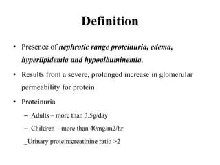
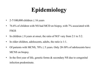
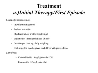
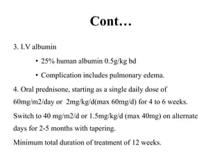
NEPHROTIC SYNDROME
DR. MATE SHILULI
Definition
• Presence of nephrotic range proteinuria, edema,
hyperlipidemia and hypoalbuminemia.
• Results from a severe, prolonged increase in glomerular
permeability for protein
• Proteinuria
– Adults – more than 3.5g/day
– Children – more than 40mg/m2/hr
_Urinary protein:creatinine ratio >2
Classical features
• Heavy proteinuria
• Hypoalbuminemia(<2.5g/dL)
• Generalized edema
• Hyperlipidemia and lipiduria
Epidemiology
• 2-7/100,000 children ≤ 16 years
• 76.6% of children with NS had MCD on biopsy, with 7% associated with
FSGS
• In children ≤ 8 years at onset, the ratio of M:F vary from 2:1 to 3:2.
• In older children, adolescents, adults, the ratio is 1:1.
• Of patients with MCNS, 70% ≤ 5 years. Only 20-30% of adolescents have
MCNS on biopsy.
• In the first year of life, genetic forms & secondary NS due to congenital
infection predominate.
Basic Physiology
Basic Physiology cont.…
Histology
Pathophysiology
Proteinuria & hypoalbuminemia
– Pathogenesis is believed to be immune-related
– Various cytokines have been implicated – IL-2, IL-4,
IL-12, IL-15
– The nephron is characteristically found to have fused
podocytes - alteration of GBM plays a role
Cont.…
– The glomerular capillary permeability to albumin is
selectively increased, this overcomes the modest
ability of the tubules to reabsorb proteins causing
proteinuria (protein leak)
– There is urinary loss of albumin, immunoglobulins,
metal binding proteins, erythropoietin, transferrin,
complement & coagulation components.
Cont...
• The largest proportion of protein lost in the urine is
albumin, but globulins are also excreted in some diseases.
• Normally the GBM is –vely charged due to presence of
polyanions e.g. Heparan sulfate, chondroitin sulfate and
sialic acid.
• This negative charge deters filtration of –vely charged
proteins e.g. albumin
Cont…
• A highly selective proteinuria consists mostly
of low molecular weight proteins (albumin,
transferrin)
• A poorly selective proteinuria consists of
higher molecular weight globulins as well as
albumin
Pathophysiology cont…
• Edema
– Proteinuria causes a decrease in plasma oncotic
pressure(due to low albumin), causing extravasation of
plasma water into the interstitial space.
– Reduced plasma volume causes activation of the renin-
angiotensin aldosterone axis and ADH.
– This causes a retention of sodium and water respectively by
renal tubules and extension of edema.
Cont…
– Another theory of edema states that massive
proteinuria leads to tubulointerstitial inflammation
and release of local vasoconstrictors and inhibition
of vasodilation.
– This leads to a reduction in GFR and thus sodium
and water retention.
Cont.…
– Is characteristically soft & pitting.
– Most marked in the periorbital regions &
dependent portions of the body.
– It may be massive, with pleural effusions &
ascites.
Pathophysiology cont…
• Hyperlipidemia
– NS is accompanied by disordered lipid metabolism.
– Apolipoprotein (apo)-B containing lipoproteins are elevated,
including VLDL, IDL, LDL & lipoprotein (a), with resultant
increases in total cholesterol & LDL-cholesterol.
– HDL-cholesterol is normal or low.
– Mechanism – increased hepatic synthesis of LDL, VLDL &
lipoprotein (a) in response to hypoalbuminemia.
Hyperlipidemia Cont…
– Decreased plasma oncotic pressure may play a role
– reduction in hyperlipidemia in patients receiving
either albumin or dextran infusions.
– Also contributing are abnormalities in regulatory
enzymes, such as lecithin-cholesterol
acyltransferase, lipoprotein lipase and cholesterol
ester transfer protein.
Pathophysiology cont…
• Lipiduria
– Due to leakage of lipoproteins across the GBM.
– The lipid appears in the urine as free or as oval fat
bodies(represent lipoprotein resorbed by tubular
epithelial cells and then shed along with the
degenerated cells).
Pathophysiology cont…
• Thrombosis
– Patients have increased risk of thrombosis.
– Could be due to loss of anticoagulant factors (eg
antithrombin III) and antiplasmin activity through
the leaky glomerulus.
Pathophysiology cont…
• Thrombosis
– There are abnormalities such as increased platelet activation and
aggregation: elevation in factors V, VII, VIII and XIII and
fibrionogen, and decreased antithrombin III, proteins C and S,
and factors XI and XIII; and increased activities of tissue
plasminogen activator and plasminogen activator inhibitor-1.
– These factors combined with potential hypovolemia, immobility
and increased incidence of infection lead to a hypercoagulable
state.
Pathophysiology cont…
• Infection
– Patients are at risk of infection, especially Streptococcus
pneumoniae.
– May be due to low IgG levels.
– Increased urinary losses of factor B, a cofactor of C3b in
the alternative complement pathway.
– Medications used to treat NS, such as corticosteroids and
alkylating agents also suppress the immune system.
Classification
i. Based on steroid sensitivity
• Steroid –sensitive
• Steroid –resistant
• Relapsing
• Steroid dependent
Classification cont…
ii. Based on etiology
– Secondary causes – etiology extrinsic to the kidney
• Infections – HIV, malaria, syphillis, Hep B, Hep C
• Systemic diseases – DM, SLE
• Drugs and toxins – NSAIDs, penicillin derivatives (penicillamine),
ACE-I (captopril), street heroin
• Mechanical factors – Renal vein thrombosis
• Tumors – Hodgkin disease, lymphoproliferative disorders
• Miscellaneous – bee-sting allergy, hereditary nephritis
Classification cont…
– Primary causes (Idiopathic NS) – glomerular
diseases intrinsic to the kidney
• Minimal change disease
• Proliferative changes
• Focal Segmental Glomerulosclerosis
• Membranous nephropathy
Minimal Change NS
• Most common cause in childhood (65%).
• Usually idiopathic, but may occur in association with
malignancies (Hodgkin lymphoma & other T-cell
malignancies), exposure to nonsteroidal anti-inflammatory
drugs, or in association with IgA nephropathy or immune-
mediated diseases (SLE, Kimura disease).
• MCNS is characterized by response to corticosteroids (>90%),
a chronic relapsing course (60-80%), and an excellent long-
term prognosis.
MCD Cont…
• Macroscopic hematuria is rare.
• Characterized by: oval fat bodies and hyaline
casts in urine.
• Pathogenesis – A soluble factor released by T
cells causes damage to the GBM (hypothesis)
Membranoproliferative
Glomerulonephritis
• In some patients with NS, mild non-sclerotic
widening of the mesangial matrix occurs, and
may include deposits (mesangial proliferative
glomerulonephritis)
Focal Segmental Glomerulosclerosis
• Characterised by complete collapse of a segment of the
glomerulus with mesangial sclerosis.
• Affects the deep juxtamedullary glomeruli first.
• Typically occurs between age 2 and 7.
• Macroscopic hematuria is rare.
• Poor response to immunosuppressive therapy and a
high rate of recurrence in renal allografts.
Membranous Nephropathy
• Most common cause of NS in adults.
• Is a noninflammatory glomerular disease characterized by the presence of
immune deposits (usually containing IgG and C3) within the basement
membrane.
• Leads to diffuse thickening of the glomerular capillary wall.
• Characterized by proteinuria.
• Insidious in onset and progression.
• Appears to be antibody-mediated.
• Immune complexes form in situ with antigens of exogenous or endogenous
origin.
Membranous Nephropathy
• It may occur in association with other systemic diseases, where it is
referred to as secondary membranous glomerulopathy
• Most commonly associated with:
– Drugs – penicillamine, captopril, NSAIDs
– Underlying malignant tumors – esp carcinoma of the lung and colon and
melanoma
– SLE
– Infections – chronic Hep B, Hep C, syphilis, schistosomiasis, malaria
– Other autoimmune disorders such as thyroiditis
Management
History
• Age of onset
• Infections e.g. Malaria, schistosomiasis, filariasis, Hepatitis B,
C; HIV, Leprosy
• Malignancy e.g. Solid tumours, Lymphomas
• Drugs e.g. NSAIDs, penincillamine, captopril, gold, mercury,
lithium, probenecid
• History of URTI
• History of recent hypersensitivity reaction or allergy.
– Abdominal swelling and/or pain
– Weight gain, general body swelling-dependent areas
– Oliguria
– Urine that is foamy
– Hematuria-more common in MPGN
– Hypertension
– Dyspnoea
– Facial puffiness–worse in the morning
– Signs of shock
– Symptoms of infection such as fever and irritability
– Anorexia,easy fatiguabilty,abdominal discomfort and diarrhea are common.
Physical Examination
• Oedema- Pitting, mostly found in the lower extremities, face
and periorbital regions, scrotum or labia and abdomen (ascites)
• There maybe palor
• Tachypnea (mechanical restriction to breathing due to marked
ascites)
• Pulmonary effusion and edema can also respiratory distress.
• Nonspecific- headache, irritability, malaise, fatigue
• Hepatomegaly
• Tender abdomen may indicate peritonitis
• Hypotension and signs of shock
Differential Diagnosis
• Congestive cardiac failure
• Liver failure
• Protein-losing enteropathy
• Protein energy malnutrition (Kwashiorkor)
• Hereditary angioedema
• Allergic reaction
• Acute Poststreptococcal Glomerulonephritis
• Acute Renal Failure
Investigations
Urinalysis
– Spot
– Dipstick : Proteinuria 3+ or >300mg/dl
– Protein/ creatinine ratio >2
– Microscopy
– 24-hour collection: quantitative proteinuria (50mg/kg/d or
40mg/m2/hr)
Cont…
Blood tests.
-serum proteins-hypoalbuminemia <2.5g/dl.
-lipid profiles-increased cholesterol,LDL.Elevated triglycerides with severe
hypoalbuminemia.Low or Normal HDL
- U/E/Cs- hyponatremia ~ dilutional.
-others:, LFTs, Ca2+,RBS & HbA1c
-CBC-HB, hct increased ~ plasma volume contraction
-Serologic studies( if necessary):
ANA, C3 C4, Hep B/C SAgs, ASOT, Anti-GBM
-Genetic screening
Cont…
Imaging
Renal Ultrasound
• normal or large kidneys in acute disease
• Small kidneys in chronic disease
• Parenchymal architecture
Surgical
Renal biopsy.
– Define the pattern of glomerular involvement
– Hence make definitive diagnosis
– Make management decisions
Indications For Renal Biopsy
-family history suggestive of disease other than MCD.
-1st yr of life or >8 yrs.
-frequently relapsing NS.
-steroid dependent.
-steroid resistant.
-change in clinical course.
-failure to respond to adequate Steroid therapy in 28 days.
Contraindications To Biopsy
• Bleeding diathesis (uncorrectable)
• Small kidneys- may indicate chronic irreversible d’se
• Severe HPTN- can’t be controlled
• Multiple bilateral cysts
• Renal tumor
• Hydronephrosis
• Active renal/ perirenal infection
• One kidney.
Treatment
a.)Initial Therapy/First Episode
1.Supportive management
– In-patient management
– Sodium restriction
– Fluid restriction( if pt hyponatremic)
– Elevation of limbs/genital area (pillow)
– Input/output charting, daily weighing
– Oral penicillin may be given to children with gross edema
2. Diuretics
• Chlorothiazide 10mg/kg/dose bd OR
• Furosemide 1-2mg/kg/does bd
Cont…
3. I.V albumin
• 25% human albumin 0.5g/kg bd
• Complication includes pulmonary edema.
4. Oral prednisone, starting as a single daily dose of
60mg/m2/day or 2mg/kg/d(max 60mg/d) for 4 to 6 weeks.
Switch to 40 mg/m2/d or 1.5mg/kg/d (max 40mg) on alternate
days for 2-5 months with tapering.
Minimum total duration of treatment of 12 weeks.
b.) Infrequent Relapses
For infrequent relapses (one relapse within 6 months of initial
response, or 1-3 relapses in any 12-month period):Administer
initial treatment dose.
Prednisone, 2 mg/kg/d or 60 mg/m2/d as a single dose until
remission for at least 3 days; then 1.5 mg/kg/d or 40 mg/m2/d as a
single dose on alternate days for 4 weeks. Steroids may then be
stopped or gradually tapered.
c.) Frequent Relapses/Steroid
Dependent
• Frequently relapsing nephrotic syndrome (FRNS) is
defined as steroid-sensitive nephrotic syndrome (SSNS) with 2
or more relapses within 6 months, or 4 or more relapses within
a 12-month period.
• Steroid-dependent nephrotic syndrome (SDNS) is defined
as SSNS with 2 or more consecutive relapses during tapering
or within 14 days of stopping steroids
cont…
Frequent relapser/dependent
change therapy to a steroid-sparing agent once remission of
proteinuria has been achieved.
• Prednisone 2 mg/kg/d or 60 mg/m2/d as a single morning
dose until remission then 1.5 mg/kg or 40 mg/m2/d single
dose on alternate days and tapered over 3 or more months.
• A steroid-sparing agent can be considered on remission.
Alkylating agent.
• Cyclophosphamide 2-3mg/kg/day OD for 8-12 wks. Continue
alt. day prednisone
• Cyclophosphamide should not be started until child is in
remission
• Chlorambucil 0.1- 0.2mg/ kg/day for 8wks 3-6mg/kg/day
• Methylprednisolone
– 30mg/kg bolus (max 1gm)
– Alternate days x 6 doses
– Taper regimen up to 18 months
• Other drugs include tacrolimus and levamisole
d.) Steroid Resistant
• A minimum of 8 wks treatment with corticosteroids
• The following are required to evaluate a child with SRNS, Diagnostic
kidney biopsy, Evaluation of kidney functions by GFR and quantitative
urine protein analysis
• Calcineurin inhibitor as the initial Rx and should be continued for a
minimum of 6 months until full or partial remission is achieved should
be combined with low dose steroids
• In children who fail to achieve remission with CNI, give
Mycophenolate mofetil, high dose steroids or a combination of these.
Cont..
Adjuncts
1. ACE inhibitors and ARBs reduce proteinuria in resistant patients
2. Antibiotics and vaccination
3. Anticoagulation
E. 20 NEPHROTIC SYNDROME
Treat the cause!!!
F. Congenital- Give albumin as you await transplant
References
• Wang, C. S., & Greenbaum, L. A. (2019). Nephrotic
syndrome. Pediatric Clinics, 66(1), 73-85.
• Tullus, K., Webb, H., & Bagga, A. (2018). Management of
steroid-resistant nephrotic syndrome in children and
adolescents. The Lancet Child & Adolescent Health, 2(12),
880-890.
• Parthasarathy, A., Menon, P. S. N., & Nair, M. K. C.
(2019). IAP Textbook of pediatrics. JP Medical Ltd.

Nephrotic syndrome

  • 1.
  • 2.
    Definition • Presence ofnephrotic range proteinuria, edema, hyperlipidemia and hypoalbuminemia. • Results from a severe, prolonged increase in glomerular permeability for protein • Proteinuria – Adults – more than 3.5g/day – Children – more than 40mg/m2/hr _Urinary protein:creatinine ratio >2
  • 3.
    Classical features • Heavyproteinuria • Hypoalbuminemia(<2.5g/dL) • Generalized edema • Hyperlipidemia and lipiduria
  • 4.
    Epidemiology • 2-7/100,000 children≤ 16 years • 76.6% of children with NS had MCD on biopsy, with 7% associated with FSGS • In children ≤ 8 years at onset, the ratio of M:F vary from 2:1 to 3:2. • In older children, adolescents, adults, the ratio is 1:1. • Of patients with MCNS, 70% ≤ 5 years. Only 20-30% of adolescents have MCNS on biopsy. • In the first year of life, genetic forms & secondary NS due to congenital infection predominate.
  • 5.
  • 6.
  • 7.
  • 8.
    Pathophysiology Proteinuria & hypoalbuminemia –Pathogenesis is believed to be immune-related – Various cytokines have been implicated – IL-2, IL-4, IL-12, IL-15 – The nephron is characteristically found to have fused podocytes - alteration of GBM plays a role
  • 9.
    Cont.… – The glomerularcapillary permeability to albumin is selectively increased, this overcomes the modest ability of the tubules to reabsorb proteins causing proteinuria (protein leak) – There is urinary loss of albumin, immunoglobulins, metal binding proteins, erythropoietin, transferrin, complement & coagulation components.
  • 10.
    Cont... • The largestproportion of protein lost in the urine is albumin, but globulins are also excreted in some diseases. • Normally the GBM is –vely charged due to presence of polyanions e.g. Heparan sulfate, chondroitin sulfate and sialic acid. • This negative charge deters filtration of –vely charged proteins e.g. albumin
  • 11.
    Cont… • A highlyselective proteinuria consists mostly of low molecular weight proteins (albumin, transferrin) • A poorly selective proteinuria consists of higher molecular weight globulins as well as albumin
  • 12.
    Pathophysiology cont… • Edema –Proteinuria causes a decrease in plasma oncotic pressure(due to low albumin), causing extravasation of plasma water into the interstitial space. – Reduced plasma volume causes activation of the renin- angiotensin aldosterone axis and ADH. – This causes a retention of sodium and water respectively by renal tubules and extension of edema.
  • 13.
    Cont… – Another theoryof edema states that massive proteinuria leads to tubulointerstitial inflammation and release of local vasoconstrictors and inhibition of vasodilation. – This leads to a reduction in GFR and thus sodium and water retention.
  • 14.
    Cont.… – Is characteristicallysoft & pitting. – Most marked in the periorbital regions & dependent portions of the body. – It may be massive, with pleural effusions & ascites.
  • 15.
    Pathophysiology cont… • Hyperlipidemia –NS is accompanied by disordered lipid metabolism. – Apolipoprotein (apo)-B containing lipoproteins are elevated, including VLDL, IDL, LDL & lipoprotein (a), with resultant increases in total cholesterol & LDL-cholesterol. – HDL-cholesterol is normal or low. – Mechanism – increased hepatic synthesis of LDL, VLDL & lipoprotein (a) in response to hypoalbuminemia.
  • 16.
    Hyperlipidemia Cont… – Decreasedplasma oncotic pressure may play a role – reduction in hyperlipidemia in patients receiving either albumin or dextran infusions. – Also contributing are abnormalities in regulatory enzymes, such as lecithin-cholesterol acyltransferase, lipoprotein lipase and cholesterol ester transfer protein.
  • 17.
    Pathophysiology cont… • Lipiduria –Due to leakage of lipoproteins across the GBM. – The lipid appears in the urine as free or as oval fat bodies(represent lipoprotein resorbed by tubular epithelial cells and then shed along with the degenerated cells).
  • 18.
    Pathophysiology cont… • Thrombosis –Patients have increased risk of thrombosis. – Could be due to loss of anticoagulant factors (eg antithrombin III) and antiplasmin activity through the leaky glomerulus.
  • 19.
    Pathophysiology cont… • Thrombosis –There are abnormalities such as increased platelet activation and aggregation: elevation in factors V, VII, VIII and XIII and fibrionogen, and decreased antithrombin III, proteins C and S, and factors XI and XIII; and increased activities of tissue plasminogen activator and plasminogen activator inhibitor-1. – These factors combined with potential hypovolemia, immobility and increased incidence of infection lead to a hypercoagulable state.
  • 20.
    Pathophysiology cont… • Infection –Patients are at risk of infection, especially Streptococcus pneumoniae. – May be due to low IgG levels. – Increased urinary losses of factor B, a cofactor of C3b in the alternative complement pathway. – Medications used to treat NS, such as corticosteroids and alkylating agents also suppress the immune system.
  • 21.
    Classification i. Based onsteroid sensitivity • Steroid –sensitive • Steroid –resistant • Relapsing • Steroid dependent
  • 22.
    Classification cont… ii. Basedon etiology – Secondary causes – etiology extrinsic to the kidney • Infections – HIV, malaria, syphillis, Hep B, Hep C • Systemic diseases – DM, SLE • Drugs and toxins – NSAIDs, penicillin derivatives (penicillamine), ACE-I (captopril), street heroin • Mechanical factors – Renal vein thrombosis • Tumors – Hodgkin disease, lymphoproliferative disorders • Miscellaneous – bee-sting allergy, hereditary nephritis
  • 23.
    Classification cont… – Primarycauses (Idiopathic NS) – glomerular diseases intrinsic to the kidney • Minimal change disease • Proliferative changes • Focal Segmental Glomerulosclerosis • Membranous nephropathy
  • 24.
    Minimal Change NS •Most common cause in childhood (65%). • Usually idiopathic, but may occur in association with malignancies (Hodgkin lymphoma & other T-cell malignancies), exposure to nonsteroidal anti-inflammatory drugs, or in association with IgA nephropathy or immune- mediated diseases (SLE, Kimura disease). • MCNS is characterized by response to corticosteroids (>90%), a chronic relapsing course (60-80%), and an excellent long- term prognosis.
  • 25.
    MCD Cont… • Macroscopichematuria is rare. • Characterized by: oval fat bodies and hyaline casts in urine. • Pathogenesis – A soluble factor released by T cells causes damage to the GBM (hypothesis)
  • 26.
    Membranoproliferative Glomerulonephritis • In somepatients with NS, mild non-sclerotic widening of the mesangial matrix occurs, and may include deposits (mesangial proliferative glomerulonephritis)
  • 27.
    Focal Segmental Glomerulosclerosis •Characterised by complete collapse of a segment of the glomerulus with mesangial sclerosis. • Affects the deep juxtamedullary glomeruli first. • Typically occurs between age 2 and 7. • Macroscopic hematuria is rare. • Poor response to immunosuppressive therapy and a high rate of recurrence in renal allografts.
  • 28.
    Membranous Nephropathy • Mostcommon cause of NS in adults. • Is a noninflammatory glomerular disease characterized by the presence of immune deposits (usually containing IgG and C3) within the basement membrane. • Leads to diffuse thickening of the glomerular capillary wall. • Characterized by proteinuria. • Insidious in onset and progression. • Appears to be antibody-mediated. • Immune complexes form in situ with antigens of exogenous or endogenous origin.
  • 29.
    Membranous Nephropathy • Itmay occur in association with other systemic diseases, where it is referred to as secondary membranous glomerulopathy • Most commonly associated with: – Drugs – penicillamine, captopril, NSAIDs – Underlying malignant tumors – esp carcinoma of the lung and colon and melanoma – SLE – Infections – chronic Hep B, Hep C, syphilis, schistosomiasis, malaria – Other autoimmune disorders such as thyroiditis
  • 30.
    Management History • Age ofonset • Infections e.g. Malaria, schistosomiasis, filariasis, Hepatitis B, C; HIV, Leprosy • Malignancy e.g. Solid tumours, Lymphomas • Drugs e.g. NSAIDs, penincillamine, captopril, gold, mercury, lithium, probenecid • History of URTI • History of recent hypersensitivity reaction or allergy.
  • 31.
    – Abdominal swellingand/or pain – Weight gain, general body swelling-dependent areas – Oliguria – Urine that is foamy – Hematuria-more common in MPGN – Hypertension – Dyspnoea – Facial puffiness–worse in the morning – Signs of shock – Symptoms of infection such as fever and irritability – Anorexia,easy fatiguabilty,abdominal discomfort and diarrhea are common.
  • 32.
    Physical Examination • Oedema-Pitting, mostly found in the lower extremities, face and periorbital regions, scrotum or labia and abdomen (ascites) • There maybe palor • Tachypnea (mechanical restriction to breathing due to marked ascites) • Pulmonary effusion and edema can also respiratory distress. • Nonspecific- headache, irritability, malaise, fatigue
  • 33.
    • Hepatomegaly • Tenderabdomen may indicate peritonitis • Hypotension and signs of shock
  • 34.
    Differential Diagnosis • Congestivecardiac failure • Liver failure • Protein-losing enteropathy • Protein energy malnutrition (Kwashiorkor) • Hereditary angioedema • Allergic reaction • Acute Poststreptococcal Glomerulonephritis • Acute Renal Failure
  • 35.
    Investigations Urinalysis – Spot – Dipstick: Proteinuria 3+ or >300mg/dl – Protein/ creatinine ratio >2 – Microscopy – 24-hour collection: quantitative proteinuria (50mg/kg/d or 40mg/m2/hr)
  • 36.
    Cont… Blood tests. -serum proteins-hypoalbuminemia<2.5g/dl. -lipid profiles-increased cholesterol,LDL.Elevated triglycerides with severe hypoalbuminemia.Low or Normal HDL - U/E/Cs- hyponatremia ~ dilutional. -others:, LFTs, Ca2+,RBS & HbA1c -CBC-HB, hct increased ~ plasma volume contraction -Serologic studies( if necessary): ANA, C3 C4, Hep B/C SAgs, ASOT, Anti-GBM -Genetic screening
  • 37.
    Cont… Imaging Renal Ultrasound • normalor large kidneys in acute disease • Small kidneys in chronic disease • Parenchymal architecture Surgical Renal biopsy. – Define the pattern of glomerular involvement – Hence make definitive diagnosis – Make management decisions
  • 38.
    Indications For RenalBiopsy -family history suggestive of disease other than MCD. -1st yr of life or >8 yrs. -frequently relapsing NS. -steroid dependent. -steroid resistant. -change in clinical course. -failure to respond to adequate Steroid therapy in 28 days.
  • 39.
    Contraindications To Biopsy •Bleeding diathesis (uncorrectable) • Small kidneys- may indicate chronic irreversible d’se • Severe HPTN- can’t be controlled • Multiple bilateral cysts • Renal tumor • Hydronephrosis • Active renal/ perirenal infection • One kidney.
  • 40.
    Treatment a.)Initial Therapy/First Episode 1.Supportivemanagement – In-patient management – Sodium restriction – Fluid restriction( if pt hyponatremic) – Elevation of limbs/genital area (pillow) – Input/output charting, daily weighing – Oral penicillin may be given to children with gross edema 2. Diuretics • Chlorothiazide 10mg/kg/dose bd OR • Furosemide 1-2mg/kg/does bd
  • 41.
    Cont… 3. I.V albumin •25% human albumin 0.5g/kg bd • Complication includes pulmonary edema. 4. Oral prednisone, starting as a single daily dose of 60mg/m2/day or 2mg/kg/d(max 60mg/d) for 4 to 6 weeks. Switch to 40 mg/m2/d or 1.5mg/kg/d (max 40mg) on alternate days for 2-5 months with tapering. Minimum total duration of treatment of 12 weeks.
  • 42.
    b.) Infrequent Relapses Forinfrequent relapses (one relapse within 6 months of initial response, or 1-3 relapses in any 12-month period):Administer initial treatment dose. Prednisone, 2 mg/kg/d or 60 mg/m2/d as a single dose until remission for at least 3 days; then 1.5 mg/kg/d or 40 mg/m2/d as a single dose on alternate days for 4 weeks. Steroids may then be stopped or gradually tapered.
  • 43.
    c.) Frequent Relapses/Steroid Dependent •Frequently relapsing nephrotic syndrome (FRNS) is defined as steroid-sensitive nephrotic syndrome (SSNS) with 2 or more relapses within 6 months, or 4 or more relapses within a 12-month period. • Steroid-dependent nephrotic syndrome (SDNS) is defined as SSNS with 2 or more consecutive relapses during tapering or within 14 days of stopping steroids
  • 44.
    cont… Frequent relapser/dependent change therapyto a steroid-sparing agent once remission of proteinuria has been achieved. • Prednisone 2 mg/kg/d or 60 mg/m2/d as a single morning dose until remission then 1.5 mg/kg or 40 mg/m2/d single dose on alternate days and tapered over 3 or more months. • A steroid-sparing agent can be considered on remission. Alkylating agent.
  • 45.
    • Cyclophosphamide 2-3mg/kg/dayOD for 8-12 wks. Continue alt. day prednisone • Cyclophosphamide should not be started until child is in remission • Chlorambucil 0.1- 0.2mg/ kg/day for 8wks 3-6mg/kg/day • Methylprednisolone – 30mg/kg bolus (max 1gm) – Alternate days x 6 doses – Taper regimen up to 18 months • Other drugs include tacrolimus and levamisole
  • 46.
    d.) Steroid Resistant •A minimum of 8 wks treatment with corticosteroids • The following are required to evaluate a child with SRNS, Diagnostic kidney biopsy, Evaluation of kidney functions by GFR and quantitative urine protein analysis • Calcineurin inhibitor as the initial Rx and should be continued for a minimum of 6 months until full or partial remission is achieved should be combined with low dose steroids • In children who fail to achieve remission with CNI, give Mycophenolate mofetil, high dose steroids or a combination of these.
  • 47.
    Cont.. Adjuncts 1. ACE inhibitorsand ARBs reduce proteinuria in resistant patients 2. Antibiotics and vaccination 3. Anticoagulation E. 20 NEPHROTIC SYNDROME Treat the cause!!! F. Congenital- Give albumin as you await transplant
  • 48.
    References • Wang, C.S., & Greenbaum, L. A. (2019). Nephrotic syndrome. Pediatric Clinics, 66(1), 73-85. • Tullus, K., Webb, H., & Bagga, A. (2018). Management of steroid-resistant nephrotic syndrome in children and adolescents. The Lancet Child & Adolescent Health, 2(12), 880-890. • Parthasarathy, A., Menon, P. S. N., & Nair, M. K. C. (2019). IAP Textbook of pediatrics. JP Medical Ltd.