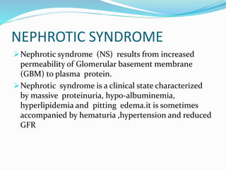
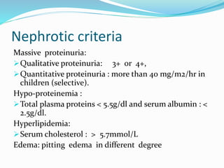
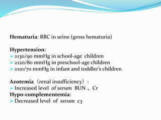
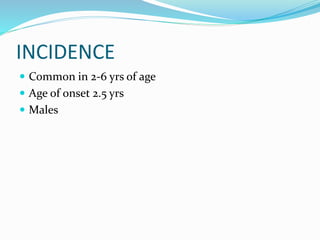
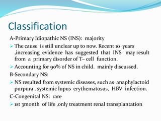
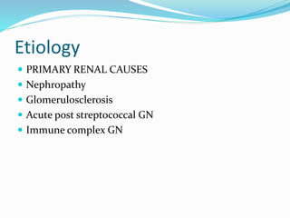
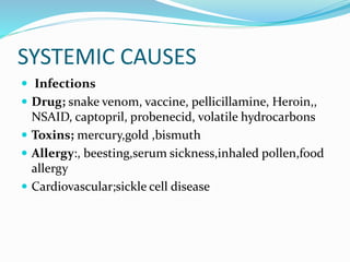
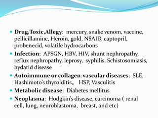
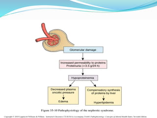
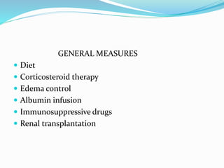
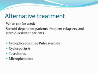
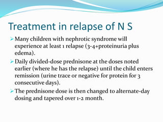
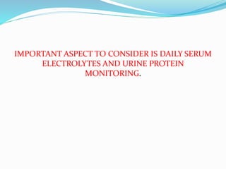
NEPHROTIC SYNDROME
Nephrotic syndrome (NS) results from increased
permeability of Glomerular basement membrane
(GBM) to plasma protein.
Nephrotic syndrome is a clinical state characterized
by massive proteinuria, hypo-albuminemia,
hyperlipidemia and pitting edema.it is sometimes
accompanied by hematuria ,hypertension and reduced
GFR
Nephrotic criteria
Massive proteinuria:
Qualitative proteinuria: 3+ or 4+,
Quantitative proteinuria : more than 40 mg/m2/hr in
children (selective).
Hypo-proteinemia :
Total plasma proteins < 5.5g/dl and serum albumin : <
2.5g/dl.
Hyperlipidemia:
Serum cholesterol : > 5.7mmol/L
Edema: pitting edema in different degree
Hematuria: RBC in urine (gross hematuria)
Hypertension:
 ≥130/90 mmHg in school-age children
 ≥120/80 mmHg in preschool-age children
 ≥110/70 mmHg in infant and toddler’s children
Azotemia(renal insufficiency):
 Increased level of serum BUN 、Cr
Hypo-complementemia:
 Decreased level of serum c3
INCIDENCE
 Common in 2-6 yrs of age
 Age of onset 2.5 yrs
 Males
Classification
A-Primary Idiopathic NS (INS): majority
 The cause is still unclear up to now. Recent 10 years
,increasing evidence has suggested that INS may result
from a primary disorder of T– cell function.
 Accounting for 90% of NS in child. mainly discussed.
B-Secondary NS:
 NS resulted from systemic diseases, such as anaphylactoid
purpura , systemic lupus erythematosus, HBV infection.
C-Congenital NS: rare
 1st 3month of life ,only treatment renal transplantation
Etiology
 PRIMARY RENAL CAUSES
 Nephropathy
 Glomerulosclerosis
 Acute post streptococcal GN
 Immune complex GN
SYSTEMIC CAUSES
 Infections
 Drug; snake venom, vaccine, pellicillamine, Heroin,,
NSAID, captopril, probenecid, volatile hydrocarbons
 Toxins; mercury,gold ,bismuth
 Allergy:, beesting,serum sickness,inhaled pollen,food
allergy
 Cardiovascular;sickle cell disease
 Drug,Toxic,Allegy: mercury, snake venom, vaccine,
pellicillamine, Heroin, gold, NSAID, captopril,
probenecid, volatile hydrocarbons
 Infection: APSGN, HBV, HIV, shunt nephropathy,
reflux nephropathy, leprosy, syphilis, Schistosomiasis,
hydatid disease
 Autoimmune or collagen-vascular diseases: SLE,
Hashimoto’s thyroiditis,, HSP, Vasculitis
 Metabolic disease: Diabetes mellitus
 Neoplasma: Hodgkin’s disease, carcinoma ( renal
cell, lung, neuroblastoma, breast, and etc)
 Genetic Disease: Alport syn, Sickle cell disease,
Amyloidosis, Congenital nephropathy
 Others: Chronic transplant rejection, congenital
nephrosclerosis
IDIOPATHIC NS PATHOLOGY
Minimal Change Nephropathy (MCN): <80%
 The glomeruli appear normal basically Under Light
microscopy, and Under Immunofluorescence under
Electron microscopy
(2) Non—MCN: <20%
 Mesangial proliferative glomerulonephritis (MsPGN):
about 10%, Focal segmental glomerulosclerosis (FSGS): 5%
 Membranous Nephropathy (MN) : 2%
 Membrane proliferative glomerulonephritis (MPGN) : 1%
pathophysiology
 The Main Trigger Of primary Nephrotic Syndrome
and Fundamental and highly important change of
pathophysiology :-
Proteinuria
Pathophysiology-protenuria
Increase glomerular permeability for proteins due to
loss of negative charged glycoprotein
Degree of protineuria:-
Mild less than 0.5g/m2/day
Moderate 0.5 – 2g/m2/day
Severe more than 2g/m2/day
Type of proteinuria:-
A-Selective proteinuria: where proteins of low
molecular weight .such as albumin, are excreted more
readily than protein of HMW
B-Non selective :
LMW+HMW are lost in urine
Pathogenesis-Hypoalbenemia
Due to hyperproteinuria----- Loss of plasma protein in
urine mainly the albumin.
Pathogenesis-Hyperlipidemia
 Response to Hypoalbuminemia → reflex to liver --→
synthesis of generalize protein ( including
lipoprotein ) and lipid in the liver ,the lipoprotein
high molecular weight no loss in urine →
hyperlipidemia
 Diminished catabolism of lipoprotein
Pathogenesis of edema
Reduction plasma colloid osmotic pressure↓
secondary to hypoalbuminemia Edema and
hypovolemia
Intravascular volume↓ antidiuretic hormone
(ADH ) and aldosterone(ALD) water and
sodium retention Edema
Intravascular volume↓ glomerular filtration rate
(GFR)↓ water and sodium retention Edema
Clinical manifestation
1.Main manifestations:
 Edema (varying degrees) is the common symptom
 Local edema: edema in face , around eyes( Periorbital swelling) ,
in lower extremities.
 Generalized edema (anasarca), edema in penis and scrotum.
2-Non-specific symptoms:
 Fever,Fatigue and lethargy
 loss of appetite, nausea and vomiting ,abdominal pain , diarrhea
 body weight increase, urine output decrease
 pleural effusion (respiratory distress)
 Susceptible to infections
Diagnosis
1.History
2.Physical examination
3.Urine analysis
gold standard in diagnosis-24 hr urine protein
meaurement
Reveals +3 or +4 proteinuria.
Protein excetion more than 2g/24 hr
-Microscopic hematuria may be present.
-Low creatine clearance
4.Blood analysis
a. Hypoproteinemia(hypoalbuminemia) below 2.5g/dl
b. Serum albumin/globulin ratio is reversed
c. Hypogammaglobulinemia with increase in
lipoprotein(alpha2 and beta globulins)
d. Serum cholesterol and triglyceride level increases.
e. Hypomagnesemia
f. Increased BUN
g. Increased platelet count
5.USG abdomen
6.Kidney biopsy
Considered in:
1-Secondary N.S
2-Frequent relapsing N.S
3-Steroid resistant N.
4- Hematuria
5-Hypertension
6- Low GFR
Complications
1.Ascites
2.Pleural effusion
3.Generalised edema
4.Coagulation disorder
5.Thrombosis
6.Recurrent infections- sepsis, pneumonia,cellulitis,
urinary tract infections
7.Renal failure
8.Reduced serum vitamin-D, calcium.
Management
The primary objective is to reduce the excretion of
urinary protein and maintain a protein free diet.
 Additional objective include:
1. Prevention or treatment of acute infection.
2. Control of edema with diuretics such as frusemide and
spirinolactone
3. Good nutrition.-balanced diet rich in protein and sodium
and water intake is restricted
4. Readjustment of altered metabolic activities.
5. Children with severe symptoms and newly diagnosed cases
are hospitalised for assessment and observation for evidence
for infection, response to therapy and parental education.
GENERAL MEASURES
 Diet
 Corticosteroid therapy
 Edema control
 Albumin infusion
 Immunosuppressive drugs
 Renal transplantation
Alternative treatment
When can be used:
Steroid-dependent patients, frequent relapsers, and
steroid-resistant patients.
 Cyclophosphamide Pulse steroids
 Cyclosporin A
 Tacrolimus
 Microphenolate
Treatment in relapse of N S
Many children with nephrotic syndrome will
experience at least 1 relapse (3-4+proteinuria plus
edema).
Daily divided-dose prednisone at the doses noted
earlier (where he has the relapse) until the child enters
remission (urine trace or negative for protein for 3
consecutive days).
The prednisone dose is then changed to alternate-day
dosing and tapered over 1-2 month.
IMPORTANT ASPECT TO CONSIDER IS DAILY SERUM
ELECTROLYTES AND URINE PROTEIN
MONITORING.
Steroid toxicity
 Stunted growth
 Cataracts
 Pseudotumor cerebri
 Psycosis
 Osteoporosis
 Cushingoid features
 Adrenal gland suppression
 hyperglycemia
 myopathy
 peptic ulcer
 poor healing of wound.
 Hirsutism
 Thromboembolism
Nursing management
A.Care during hospitalisation
Daily monitoring of intake and output,wt,vitalsigns .
Infants and neonates- diapers weight.
Examination of daily urine -protein, albumin, abdominal
girth, edema- degree or pitting, colour and texture of
skin,infection
B.Administer medications
C.Maintain fluid and electrolyte balance
D.Prevention of infection
E. Promote rest
F.Emotional support.
 Edema
 Infection prevention
 Nutrition
 Family support and home care
Dietry modification
 One of the aims of nutritional management is to
replace as much of the protein loss in the urine by a
intake of good quality protein
 Protein (1-2gm per kilogram body weight) should
given.
 Sodium should be restricted to 500mg.
 Water intake 24hr urine output + 500ml.
Nursing diagnosis
1.Fluid volume excess related to fluid accumulation in
tissues.
2.Impaired nutrition less than body requirement.
3.Impaired family process
4.Knowledge deficit
5.Self esteem
6.Risk for infection.
nephrotic syndrome.pptx

nephrotic syndrome.pptx

  • 2.
    NEPHROTIC SYNDROME Nephrotic syndrome(NS) results from increased permeability of Glomerular basement membrane (GBM) to plasma protein. Nephrotic syndrome is a clinical state characterized by massive proteinuria, hypo-albuminemia, hyperlipidemia and pitting edema.it is sometimes accompanied by hematuria ,hypertension and reduced GFR
  • 3.
    Nephrotic criteria Massive proteinuria: Qualitativeproteinuria: 3+ or 4+, Quantitative proteinuria : more than 40 mg/m2/hr in children (selective). Hypo-proteinemia : Total plasma proteins < 5.5g/dl and serum albumin : < 2.5g/dl. Hyperlipidemia: Serum cholesterol : > 5.7mmol/L Edema: pitting edema in different degree
  • 4.
    Hematuria: RBC inurine (gross hematuria) Hypertension:  ≥130/90 mmHg in school-age children  ≥120/80 mmHg in preschool-age children  ≥110/70 mmHg in infant and toddler’s children Azotemia(renal insufficiency):  Increased level of serum BUN 、Cr Hypo-complementemia:  Decreased level of serum c3
  • 5.
    INCIDENCE  Common in2-6 yrs of age  Age of onset 2.5 yrs  Males
  • 6.
    Classification A-Primary Idiopathic NS(INS): majority  The cause is still unclear up to now. Recent 10 years ,increasing evidence has suggested that INS may result from a primary disorder of T– cell function.  Accounting for 90% of NS in child. mainly discussed. B-Secondary NS:  NS resulted from systemic diseases, such as anaphylactoid purpura , systemic lupus erythematosus, HBV infection. C-Congenital NS: rare  1st 3month of life ,only treatment renal transplantation
  • 7.
    Etiology  PRIMARY RENALCAUSES  Nephropathy  Glomerulosclerosis  Acute post streptococcal GN  Immune complex GN
  • 8.
    SYSTEMIC CAUSES  Infections Drug; snake venom, vaccine, pellicillamine, Heroin,, NSAID, captopril, probenecid, volatile hydrocarbons  Toxins; mercury,gold ,bismuth  Allergy:, beesting,serum sickness,inhaled pollen,food allergy  Cardiovascular;sickle cell disease
  • 9.
     Drug,Toxic,Allegy: mercury,snake venom, vaccine, pellicillamine, Heroin, gold, NSAID, captopril, probenecid, volatile hydrocarbons  Infection: APSGN, HBV, HIV, shunt nephropathy, reflux nephropathy, leprosy, syphilis, Schistosomiasis, hydatid disease  Autoimmune or collagen-vascular diseases: SLE, Hashimoto’s thyroiditis,, HSP, Vasculitis  Metabolic disease: Diabetes mellitus  Neoplasma: Hodgkin’s disease, carcinoma ( renal cell, lung, neuroblastoma, breast, and etc)
  • 10.
     Genetic Disease:Alport syn, Sickle cell disease, Amyloidosis, Congenital nephropathy  Others: Chronic transplant rejection, congenital nephrosclerosis
  • 11.
    IDIOPATHIC NS PATHOLOGY MinimalChange Nephropathy (MCN): <80%  The glomeruli appear normal basically Under Light microscopy, and Under Immunofluorescence under Electron microscopy (2) Non—MCN: <20%  Mesangial proliferative glomerulonephritis (MsPGN): about 10%, Focal segmental glomerulosclerosis (FSGS): 5%  Membranous Nephropathy (MN) : 2%  Membrane proliferative glomerulonephritis (MPGN) : 1%
  • 12.
    pathophysiology  The MainTrigger Of primary Nephrotic Syndrome and Fundamental and highly important change of pathophysiology :- Proteinuria
  • 13.
    Pathophysiology-protenuria Increase glomerular permeabilityfor proteins due to loss of negative charged glycoprotein Degree of protineuria:- Mild less than 0.5g/m2/day Moderate 0.5 – 2g/m2/day Severe more than 2g/m2/day Type of proteinuria:- A-Selective proteinuria: where proteins of low molecular weight .such as albumin, are excreted more readily than protein of HMW B-Non selective : LMW+HMW are lost in urine
  • 14.
    Pathogenesis-Hypoalbenemia Due to hyperproteinuria-----Loss of plasma protein in urine mainly the albumin.
  • 15.
    Pathogenesis-Hyperlipidemia  Response toHypoalbuminemia → reflex to liver --→ synthesis of generalize protein ( including lipoprotein ) and lipid in the liver ,the lipoprotein high molecular weight no loss in urine → hyperlipidemia  Diminished catabolism of lipoprotein
  • 16.
    Pathogenesis of edema Reductionplasma colloid osmotic pressure↓ secondary to hypoalbuminemia Edema and hypovolemia Intravascular volume↓ antidiuretic hormone (ADH ) and aldosterone(ALD) water and sodium retention Edema Intravascular volume↓ glomerular filtration rate (GFR)↓ water and sodium retention Edema
  • 18.
    Clinical manifestation 1.Main manifestations: Edema (varying degrees) is the common symptom  Local edema: edema in face , around eyes( Periorbital swelling) , in lower extremities.  Generalized edema (anasarca), edema in penis and scrotum. 2-Non-specific symptoms:  Fever,Fatigue and lethargy  loss of appetite, nausea and vomiting ,abdominal pain , diarrhea  body weight increase, urine output decrease  pleural effusion (respiratory distress)  Susceptible to infections
  • 20.
    Diagnosis 1.History 2.Physical examination 3.Urine analysis goldstandard in diagnosis-24 hr urine protein meaurement Reveals +3 or +4 proteinuria. Protein excetion more than 2g/24 hr -Microscopic hematuria may be present. -Low creatine clearance
  • 21.
    4.Blood analysis a. Hypoproteinemia(hypoalbuminemia)below 2.5g/dl b. Serum albumin/globulin ratio is reversed c. Hypogammaglobulinemia with increase in lipoprotein(alpha2 and beta globulins) d. Serum cholesterol and triglyceride level increases. e. Hypomagnesemia f. Increased BUN g. Increased platelet count 5.USG abdomen
  • 22.
    6.Kidney biopsy Considered in: 1-SecondaryN.S 2-Frequent relapsing N.S 3-Steroid resistant N. 4- Hematuria 5-Hypertension 6- Low GFR
  • 23.
    Complications 1.Ascites 2.Pleural effusion 3.Generalised edema 4.Coagulationdisorder 5.Thrombosis 6.Recurrent infections- sepsis, pneumonia,cellulitis, urinary tract infections 7.Renal failure 8.Reduced serum vitamin-D, calcium.
  • 24.
    Management The primary objectiveis to reduce the excretion of urinary protein and maintain a protein free diet.
  • 25.
     Additional objectiveinclude: 1. Prevention or treatment of acute infection. 2. Control of edema with diuretics such as frusemide and spirinolactone 3. Good nutrition.-balanced diet rich in protein and sodium and water intake is restricted 4. Readjustment of altered metabolic activities. 5. Children with severe symptoms and newly diagnosed cases are hospitalised for assessment and observation for evidence for infection, response to therapy and parental education.
  • 26.
    GENERAL MEASURES  Diet Corticosteroid therapy  Edema control  Albumin infusion  Immunosuppressive drugs  Renal transplantation
  • 27.
    Alternative treatment When canbe used: Steroid-dependent patients, frequent relapsers, and steroid-resistant patients.  Cyclophosphamide Pulse steroids  Cyclosporin A  Tacrolimus  Microphenolate
  • 28.
    Treatment in relapseof N S Many children with nephrotic syndrome will experience at least 1 relapse (3-4+proteinuria plus edema). Daily divided-dose prednisone at the doses noted earlier (where he has the relapse) until the child enters remission (urine trace or negative for protein for 3 consecutive days). The prednisone dose is then changed to alternate-day dosing and tapered over 1-2 month.
  • 29.
    IMPORTANT ASPECT TOCONSIDER IS DAILY SERUM ELECTROLYTES AND URINE PROTEIN MONITORING.
  • 30.
    Steroid toxicity  Stuntedgrowth  Cataracts  Pseudotumor cerebri  Psycosis  Osteoporosis  Cushingoid features  Adrenal gland suppression  hyperglycemia  myopathy  peptic ulcer  poor healing of wound.  Hirsutism  Thromboembolism
  • 31.
    Nursing management A.Care duringhospitalisation Daily monitoring of intake and output,wt,vitalsigns . Infants and neonates- diapers weight. Examination of daily urine -protein, albumin, abdominal girth, edema- degree or pitting, colour and texture of skin,infection B.Administer medications C.Maintain fluid and electrolyte balance D.Prevention of infection E. Promote rest F.Emotional support.
  • 32.
     Edema  Infectionprevention  Nutrition  Family support and home care
  • 33.
    Dietry modification  Oneof the aims of nutritional management is to replace as much of the protein loss in the urine by a intake of good quality protein  Protein (1-2gm per kilogram body weight) should given.  Sodium should be restricted to 500mg.  Water intake 24hr urine output + 500ml.
  • 34.
    Nursing diagnosis 1.Fluid volumeexcess related to fluid accumulation in tissues. 2.Impaired nutrition less than body requirement. 3.Impaired family process 4.Knowledge deficit 5.Self esteem 6.Risk for infection.