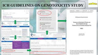
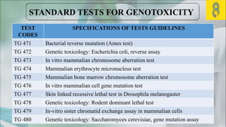
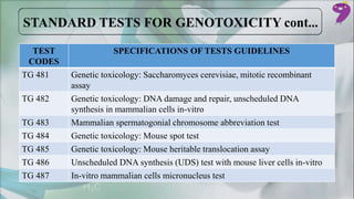
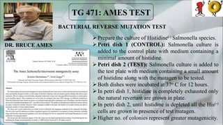
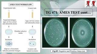
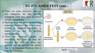
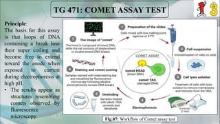
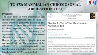
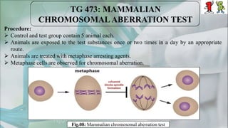
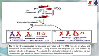
GENOTOXICITY STUDY
Under the guidance of:
DR. SHAMSHER SINGH BAJWA
Professor and Head of
Department of Pharmacology
ISF College of Pharmacy, Punjab
Presented By:
PRATYUSH POREL
M.Pharm, 2nd Sem.
Department of Pharmacology
ISF College of Pharmacy, Punjab
SL NO. TOPIC NAME SLIDE NO.
01 INTRODUCTION: GENE 01
02 ALLELES, HOMOZYGOCITY & HETEROZYGOCITY 02
03 PHENOTYPIC & GENOTYPIC CHARACTERISTICS 03-04
04 ICH GUIDELINES ON GENOTOXICITY STUDY 05
05 CAUSES, MECHANISM AND STANDARD TESTS 06-09
06 AMES TEST (BACTERIAL REVERSE MUTATION TEST) 10-12
07 COMET ASSAY TEST 13
08 MAMMALIAN CHROMOSOMAL ABERRATION TEST 14-16
09 MAMMALIAN ERYTHROCYTE MICRONUCLEUS TEST 17-18
10 APPLICATIONS, LIMITATIONS AND FUTURE ASPECTS 19-20
Genes are like a story and DNA is the language that the story is written in.
- Sam Kean
Gene1, unit of hereditary information that
occupies a fixed position (locus) on
a chromosome. Genes achieve their effects
by directing the synthesis of proteins.
Allele2, any one of two or more genes that may
occur alternatively at a given site (locus) on
a chromosome. Alleles may occur in pairs, or
there may be multiple alleles affecting the
expression (phenotype) of a particular trait.
The combination of alleles that an organism
carries constitutes its genotype.
 Homozygous: Organism has the paired
alleles for any particular traits.
 Heterozygous: Organism has the different
pair of alleles for any particular traits.
Fig.02: Homozygosity and heterozygosity
Fig.01: Alleles on chromosome
Allele for phenotype A
Allele for phenotype B
Chromo
-somes
Alleles
Phenotype3, all the observable
characteristics of an organism that
result from the interaction of its
genotype (total genetic inheritance)
with the environment. Examples of
observable characteristics include
behavior, color, shape, and size.
Fig.03: Different phenotypic characteristics
Genotype3, is the genetic makeup
responsible for the all the observable
characteristics of an organism (total
genetic inheritance).
Fig.04: Monohybrid cross in genetics
 Monohybrid cross: The cross between two
monohybrid traits (TT and tt) is called a Monohybrid
Cross.
E.g., Height of the plant, position of the flower etc.
 Dihybrid cross: Dihybrid cross is the cross between
two different genes that differ in two observed traits.
E.g., Shape and color of the seed, shape and position of
petals of the same flower etc.
GREGOR JOHANN
MENDEL
 Genotoxicity is a word used in genetics, that has the destructive effects on the genetic
material of the cell (DNA, RNA), thus affecting the integrity of the cell.
 Genotoxins are one type of mutagens that causes genotoxicity followed by mutation.
 Genetic toxicology is the branch of toxicology that deals with those substances that can
damage the genetic makeup even a chromosome.
Genotoxic risk
Somatic cells Germ cells
Aging Cancer Sterility Genetic diseases Multifactorial
diseases
Cystic fibrosis Sickle cell anaemia Haemophilia
Diabetes mellitus Cardio-vascular diseases Psychosis
Depending on their effects genotoxins can be classified as :
1. Carcinogenic or cancer causing agents
2. Mutagens or mutation causing agents
3. Teratogens or birth defect causing agents
The damage to the genetic material is caused by the interactions of the genotoxic
substance with the DNA structure and sequence. These genotoxic substance interact at a
specific location or base sequence of the DNA structure causing lesions, breakage, fusion,
deletion, alteration, flip-flop movements of nucleotides, mis-segregation or non-
disjunction leading to damage of DNA and mutation. These types of chromosomal
aberration may leads to abnormal cell growth, sometimes tumorigenic. Generally, ROS,
UV rays, topoisomerase inhibitor and protein synthesis inhibitors are the causative agents.
TEST
CODES
SPECIFICATIONS OF TESTS GUIDELINES
TG 471 Bacterial reverse mutation (Ames test)
TG 472 Genetic toxicology: Escherichia coli, reverse assay
TG 473 In vitro mammalian chromosome aberration test
TG 474 Mammalian erythrocyte micronucleus test
TG 475 Mammalian bone marrow chromosome aberration test
TG 476 In vitro mammalian cell gene mutation test
TG 477 Skin linked recessive lethal test in Drosophila melanogaster
TG 478 Genetic toxicology: Rodent dominant lethal test
TG 479 In-vitro sister chromatid exchange assay in mammalian cells
TG 480 Genetic toxicology: Saccharomyces cerevisiae, gene mutation assay
TEST
CODES
SPECIFICATIONS OF TESTS GUIDELINES
TG 481 Genetic toxicology: Saccharomyces cerevisiae, mitotic recombinant
assay
TG 482 Genetic toxicology: DNA damage and repair, unscheduled DNA
synthesis in mammalian cells in-vitro
TG 483 Mammalian spermatogonial chromosome abbreviation test
TG 484 Genetic toxicology: Mouse spot test
TG 485 Genetic toxicology: Mouse heritable translocation assay
TG 486 Unscheduled DNA synthesis (UDS) test with mouse liver cells in-vitro
TG 487 In-vitro mammalian cells micronucleus test
BACTERIAL REVERSE MUTATION TEST
Prepare the culture of Histidine(-) Salmonella species.
Petri dish 1 (CONTROL): Salmonella culture is
added to the control plate with medium containing a
minimal amount of histidine.
Petri dish 2 (TEST): Salmonella culture is added to
the test plate with medium containing a small amount
of histidine along with the mutagen to be tested.
Both dishes were incubated at 37o C for 12 hours.
In petri dish 1, histidine is completely exhausted only
the natural revertant are grown in plate.
In petri dish 2, until histidine is depleted all the His(-)
cells are grown in presence of test mutagen.
Higher no. of colonies represent greater mutagenicity.
Control
DR. BRUCE AMES
AMES TEST WORKFLOW
Experimental tube Control tube
Histidine selective
media
Incubation Incubation
Control with
natural revertants
High no. of revertants
indicates potential mutagen
Suspected mutagen,
agar, rat liver extract
and salmonella strains
Agar, rat liver extract
and salmonella strains
Fig.05: Negative and Positive Ames test
 There are some chemicals that are
non mutagenic, but they become
mutagenic when they come in contact
in body metabolism.
 Bacteria does not have metabolizing
capacity, therefore, the liver extract
(called as metabolic activator) is
added to the test, to perform
transformation.
 Then the bacterial sample is
inoculated. But from the idea the
negative test will not show any
growth, but the growth can occurs
due to spontaneous mutation, this is
only the limitation .
Fig.06: Ames test
Principle:
The basis for this assay
is that loops of DNA
containing a break lose
their super coiling and
become free to extend
toward the anode when
exposed to current
during electrophoresis at
high pH.
• The results appear as
structures resembling
comets observed by
fluorescence
microscopy. Fig.07: Workflow of Comet assay test
Principle:
The short-term in vitro mammalian cell
chromosome aberration test is used to
assess potential genotoxic hazard of test
substances. Mammalian cells are cultured
in vitro, exposed to a test substance,
harvested, and the frequency of
asymmetrical structural chromosome
aberrations is measured.
Human peripheral blood lymphocytes do
not normally divide. The assessment of the
effects of cyclophosphamide on
lymphocytes, stimulated to divide in whole
blood cultures in vitro, is described.
Procedure:
 Control and test group contain 5 animal each.
 Animals are exposed to the test substances once or two times in a day by an appropriate
route.
 Animals are treated with metaphase arresting agents.
 Metaphase cells are observed for chromosomal aberration.
Fig.08: Mammalian chromosomal aberration test
Fig.10: In vitro mammalian chromosome aberration test (TG 473) The cells are plated and
treated with the metabolic activator (A), along with the test compound (B). This followed by
exposure of cells to Colcemide / Nocodazole (C), which allows the arrest at metaphase. Various
chromosomal type aberration (D) and chromatid type aberration are observed.
Principle:
• Animals are exposed to the test
substance by an appropriate route
• If the bone marrow > the animals
are sacrificed, bone marrow
extracted and preparations are
made and stained.
• If peripheral blood > the blood is
collected at appropriate times
after treatment and smear
preparations are made and stained.
• Preparations are analysed for the
presence of micronuclei
Procedure:
 Animals are exposed to the test substance by an
appropriate route.
 Bone marrow / blood cells are collected, prepared
and stained.
 Preparations are analysed for the presence of
micronuclei
 Each treated and control group must include at least
5 analysable animals per sex.
 Administration of the treatments consists of a single
dose or two daily doses.
 The limit dose is 200mg/kg/body weight /day for
treatment up to 14 days, and 1000 mg/kg/body
weight/day for treatment longer than 14 days.
1. Assessment of genotoxicity in occupational exposed individual.
2. A study with 838 drugs, analyzed by the different assays, pointed out that 56.3% of them
were genotoxic. So, it is used for testing of new pharmaceutical product for its safety.
3. Testing of various products used by humans cosmetics, food products.
1. Nowadays, most of the toxicological studies are based on traditional methods, they are not
always the best choice for predicting human health effects.
2. Cell culture methodologies have substantially improved resulting in a number of promising
cell models composed of relevant morphological and biochemical signaling processes that
may reproduce the in vivo environment.
1. The genotoxicity tests are an important for the safety assessment of chemicals and drugs
according to the guidelines set by international regulatory authorities.
2. The novel in vitro model system along with other emerging technologies such as high-
throughput assay system, imaging technologies, computational approaches will help in
developing completely new integrated approaches for genotoxicity testing and assessment,
which will be more advanced and promising than the traditional methods.
3. In the current scenario, more research, development, and data integration are required to meet
the demands of regulatory testing. Only then, there is a possible likelihood for reduction and
complete replacement of traditional testing method acceptable to regulatory authorities.
1. Britannica, T. Editors of Encyclopedia. "gene." Encyclopedia Britannica, January 25,
2024. https://www.britannica.com/science/gene.
2. Britannica, The Editors of Encyclopedia. "allele". Encyclopedia Britannica, 2 Jun. 2023,
https://www.britannica.com/science/allele. Accessed 5 February 2024.
3. Britannica, The Editors of Encyclopedia. "genotype". Encyclopedia Britannica, 11 Jan.
2024, https://www.britannica.com/science/genotype. Accessed 7 February 2024.
4. Edward E. Gahres, Tongue rolling and tongue folding: and other Hereditary Movements of
the Tongue, Journal of Heredity, Volume 43, Issue 5, September 1952, Pages 221–
225, https://doi.org/10.1093/oxfordjournals.jhered.a106313
5. Zhang XJ, He PP, Liang YH, Yang S, Yuan WT, Xu SJ, Huang W. A gene for freckles
maps to chromosome 4q32–q34. Journal of investigative dermatology. 2004 Feb
1;122(2):286-90.
6. Ordu KS, Agi CE. Inheritance pattern of hairline shape amongst Nigerian population. Int J
Curr Microbio App Sci. 2014;3:61-5.
7. Barton NH. Genetic hitchhiking. Philosophical Transactions of the Royal Society of
London. Series B: Biological Sciences. 2000 Nov 29;355(1403):1553-62.
8. Britannica, The Editors of Encyclopedia. "genotype". Encyclopedia Britannica, 11 Jan.
2024, https://www.britannica.com/science/genotype. Accessed 7 February 2024.
9. Mallick A, Gautam A. Mendelian Crosses.
10. Kamath GH, Rao KS. Genotoxicity guidelines recommended by International Conference
of Harmonization (ICH). Genotoxicity Assessment: Methods and Protocols. 2013:431-58.
11. Mortelmans K, Zeiger E. The Ames Salmonella/microsome mutagenicity assay. Mutation
research/fundamental and molecular mechanisms of mutagenesis. 2000 Nov 20;455(1-2):29-
60.
12. Speit G, Vasquez M, Hartmann A. The comet assay as an indicator test for germ cell
genotoxicity. Mutation Research/Reviews in Mutation Research. 2009 Jan 1;681(1):3-12.
13. Araldi RP, de Melo TC, Mendes TB, de Sá Júnior PL, Nozima BH, Ito ET, de Carvalho
RF, de Souza EB, de Cassia Stocco R. Using the comet and micronucleus assays for
genotoxicity studies: A review. Biomedicine & Pharmacotherapy. 2015 May 1;72:74-82.
GENOTOXICITY STUDY.pptx

GENOTOXICITY STUDY.pptx

  • 1.
    GENOTOXICITY STUDY Under theguidance of: DR. SHAMSHER SINGH BAJWA Professor and Head of Department of Pharmacology ISF College of Pharmacy, Punjab Presented By: PRATYUSH POREL M.Pharm, 2nd Sem. Department of Pharmacology ISF College of Pharmacy, Punjab
  • 2.
    SL NO. TOPICNAME SLIDE NO. 01 INTRODUCTION: GENE 01 02 ALLELES, HOMOZYGOCITY & HETEROZYGOCITY 02 03 PHENOTYPIC & GENOTYPIC CHARACTERISTICS 03-04 04 ICH GUIDELINES ON GENOTOXICITY STUDY 05 05 CAUSES, MECHANISM AND STANDARD TESTS 06-09 06 AMES TEST (BACTERIAL REVERSE MUTATION TEST) 10-12 07 COMET ASSAY TEST 13 08 MAMMALIAN CHROMOSOMAL ABERRATION TEST 14-16 09 MAMMALIAN ERYTHROCYTE MICRONUCLEUS TEST 17-18 10 APPLICATIONS, LIMITATIONS AND FUTURE ASPECTS 19-20
  • 3.
    Genes are likea story and DNA is the language that the story is written in. - Sam Kean Gene1, unit of hereditary information that occupies a fixed position (locus) on a chromosome. Genes achieve their effects by directing the synthesis of proteins.
  • 4.
    Allele2, any oneof two or more genes that may occur alternatively at a given site (locus) on a chromosome. Alleles may occur in pairs, or there may be multiple alleles affecting the expression (phenotype) of a particular trait. The combination of alleles that an organism carries constitutes its genotype.  Homozygous: Organism has the paired alleles for any particular traits.  Heterozygous: Organism has the different pair of alleles for any particular traits. Fig.02: Homozygosity and heterozygosity Fig.01: Alleles on chromosome Allele for phenotype A Allele for phenotype B Chromo -somes Alleles
  • 5.
    Phenotype3, all theobservable characteristics of an organism that result from the interaction of its genotype (total genetic inheritance) with the environment. Examples of observable characteristics include behavior, color, shape, and size. Fig.03: Different phenotypic characteristics
  • 6.
    Genotype3, is thegenetic makeup responsible for the all the observable characteristics of an organism (total genetic inheritance). Fig.04: Monohybrid cross in genetics  Monohybrid cross: The cross between two monohybrid traits (TT and tt) is called a Monohybrid Cross. E.g., Height of the plant, position of the flower etc.  Dihybrid cross: Dihybrid cross is the cross between two different genes that differ in two observed traits. E.g., Shape and color of the seed, shape and position of petals of the same flower etc. GREGOR JOHANN MENDEL
  • 8.
     Genotoxicity isa word used in genetics, that has the destructive effects on the genetic material of the cell (DNA, RNA), thus affecting the integrity of the cell.  Genotoxins are one type of mutagens that causes genotoxicity followed by mutation.  Genetic toxicology is the branch of toxicology that deals with those substances that can damage the genetic makeup even a chromosome. Genotoxic risk Somatic cells Germ cells Aging Cancer Sterility Genetic diseases Multifactorial diseases Cystic fibrosis Sickle cell anaemia Haemophilia Diabetes mellitus Cardio-vascular diseases Psychosis
  • 9.
    Depending on theireffects genotoxins can be classified as : 1. Carcinogenic or cancer causing agents 2. Mutagens or mutation causing agents 3. Teratogens or birth defect causing agents The damage to the genetic material is caused by the interactions of the genotoxic substance with the DNA structure and sequence. These genotoxic substance interact at a specific location or base sequence of the DNA structure causing lesions, breakage, fusion, deletion, alteration, flip-flop movements of nucleotides, mis-segregation or non- disjunction leading to damage of DNA and mutation. These types of chromosomal aberration may leads to abnormal cell growth, sometimes tumorigenic. Generally, ROS, UV rays, topoisomerase inhibitor and protein synthesis inhibitors are the causative agents.
  • 10.
    TEST CODES SPECIFICATIONS OF TESTSGUIDELINES TG 471 Bacterial reverse mutation (Ames test) TG 472 Genetic toxicology: Escherichia coli, reverse assay TG 473 In vitro mammalian chromosome aberration test TG 474 Mammalian erythrocyte micronucleus test TG 475 Mammalian bone marrow chromosome aberration test TG 476 In vitro mammalian cell gene mutation test TG 477 Skin linked recessive lethal test in Drosophila melanogaster TG 478 Genetic toxicology: Rodent dominant lethal test TG 479 In-vitro sister chromatid exchange assay in mammalian cells TG 480 Genetic toxicology: Saccharomyces cerevisiae, gene mutation assay
  • 11.
    TEST CODES SPECIFICATIONS OF TESTSGUIDELINES TG 481 Genetic toxicology: Saccharomyces cerevisiae, mitotic recombinant assay TG 482 Genetic toxicology: DNA damage and repair, unscheduled DNA synthesis in mammalian cells in-vitro TG 483 Mammalian spermatogonial chromosome abbreviation test TG 484 Genetic toxicology: Mouse spot test TG 485 Genetic toxicology: Mouse heritable translocation assay TG 486 Unscheduled DNA synthesis (UDS) test with mouse liver cells in-vitro TG 487 In-vitro mammalian cells micronucleus test
  • 12.
    BACTERIAL REVERSE MUTATIONTEST Prepare the culture of Histidine(-) Salmonella species. Petri dish 1 (CONTROL): Salmonella culture is added to the control plate with medium containing a minimal amount of histidine. Petri dish 2 (TEST): Salmonella culture is added to the test plate with medium containing a small amount of histidine along with the mutagen to be tested. Both dishes were incubated at 37o C for 12 hours. In petri dish 1, histidine is completely exhausted only the natural revertant are grown in plate. In petri dish 2, until histidine is depleted all the His(-) cells are grown in presence of test mutagen. Higher no. of colonies represent greater mutagenicity. Control DR. BRUCE AMES
  • 13.
    AMES TEST WORKFLOW Experimentaltube Control tube Histidine selective media Incubation Incubation Control with natural revertants High no. of revertants indicates potential mutagen Suspected mutagen, agar, rat liver extract and salmonella strains Agar, rat liver extract and salmonella strains Fig.05: Negative and Positive Ames test
  • 14.
     There aresome chemicals that are non mutagenic, but they become mutagenic when they come in contact in body metabolism.  Bacteria does not have metabolizing capacity, therefore, the liver extract (called as metabolic activator) is added to the test, to perform transformation.  Then the bacterial sample is inoculated. But from the idea the negative test will not show any growth, but the growth can occurs due to spontaneous mutation, this is only the limitation . Fig.06: Ames test
  • 15.
    Principle: The basis forthis assay is that loops of DNA containing a break lose their super coiling and become free to extend toward the anode when exposed to current during electrophoresis at high pH. • The results appear as structures resembling comets observed by fluorescence microscopy. Fig.07: Workflow of Comet assay test
  • 16.
    Principle: The short-term invitro mammalian cell chromosome aberration test is used to assess potential genotoxic hazard of test substances. Mammalian cells are cultured in vitro, exposed to a test substance, harvested, and the frequency of asymmetrical structural chromosome aberrations is measured. Human peripheral blood lymphocytes do not normally divide. The assessment of the effects of cyclophosphamide on lymphocytes, stimulated to divide in whole blood cultures in vitro, is described.
  • 17.
    Procedure:  Control andtest group contain 5 animal each.  Animals are exposed to the test substances once or two times in a day by an appropriate route.  Animals are treated with metaphase arresting agents.  Metaphase cells are observed for chromosomal aberration. Fig.08: Mammalian chromosomal aberration test
  • 18.
    Fig.10: In vitromammalian chromosome aberration test (TG 473) The cells are plated and treated with the metabolic activator (A), along with the test compound (B). This followed by exposure of cells to Colcemide / Nocodazole (C), which allows the arrest at metaphase. Various chromosomal type aberration (D) and chromatid type aberration are observed.
  • 19.
    Principle: • Animals areexposed to the test substance by an appropriate route • If the bone marrow > the animals are sacrificed, bone marrow extracted and preparations are made and stained. • If peripheral blood > the blood is collected at appropriate times after treatment and smear preparations are made and stained. • Preparations are analysed for the presence of micronuclei Procedure:  Animals are exposed to the test substance by an appropriate route.  Bone marrow / blood cells are collected, prepared and stained.  Preparations are analysed for the presence of micronuclei  Each treated and control group must include at least 5 analysable animals per sex.  Administration of the treatments consists of a single dose or two daily doses.  The limit dose is 200mg/kg/body weight /day for treatment up to 14 days, and 1000 mg/kg/body weight/day for treatment longer than 14 days.
  • 21.
    1. Assessment ofgenotoxicity in occupational exposed individual. 2. A study with 838 drugs, analyzed by the different assays, pointed out that 56.3% of them were genotoxic. So, it is used for testing of new pharmaceutical product for its safety. 3. Testing of various products used by humans cosmetics, food products. 1. Nowadays, most of the toxicological studies are based on traditional methods, they are not always the best choice for predicting human health effects. 2. Cell culture methodologies have substantially improved resulting in a number of promising cell models composed of relevant morphological and biochemical signaling processes that may reproduce the in vivo environment.
  • 22.
    1. The genotoxicitytests are an important for the safety assessment of chemicals and drugs according to the guidelines set by international regulatory authorities. 2. The novel in vitro model system along with other emerging technologies such as high- throughput assay system, imaging technologies, computational approaches will help in developing completely new integrated approaches for genotoxicity testing and assessment, which will be more advanced and promising than the traditional methods. 3. In the current scenario, more research, development, and data integration are required to meet the demands of regulatory testing. Only then, there is a possible likelihood for reduction and complete replacement of traditional testing method acceptable to regulatory authorities.
  • 23.
    1. Britannica, T.Editors of Encyclopedia. "gene." Encyclopedia Britannica, January 25, 2024. https://www.britannica.com/science/gene. 2. Britannica, The Editors of Encyclopedia. "allele". Encyclopedia Britannica, 2 Jun. 2023, https://www.britannica.com/science/allele. Accessed 5 February 2024. 3. Britannica, The Editors of Encyclopedia. "genotype". Encyclopedia Britannica, 11 Jan. 2024, https://www.britannica.com/science/genotype. Accessed 7 February 2024. 4. Edward E. Gahres, Tongue rolling and tongue folding: and other Hereditary Movements of the Tongue, Journal of Heredity, Volume 43, Issue 5, September 1952, Pages 221– 225, https://doi.org/10.1093/oxfordjournals.jhered.a106313 5. Zhang XJ, He PP, Liang YH, Yang S, Yuan WT, Xu SJ, Huang W. A gene for freckles maps to chromosome 4q32–q34. Journal of investigative dermatology. 2004 Feb 1;122(2):286-90. 6. Ordu KS, Agi CE. Inheritance pattern of hairline shape amongst Nigerian population. Int J Curr Microbio App Sci. 2014;3:61-5.
  • 24.
    7. Barton NH.Genetic hitchhiking. Philosophical Transactions of the Royal Society of London. Series B: Biological Sciences. 2000 Nov 29;355(1403):1553-62. 8. Britannica, The Editors of Encyclopedia. "genotype". Encyclopedia Britannica, 11 Jan. 2024, https://www.britannica.com/science/genotype. Accessed 7 February 2024. 9. Mallick A, Gautam A. Mendelian Crosses. 10. Kamath GH, Rao KS. Genotoxicity guidelines recommended by International Conference of Harmonization (ICH). Genotoxicity Assessment: Methods and Protocols. 2013:431-58. 11. Mortelmans K, Zeiger E. The Ames Salmonella/microsome mutagenicity assay. Mutation research/fundamental and molecular mechanisms of mutagenesis. 2000 Nov 20;455(1-2):29- 60. 12. Speit G, Vasquez M, Hartmann A. The comet assay as an indicator test for germ cell genotoxicity. Mutation Research/Reviews in Mutation Research. 2009 Jan 1;681(1):3-12. 13. Araldi RP, de Melo TC, Mendes TB, de Sá Júnior PL, Nozima BH, Ito ET, de Carvalho RF, de Souza EB, de Cassia Stocco R. Using the comet and micronucleus assays for genotoxicity studies: A review. Biomedicine & Pharmacotherapy. 2015 May 1;72:74-82.