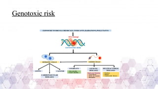
Genotoxicity Studies: Understanding
OECD 471 & 473
Submitted by: Submitted to:
Tushali Khanna Dr. Manmohan Singh
Mpharm Pharmacology ( Pharmacology Department)
1000024972
Genotoxicity
• It is the ability of drug substances/chemicals which can cause genetic
alterations in somatic/ germ cells.
• These effects may occur after a long exposure period.
• Genotoxins are mutagens capable of causing DNA or chromosomal
damage, leading to genetic mutations.
Types of genotoxins
• Topoisomerase inhibitor:- Inhibits the functions of topoisomerase
enzyme responsible for DNA replication and repair, resulting in
double-stranded DNA break formation that can cause mutation and
chromosomal abnormalities. (Topotecan)
• UV radiation:- cause skin cancer
• Protein synthesis inhibitor:- disrupts production of protein required for
DNA repairing.
• Cytotoxic drugs
Genotoxic risk
History
• Gregor Mendel father of Genetics
• 1900 Origin of genetic toxicology
• Muller (1927) observed artificial transmutation of genes in the fruit
fly Drosophila melanogaster
• 1966: Geneticist recommended testing of food additives, drugs and
chemicals for mutagenicity at a conference sponsored by the United
States National Institute of Health
• 1987 OECD test guidelines were published for genetic toxicology
Importance
• Genotoxicity assays have become an integral part of regulatory
requirements.
• Aim to identify compounds that can cause genetic alterations to
somatic/ germ cells.
• Positive compounds in this test, have the potency to be human
carcinogens and mutagens.
Genotoxicity studies
Genotoxicity studies are scientific tests conducted to evaluate whether a
substance can damage genetic material in cells, potentially leading to
mutations, cancer , or hereditary diseases.
Ames assay(TG 471)
• 1970 Dr. Bruce Ames developed this method
• The Ames assay is a bacterial reverse mutation test that evaluates the
mutagenic potential of a chemical by measuring its ability to induce
genetic mutation in Histidine-Deficient Salmonella typhimurium
strains.
• If the chemical causes DNA mutations, bacteria revert to its normal
state and grow on a histidine-deficient medium.
• By point mutation, Salmonella typhi are made histidine-deficient.
Principle
• Exposure of test substance to histidine-deficient Salmonella typhi
bacterial cells.
• Add a metabolic activation system if the test system needs to be
activated.
• Bacterial cells are made to grow on histidine-deficient agar medium.
• If the test substance is mutagenic, it causes reverse mutation to the
histidine coding gene. Resulting in the survival of bacterial cells in a
histidine-deficient medium.
• The number of reverted colonies is counted for the test and control
group.
Description
Preparation
• Bacteria: A fresh culture of bacteria should be grown up to the late exponential or early
stationary phase of growth (approximately 109 cells per ml). Temp of culture- 37 ·C;
salmonella strain (TA1535; TA1537 or TA97a or TA97; TA98; and TA100) to be used.
• Medium: An appropriate minimal agar (e.g. containing Vogel-Bonner minimal medium
E and glucose) and an overlay agar containing histidine and biotin or tryptophan, to
allow for a few cell divisions, is used
• Metabolic activation: Bacteria should be exposed to the test substance both in the
presence and absence of an appropriate metabolic activation system. The most
commonly used system is a cofactor- supplemented post-mitochondrial fraction (S9)
prepared from the livers of rodents treated with enzyme-inducing agents such as Aroclor
1254
• Test substance preparation: solid test sample suspended or dissolved in-
app. Solvent; liquid sample added to test system
Test conditions:-
• The vehicle should not react with the test substance and should be
compatible with the survival of bacteria and S9 activity.
• Exposure concentration: The recommended maximum test concentration
for soluble non-cytotoxic substances is 5 mg/plate or 5 µl/plate. For non-
cytotoxic substances that are not soluble at 5 mg/plate or 5 µl/plate, one or
more concentrations tested should be insoluble in the final treatment mixture.
Test substances that are cytotoxic already below 5 mg/plate or 5 µl/plate
should be tested up to a cytotoxic concentration.
Procedure
Plate incorporation method:
• 0.05 ml test solution + 0.1 ml bacterial culture + 0.5 ml sterile buffer are
mixed with 2 ml of overlay agar. (without metabolic activation)
• 0.5 ml post mitochondrial fraction + 0.05 ml test solution + 0.1 ml
bacterial culture are mixed with 2 ml of overlay agar (small amount of
histidine). (with metabolic activation)
• The mixture is mixed properly and poured into the histidine-deficient agar
plate (minimal), and we wait till agar solidifies before incubation.
• All plates are incubated for 2 to 3 days at 37°C.
• After incubation, the number of reverted colonies per plate is counted.
Plate incorporation method
Pre- incubation method
• 0.5 ml test solution + 0.1 ml bacterial culture + 0.5 ml sterile buffer
are mixed and preincubated for 20 mins at 37° C.
• 0.5 ml post mitochondrial fraction + 0.05 ml test solution + 0.1 ml
bacterial culture are mixed for 20 mins at 37°C.
• After preincubation, the mixture is mixed with overlay agar and
poured into the histidine-deficient agar plates.
• The plates are incubated for 48 hours at 37°C.
• If the test substance is mutagenic, the number of reverted colonies will
increase.
Data and Reporting
• Data should be presented as the number of revertant colonies per plate
and for treated and control group.
• Report:
• Test substance
• Vehicle
• Strains
• Test conditions
• Result:
• Signs of toxicity
• Sign of precipitation
• Individual plate count
• Dose response relationship
• Standard deviation
In vitro mammalian chromosomal aberration
test(TG 473)
• It is a test method used to evaluate the ability of a chemical substance to
induce structural chromosomal aberrations in cultured mammalian cells.
• It helps assess the genotoxic potential of a substance by detecting changes
in chromosome structure, which may lead to mutations and cancer.
• Chromosomal aberration is defined as the permanent changes in the
structure of chromosomes, usually caused by DNA damage. It is of two
types:
• Chromatid type
• Chromosome type (polyploidy)
Principle
• Cell cultures of human (human lymphocytes) or other mammalian origin are
exposed to the test chemical with and without an exogenous source of
metabolic activation (S9 mix) unless cells with an adequate metabolizing
capability are used.
• The cells are exposed for 3-6 hours.
• At appropriate predetermined intervals after the start of exposure of cell
cultures to the test chemical, they are treated with a metaphase-arresting
substance (e.g. Colcemid® or colchicine), harvested, stained, and metaphase
cells are analyzed microscopically for the presence of chromatid-type and
chromosome-type aberrations.
Description
Preparation:
• Cells: human lymphocyte cells, Chinese hamster ovary, Chinese hamster lung
and primary cell culture is used. Human Lymphocyte: 18-35 years and healthy.
• Culture conditions: temperature 37°C; 5% CO2; cell lines to be checked for
mycoplasma contamination.
• Culture: cell lines are obtained from stock culture and suspended in culture
medium
• Lymphocytes: whole blood treated with heparin and lymphocytes obtained are
cultured for 48 hours in the presence of phytohaemagglutinin to induce cell
division before exposure.
• Metabolic activation:S9 mix
• Test substance: solid sample is diluted using suitable solvent whereas
liquid sample is directly introduced to test system.
Procedure
• The cell culture is treated with test substance with or without metabolic
activation system.
• Cells are exposed to test substance for 3-6 hours with or without a metabolic
activation system.
• Sampled at a time equivalent to about 1.5 normal cell cycle length after the
beginning of treatment.
• Chromosome preparation: cell cultures are treated with Colcemid® for 1-3
hours before harvesting. Each cell cultured is harvested and processed
separately {hypotonic treatment of the cells KCl (swell), fixation (methanol
acetic acid) and staining (G-banding)} for preparation of chromosome.
• Analysis: Cells are analysed under a microscope for chromosomal
aberration. Structural abnormalities like breaks, deletions, exchanges,
gaps, rings and fragments are identified.
• A minimum of 200 metaphases per concentration is usually analysed.
• The percentage of aberrant cells is compared to negative and positive
control to determine genotoxicity.
Types of chromosomal abberations
• Chromosomal aberrations are structural or numerical chromosome changes,
leading to genetic disorders or cellular abnormalities.
Types:
Structural aberrations: These involve changes in the structure of chromosomes
due to breaks and incorrect rejoining.
1. Duplication: A chromosome segment is duplicated, leading to extra genetic
material. Example: Charcot-Marie-Tooth disease (chromosome 17 duplication)
2. Inversion: it appears when there are two breaks in a chromosome and the
detached segment becomes reinserted in the reverse order. It is of two types:
• Paracentric: both breaks in one arm of chromosome.
• Pericentric: breakpoint on either side of centromere.
3. Deletion: loss of a small portion of chromosome. It occurs from a
breakage at random in both chromatids of a chromosome or only in
one chromatid.
4. Translocation: sometimes a segment of a chromosome get detached
and unites with another non-homologous chromosome.
Numerical aberration: These involve changes in chromosome
numbers and occur due to nondisjunction during cell division.
• Aneuploidy: gain or loss of chromosomes.
• Polyploidy: A complete extra set of chromosomes.
Genotoxicity studies : Understanding OECD 471 &473

Genotoxicity studies : Understanding OECD 471 &473

  • 1.
    Genotoxicity Studies: Understanding OECD471 & 473 Submitted by: Submitted to: Tushali Khanna Dr. Manmohan Singh Mpharm Pharmacology ( Pharmacology Department) 1000024972
  • 2.
    Genotoxicity • It isthe ability of drug substances/chemicals which can cause genetic alterations in somatic/ germ cells. • These effects may occur after a long exposure period. • Genotoxins are mutagens capable of causing DNA or chromosomal damage, leading to genetic mutations.
  • 3.
    Types of genotoxins •Topoisomerase inhibitor:- Inhibits the functions of topoisomerase enzyme responsible for DNA replication and repair, resulting in double-stranded DNA break formation that can cause mutation and chromosomal abnormalities. (Topotecan) • UV radiation:- cause skin cancer • Protein synthesis inhibitor:- disrupts production of protein required for DNA repairing. • Cytotoxic drugs
  • 4.
  • 5.
    History • Gregor Mendelfather of Genetics • 1900 Origin of genetic toxicology • Muller (1927) observed artificial transmutation of genes in the fruit fly Drosophila melanogaster • 1966: Geneticist recommended testing of food additives, drugs and chemicals for mutagenicity at a conference sponsored by the United States National Institute of Health • 1987 OECD test guidelines were published for genetic toxicology
  • 6.
    Importance • Genotoxicity assayshave become an integral part of regulatory requirements. • Aim to identify compounds that can cause genetic alterations to somatic/ germ cells. • Positive compounds in this test, have the potency to be human carcinogens and mutagens.
  • 7.
    Genotoxicity studies Genotoxicity studiesare scientific tests conducted to evaluate whether a substance can damage genetic material in cells, potentially leading to mutations, cancer , or hereditary diseases.
  • 8.
    Ames assay(TG 471) •1970 Dr. Bruce Ames developed this method • The Ames assay is a bacterial reverse mutation test that evaluates the mutagenic potential of a chemical by measuring its ability to induce genetic mutation in Histidine-Deficient Salmonella typhimurium strains. • If the chemical causes DNA mutations, bacteria revert to its normal state and grow on a histidine-deficient medium. • By point mutation, Salmonella typhi are made histidine-deficient.
  • 9.
    Principle • Exposure oftest substance to histidine-deficient Salmonella typhi bacterial cells. • Add a metabolic activation system if the test system needs to be activated. • Bacterial cells are made to grow on histidine-deficient agar medium. • If the test substance is mutagenic, it causes reverse mutation to the histidine coding gene. Resulting in the survival of bacterial cells in a histidine-deficient medium. • The number of reverted colonies is counted for the test and control group.
  • 10.
    Description Preparation • Bacteria: Afresh culture of bacteria should be grown up to the late exponential or early stationary phase of growth (approximately 109 cells per ml). Temp of culture- 37 ·C; salmonella strain (TA1535; TA1537 or TA97a or TA97; TA98; and TA100) to be used. • Medium: An appropriate minimal agar (e.g. containing Vogel-Bonner minimal medium E and glucose) and an overlay agar containing histidine and biotin or tryptophan, to allow for a few cell divisions, is used • Metabolic activation: Bacteria should be exposed to the test substance both in the presence and absence of an appropriate metabolic activation system. The most commonly used system is a cofactor- supplemented post-mitochondrial fraction (S9) prepared from the livers of rodents treated with enzyme-inducing agents such as Aroclor 1254
  • 11.
    • Test substancepreparation: solid test sample suspended or dissolved in- app. Solvent; liquid sample added to test system Test conditions:- • The vehicle should not react with the test substance and should be compatible with the survival of bacteria and S9 activity. • Exposure concentration: The recommended maximum test concentration for soluble non-cytotoxic substances is 5 mg/plate or 5 µl/plate. For non- cytotoxic substances that are not soluble at 5 mg/plate or 5 µl/plate, one or more concentrations tested should be insoluble in the final treatment mixture. Test substances that are cytotoxic already below 5 mg/plate or 5 µl/plate should be tested up to a cytotoxic concentration.
  • 12.
    Procedure Plate incorporation method: •0.05 ml test solution + 0.1 ml bacterial culture + 0.5 ml sterile buffer are mixed with 2 ml of overlay agar. (without metabolic activation) • 0.5 ml post mitochondrial fraction + 0.05 ml test solution + 0.1 ml bacterial culture are mixed with 2 ml of overlay agar (small amount of histidine). (with metabolic activation) • The mixture is mixed properly and poured into the histidine-deficient agar plate (minimal), and we wait till agar solidifies before incubation. • All plates are incubated for 2 to 3 days at 37°C. • After incubation, the number of reverted colonies per plate is counted.
  • 13.
  • 14.
    Pre- incubation method •0.5 ml test solution + 0.1 ml bacterial culture + 0.5 ml sterile buffer are mixed and preincubated for 20 mins at 37° C. • 0.5 ml post mitochondrial fraction + 0.05 ml test solution + 0.1 ml bacterial culture are mixed for 20 mins at 37°C. • After preincubation, the mixture is mixed with overlay agar and poured into the histidine-deficient agar plates. • The plates are incubated for 48 hours at 37°C. • If the test substance is mutagenic, the number of reverted colonies will increase.
  • 16.
    Data and Reporting •Data should be presented as the number of revertant colonies per plate and for treated and control group. • Report: • Test substance • Vehicle • Strains • Test conditions
  • 17.
    • Result: • Signsof toxicity • Sign of precipitation • Individual plate count • Dose response relationship • Standard deviation
  • 18.
    In vitro mammalianchromosomal aberration test(TG 473) • It is a test method used to evaluate the ability of a chemical substance to induce structural chromosomal aberrations in cultured mammalian cells. • It helps assess the genotoxic potential of a substance by detecting changes in chromosome structure, which may lead to mutations and cancer. • Chromosomal aberration is defined as the permanent changes in the structure of chromosomes, usually caused by DNA damage. It is of two types: • Chromatid type • Chromosome type (polyploidy)
  • 19.
    Principle • Cell culturesof human (human lymphocytes) or other mammalian origin are exposed to the test chemical with and without an exogenous source of metabolic activation (S9 mix) unless cells with an adequate metabolizing capability are used. • The cells are exposed for 3-6 hours. • At appropriate predetermined intervals after the start of exposure of cell cultures to the test chemical, they are treated with a metaphase-arresting substance (e.g. Colcemid® or colchicine), harvested, stained, and metaphase cells are analyzed microscopically for the presence of chromatid-type and chromosome-type aberrations.
  • 20.
    Description Preparation: • Cells: humanlymphocyte cells, Chinese hamster ovary, Chinese hamster lung and primary cell culture is used. Human Lymphocyte: 18-35 years and healthy. • Culture conditions: temperature 37°C; 5% CO2; cell lines to be checked for mycoplasma contamination. • Culture: cell lines are obtained from stock culture and suspended in culture medium • Lymphocytes: whole blood treated with heparin and lymphocytes obtained are cultured for 48 hours in the presence of phytohaemagglutinin to induce cell division before exposure.
  • 21.
    • Metabolic activation:S9mix • Test substance: solid sample is diluted using suitable solvent whereas liquid sample is directly introduced to test system.
  • 22.
    Procedure • The cellculture is treated with test substance with or without metabolic activation system. • Cells are exposed to test substance for 3-6 hours with or without a metabolic activation system. • Sampled at a time equivalent to about 1.5 normal cell cycle length after the beginning of treatment. • Chromosome preparation: cell cultures are treated with Colcemid® for 1-3 hours before harvesting. Each cell cultured is harvested and processed separately {hypotonic treatment of the cells KCl (swell), fixation (methanol acetic acid) and staining (G-banding)} for preparation of chromosome.
  • 23.
    • Analysis: Cellsare analysed under a microscope for chromosomal aberration. Structural abnormalities like breaks, deletions, exchanges, gaps, rings and fragments are identified. • A minimum of 200 metaphases per concentration is usually analysed. • The percentage of aberrant cells is compared to negative and positive control to determine genotoxicity.
  • 25.
    Types of chromosomalabberations • Chromosomal aberrations are structural or numerical chromosome changes, leading to genetic disorders or cellular abnormalities. Types: Structural aberrations: These involve changes in the structure of chromosomes due to breaks and incorrect rejoining. 1. Duplication: A chromosome segment is duplicated, leading to extra genetic material. Example: Charcot-Marie-Tooth disease (chromosome 17 duplication) 2. Inversion: it appears when there are two breaks in a chromosome and the detached segment becomes reinserted in the reverse order. It is of two types: • Paracentric: both breaks in one arm of chromosome. • Pericentric: breakpoint on either side of centromere.
  • 26.
    3. Deletion: lossof a small portion of chromosome. It occurs from a breakage at random in both chromatids of a chromosome or only in one chromatid. 4. Translocation: sometimes a segment of a chromosome get detached and unites with another non-homologous chromosome.
  • 27.
    Numerical aberration: Theseinvolve changes in chromosome numbers and occur due to nondisjunction during cell division. • Aneuploidy: gain or loss of chromosomes. • Polyploidy: A complete extra set of chromosomes.