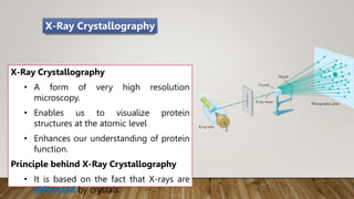
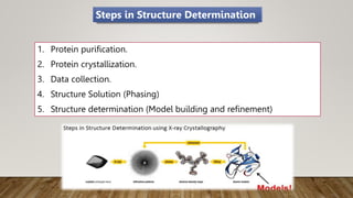
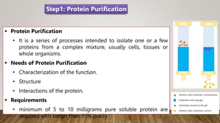
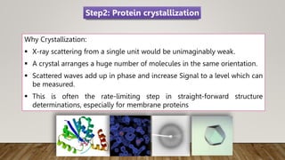
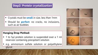
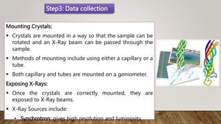
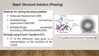
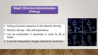
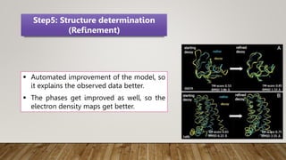
Application X-Ray crystallography
in protein structure prediction
SIRAJUDDIN MOLLA
Research scholar
DEPARTMENT OF PHARMACOLOGY
SPER, JAMIA HAMDARD
 Various functions of biological system depend upon the structure and
function of proteins.
 Determination of structure and functions of proteins assist in
scrutinizing the dynamics of proteins.
 To understand the functions of proteins at a molecular level, it is often
necessary to determine their three-dimensional structure.
Introduction
It helps us in Understanding:
i. How proteins interact with other molecules.
ii. How they perform catalysis in the case of enzymes.
iii. Interaction of protein with other molecules including protein
itself.
iv. Miscoding and/or misfolding of proteins associated with
diseases.
Why Structure Determination
X-Ray Crystallography
• A form of very high resolution
microscopy.
• Enables us to visualize protein
structures at the atomic level
• Enhances our understanding of protein
function.
Principle behind X-Ray Crystallography
• It is based on the fact that X-rays are
diffracted by crystals.
X-Ray Crystallography
Why X-Rays? Not Others?
No. Microscopy Wavelength Visualization
1 Light 300 nm Individual cells and sub-
cellular organelles
2 Electron 10 nm Cellular architecture
Shapes of large protein
molecules
3 X-Rays 0.1nm or 1Å Atomic detail of protein
 X-rays is in the order of atom diameter and bond
lengths, allowing these to be individually resolved.
 No lenses available to focus X-rays.
 Crystal acts as a magnifier of the scattering of X-
rays.
Why use X-rays and crystals?
1. Protein purification.
2. Protein crystallization.
3. Data collection.
4. Structure Solution (Phasing)
5. Structure determination (Model building and refinement)
Steps in Structure Determination
 Protein Purification
• It is a series of processes intended to isolate one or a few
proteins from a complex mixture, usually cells, tissues or
whole organisms.
 Needs of Protein Purification
• Characterization of the function.
• Structure
• Interactions of the protein.
 Requirements
• minimum of 5 to 10 milligrams pure soluble protein are
required with better than 95% purity
Step1: Protein Purification
Why Crystallization:
 X-ray scattering from a single unit would be unimaginably weak.
 A crystal arranges a huge number of molecules in the same orientation.
 Scattered waves add up in phase and increase Signal to a level which can
be measured.
 This is often the rate-limiting step in straight-forward structure
determinations, especially for membrane proteins
Step2: Protein crystallization
 Crystals must be small in size, less than 1mm
 Should be perfect: no cracks, no inclusions,
such as air bubbles
Hanging Drop Method:
 1 to 5µl protein solution is suspended over a 1 ml
reservoir containing precipitant solution
 e.g. ammonium sulfate solution or polyethylene
glycol
Step2: Protein crystallization
Mounting Crystals:
 Crystals are mounted in a way so that the sample can be
rotated and an X-Ray beam can be passed through the
sample.
 Methods of mounting include using either a capillary or a
tube.
 Both capillary and tubes are mounted on a goniometer.
Exposing X-Rays:
 Once the crystals are correctly mounted, they are
exposed to X-Ray beams.
 X-Ray Sources include:
• Synchrotron: gives high resolution and luminosity
Step3: Data collection
 The source of the X-rays is often a synchrotron.
 The typical size for a crystal for data collection may be 0.3 x 0.3 x 0.1
mm.
 The crystals are bombarded with X-rays which are scattered from the
planes of the crystal lattice.
 The scattered X-rays are captured as a diffraction pattern on a detector
such as film or an electronic device.
contd…
 Rotate crystal through 1 degree and Record XRD pattern
 If XRD pattern is very crowded, reduce the degree of rotation
 Repeat until 30 degrees were obtained
 Sometimes 180 degrees depending on crystal symmetry
 Lower the symmetry  More data are required
 For high resolution, use Synchrotron
contd…
Step4: Structure Solution (Phasing)
Diffraction
Phasing
Purification
&
Crystallization
Phasing
Phase problems
 In the measurement of data from an X-ray crystallographic experiment
only the amplitude of the wave is determined.
 To compute a structure, the phase must also be known.
 Since it cannot be determined directly, it must be determined
indirectly or by some other experiment.
Methods for solving the phase problem
 Molecular Replacement (MR)
 Multiple/Single isomorphous
replacement (MIR/SIR)
 Multiple/Single wavelength
Anomalous Diffraction(MAD/SAD)
Principle using Fourier Transform (FT):
 FT of the diffraction data gives us a
representation of the contents of the
crystal.
A crystal and its Fourier Transform
Step4: Structure Solution (Phasing)
 Fitting of protein sequence in the electron density.
 Electron density - Not self explanatory
 Can be automated, if resolution is close to 2Å or
better.
 It can be interpreted is largely defined by resolution.
Step5: Structure determination
(Fitting)
 Automated improvement of the model, so
it explains the observed data better.
 The phases get improved as well, so the
electron density maps get better.
Step5: Structure determination
(Refinement)
Advantages:
 Very routine and high potential for automation.
 Fast structure determination (From data to structure under 6 hours)
Disadvantages: -
 Crystal growth is a major unsolved problem.
 Crystals maybe obtained at irrelevant conditions.
 Crystal packing forces may influence structure.
 Aqueous proteins exist in a conformational ensemble. It can be argued
that X-ray may filter for the conformation that crystallizes.
 Glycosylation will normally render the protein hard to crystallize. Multi-
unit proteins are hard to crystallize.
X-ray Crystallography Summary
 https://www.ncbi.nlm.nih.gov/pmc/articles/PMC1186895/
 https://slideplayer.com/slide/3866535/
 https://www.slideshare.net/AmjadIbrahim/protein-structure-
determination
REFERENCES
Application X-Ray crystallography in protein structure prediction.pptx

Application X-Ray crystallography in protein structure prediction.pptx

  • 1.
    Application X-Ray crystallography inprotein structure prediction SIRAJUDDIN MOLLA Research scholar DEPARTMENT OF PHARMACOLOGY SPER, JAMIA HAMDARD
  • 2.
     Various functionsof biological system depend upon the structure and function of proteins.  Determination of structure and functions of proteins assist in scrutinizing the dynamics of proteins.  To understand the functions of proteins at a molecular level, it is often necessary to determine their three-dimensional structure. Introduction
  • 3.
    It helps usin Understanding: i. How proteins interact with other molecules. ii. How they perform catalysis in the case of enzymes. iii. Interaction of protein with other molecules including protein itself. iv. Miscoding and/or misfolding of proteins associated with diseases. Why Structure Determination
  • 4.
    X-Ray Crystallography • Aform of very high resolution microscopy. • Enables us to visualize protein structures at the atomic level • Enhances our understanding of protein function. Principle behind X-Ray Crystallography • It is based on the fact that X-rays are diffracted by crystals. X-Ray Crystallography
  • 5.
    Why X-Rays? NotOthers? No. Microscopy Wavelength Visualization 1 Light 300 nm Individual cells and sub- cellular organelles 2 Electron 10 nm Cellular architecture Shapes of large protein molecules 3 X-Rays 0.1nm or 1Å Atomic detail of protein
  • 6.
     X-rays isin the order of atom diameter and bond lengths, allowing these to be individually resolved.  No lenses available to focus X-rays.  Crystal acts as a magnifier of the scattering of X- rays. Why use X-rays and crystals?
  • 7.
    1. Protein purification. 2.Protein crystallization. 3. Data collection. 4. Structure Solution (Phasing) 5. Structure determination (Model building and refinement) Steps in Structure Determination
  • 8.
     Protein Purification •It is a series of processes intended to isolate one or a few proteins from a complex mixture, usually cells, tissues or whole organisms.  Needs of Protein Purification • Characterization of the function. • Structure • Interactions of the protein.  Requirements • minimum of 5 to 10 milligrams pure soluble protein are required with better than 95% purity Step1: Protein Purification
  • 9.
    Why Crystallization:  X-rayscattering from a single unit would be unimaginably weak.  A crystal arranges a huge number of molecules in the same orientation.  Scattered waves add up in phase and increase Signal to a level which can be measured.  This is often the rate-limiting step in straight-forward structure determinations, especially for membrane proteins Step2: Protein crystallization
  • 10.
     Crystals mustbe small in size, less than 1mm  Should be perfect: no cracks, no inclusions, such as air bubbles Hanging Drop Method:  1 to 5µl protein solution is suspended over a 1 ml reservoir containing precipitant solution  e.g. ammonium sulfate solution or polyethylene glycol Step2: Protein crystallization
  • 11.
    Mounting Crystals:  Crystalsare mounted in a way so that the sample can be rotated and an X-Ray beam can be passed through the sample.  Methods of mounting include using either a capillary or a tube.  Both capillary and tubes are mounted on a goniometer. Exposing X-Rays:  Once the crystals are correctly mounted, they are exposed to X-Ray beams.  X-Ray Sources include: • Synchrotron: gives high resolution and luminosity Step3: Data collection
  • 12.
     The sourceof the X-rays is often a synchrotron.  The typical size for a crystal for data collection may be 0.3 x 0.3 x 0.1 mm.  The crystals are bombarded with X-rays which are scattered from the planes of the crystal lattice.  The scattered X-rays are captured as a diffraction pattern on a detector such as film or an electronic device. contd…
  • 13.
     Rotate crystalthrough 1 degree and Record XRD pattern  If XRD pattern is very crowded, reduce the degree of rotation  Repeat until 30 degrees were obtained  Sometimes 180 degrees depending on crystal symmetry  Lower the symmetry  More data are required  For high resolution, use Synchrotron contd…
  • 14.
  • 15.
    Diffraction Phasing Purification & Crystallization Phasing Phase problems  Inthe measurement of data from an X-ray crystallographic experiment only the amplitude of the wave is determined.  To compute a structure, the phase must also be known.  Since it cannot be determined directly, it must be determined indirectly or by some other experiment.
  • 16.
    Methods for solvingthe phase problem  Molecular Replacement (MR)  Multiple/Single isomorphous replacement (MIR/SIR)  Multiple/Single wavelength Anomalous Diffraction(MAD/SAD) Principle using Fourier Transform (FT):  FT of the diffraction data gives us a representation of the contents of the crystal. A crystal and its Fourier Transform Step4: Structure Solution (Phasing)
  • 17.
     Fitting ofprotein sequence in the electron density.  Electron density - Not self explanatory  Can be automated, if resolution is close to 2Å or better.  It can be interpreted is largely defined by resolution. Step5: Structure determination (Fitting)
  • 18.
     Automated improvementof the model, so it explains the observed data better.  The phases get improved as well, so the electron density maps get better. Step5: Structure determination (Refinement)
  • 19.
    Advantages:  Very routineand high potential for automation.  Fast structure determination (From data to structure under 6 hours) Disadvantages: -  Crystal growth is a major unsolved problem.  Crystals maybe obtained at irrelevant conditions.  Crystal packing forces may influence structure.  Aqueous proteins exist in a conformational ensemble. It can be argued that X-ray may filter for the conformation that crystallizes.  Glycosylation will normally render the protein hard to crystallize. Multi- unit proteins are hard to crystallize. X-ray Crystallography Summary
  • 20.
     https://www.ncbi.nlm.nih.gov/pmc/articles/PMC1186895/  https://slideplayer.com/slide/3866535/ https://www.slideshare.net/AmjadIbrahim/protein-structure- determination REFERENCES