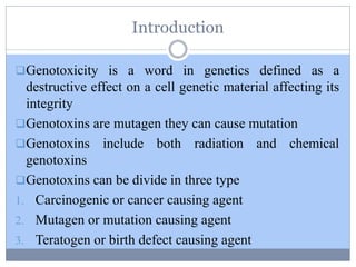
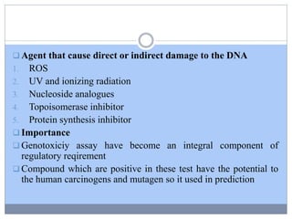
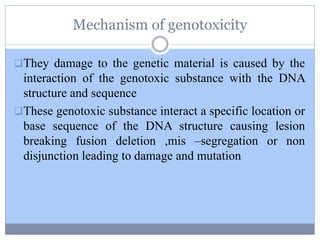
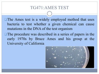
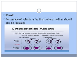
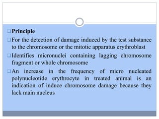
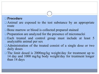
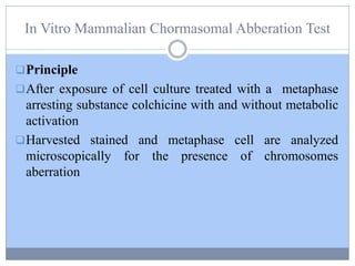
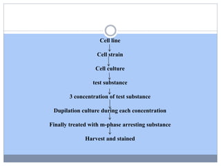
SUBMITTED BY-
SHRUTI RAJEEV GAUTAM
(M.PHARM 1 𝑠 𝑡 YEAR )
GUIDE-
DR. VIVEK PAITHANKAR
VIDYA BHARATI COLLEGE OF PHARMACY,
AMRAVATI.
2019-2020
Seminar on
Genotoxicity Studies
Index
Introduction
Mechanism of genotoxicity
Ames Test
In Vitro & In Vivo Mammalian Cell Micronucleus Test
Chromosomal Aberration Test
Reference
Introduction
Genotoxicity is a word in genetics defined as a
destructive effect on a cell genetic material affecting its
integrity
Genotoxins are mutagen they can cause mutation
Genotoxins include both radiation and chemical
genotoxins
Genotoxins can be divide in three type
1. Carcinogenic or cancer causing agent
2. Mutagen or mutation causing agent
3. Teratogen or birth defect causing agent
Genotoxic Risk
Somatic Cell Germ Cell
Cvs Cancer Multifactorial Disease
Genetic Defect
Diabetes Psychoses Cvs
Cystic Sickle Hameomphilia
Fibrosis Cell
Anaemia
 Agent that cause direct or indirect damage to the DNA
1. ROS
2. UV and ionizing radiation
3. Nucleoside analogues
4. Topoisomerase inhibitor
5. Protein synthesis inhibitor
 Importance
 Genotoxiciy assay have become an integral component of
regulatory reqirement
 Compound which are positive in these test have the potential to
the human carcinogens and mutagen so it used in prediction
Mechanism of genotoxicity
They damage to the genetic material is caused by the
interaction of the genotoxic substance with the DNA
structure and sequence
These genotoxic substance interact a specific location or
base sequence of the DNA structure causing lesion
breaking fusion deletion ,mis –segregation or non
disjunction leading to damage and mutation
TG471:AMES TEST
The Ames test is a widely employed method that uses
bacteria to test whether a given chemical can cause
mutations in the DNA of the test organism
The procedure was described in a series of papers in the
early 1970s by Bruce Ames and his group at the
University of California
Principle
Ames test uses several strains of bacteria (Salmonella,
E.coli) that carry a particular mutation.
Point mutations are made in the histidine (Salmonella
typhimurium) or the tryptophan (Escherichia coli)
operon, rendering the bacteria incapable of producing
the corresponding amino acid.
These mutations result in his- or trp- organisms that
cannot grow unless histidine or tryptophan is supplied.
Procedure
 Isolate an auxotrophic strain of Salmonella Typhimurium
for histidine. (ie. His-ve)
Prepare a test suspension of his-ve Salmonella Typhimurium
in a plain buffer with test chemical (eg. 2-aminofluorene).
Also add a small amount of histidine.
Also prepare a control suspension of His-ve Salmonella
Typhimurium but without test chemicals.
 Incubate the suspensions at 37°C for 20 minutes
 Prepare the two agar plate and spread the suspension on
agar plate.
 Incubate the plates at 37°C for 48 hours.
After48 hours count the number of colonies in each plate.
Result interpretation
 The mutagenicity of chemicals is proportional to number of
colonies observed.
 If there is a large number of colonies on the test plate in
comparison to control, then such chemical are said to be
mutagens.
 Uses
 While ames test is used to identify the revert mutations which are
present in strains, it can also be used to detect the mutagenicity of
environmental samples such as drugs, dyes, reagents, cosmetics,
waste water, pesticides and other substances which are easily
solubilized in a liquid suspension
In Vitro Mammalian Cell Micronucleus Test
The in vitro micronucleus (mnvit) test is a genotoxicity
test for the detection of micronuclei (MN) in the
cytoplasm of interphase cells. Micronuclei may originate
from acentric chromosome fragments (i.e.Lacking a
centromere), or whole chromosomes that are unable to migrate
to the poles during the anaphase stage of cell division.
 Therefore the mnvit test is an in vitromethod that
provides a comprehensive basis for investigating chromosome
damaging potential in vitro because both aneugens and
clastogens can be detected in cells that have undergone cell
division during or after exposure to the test chemical
Procedure
Detection of the frequency of micronucleus
Cell culture of human or other mammalian origin are
exposed to the test chemical formation of micronucleus in
interphase cells
Harvested and stained interphase cell are analyzed for the
presence of micronuclei treated with a cytokinesis
Assay detect the activity of clastogenic and aneugenic
chemical
Result
Percentage of vehicle in the final culture medium should
also be indicated
MAMMALIAN ERYTHROCYTE MICRONUCLEUS
TEST
Animal are exposed to the test substance by an
appropriate route
If bone marrow > the animal are scarified bone marrow
extracted and preparation made and stained
If peripheral blood > the blood is collected at appropriate
time after treatment and smear preparation are made and
stained
Preparation are analyzed for the presence of micronuclei
Principle
For the detection of damage induced by the test substance
to the chromosome or the mitotic apparatus erythroblast
Identifies micronuclei containing lagging chromosome
fragment or whole chromosome
An increase in the frequency of micro nucleated
polynucleotide erythrocyte in treated animal is an
indication of induce chromosome damage because they
lack main nucleus
Procedure
Animal are exposed to the test substance by an appropriate
route
Bone marrow or blood is collected prepared stained
Preparation are analyzed for the presence of micronuclei
Each treated and control group must include at least 5
analyzable animal per sex
Administration of the treated consist of a single dose or two
daily doses
The limit dosed is 2000mg/kg weight/day for treatment up to
14 day and 1000 mg/kg body weight/day for treatment longer
than 14 days
In Vitro Mammalian Chormasomal Abberation Test
Principle
After exposure of cell culture treated with a metaphase
arresting substance colchicine with and without metabolic
activation
Harvested stained and metaphase cell are analyzed
microscopically for the presence of chromosomes
aberration
Cell line
Cell strain
Cell culture
test substance
3 concentration of test substance
Dupilation culture during each concentration
Finally treated with m-phase arresting substance
Harvest and stained
Result
% Of cell with structural chromosome aberration
In Vitro Mammalian Bone Marrow Chromosomal
Aberration Test
Principle
For detection of structural chromosome aberration
induced test compound only in bone marrow cell of
animal
Animal exposed to the test substance metaphase arresting
agent sacrificed at appropriate times after treatment
Procedure
Animal are exposed to the test substance by an
appropriate route
Then animal are treated with a metaphase arresting
agent
Chromosome preparation are then made from the bone
marrow cell and stained
Each treated and control group must include at least 5
analyzable animal per sex
The limit dosed is 2000mg/kg weight/day for treatment
up to 14 day and 1000 mg/kg body weight/day for
treatment longer than 14 days
Prior to sacrifice animal are injected i.p with an
appropriate dose of a metaphase arresting agent ,sampled
Cell are harvested from bone marrow and analyzed from
chromosome aberration
Chromosome preparation : bone marrow in hypotonic
solution ,spread on slides and stained
Result
The mitotic index should be determined as a measure of
cytotoxicity in at least 1000 cell per animal
REFERNCE
 Genotoxicity Assessment: Methods and Protocols by
Mahima Bajpayee, Alok Dhawan
 IOSR journal of pharmacy and biological sciences
volume 1 issue 2
 https://microbiologyinfo.com/ames-test/
 https://ntp.niehs.nih.gov/iccvam/suppdocs/feddocs
/oecd/oecd-tg487-2014-508.pdf

Genotoxicity studies

  • 1.
    SUBMITTED BY- SHRUTI RAJEEVGAUTAM (M.PHARM 1 𝑠 𝑡 YEAR ) GUIDE- DR. VIVEK PAITHANKAR VIDYA BHARATI COLLEGE OF PHARMACY, AMRAVATI. 2019-2020 Seminar on Genotoxicity Studies
  • 2.
    Index Introduction Mechanism of genotoxicity AmesTest In Vitro & In Vivo Mammalian Cell Micronucleus Test Chromosomal Aberration Test Reference
  • 3.
    Introduction Genotoxicity is aword in genetics defined as a destructive effect on a cell genetic material affecting its integrity Genotoxins are mutagen they can cause mutation Genotoxins include both radiation and chemical genotoxins Genotoxins can be divide in three type 1. Carcinogenic or cancer causing agent 2. Mutagen or mutation causing agent 3. Teratogen or birth defect causing agent
  • 4.
    Genotoxic Risk Somatic CellGerm Cell Cvs Cancer Multifactorial Disease Genetic Defect Diabetes Psychoses Cvs Cystic Sickle Hameomphilia Fibrosis Cell Anaemia
  • 5.
     Agent thatcause direct or indirect damage to the DNA 1. ROS 2. UV and ionizing radiation 3. Nucleoside analogues 4. Topoisomerase inhibitor 5. Protein synthesis inhibitor  Importance  Genotoxiciy assay have become an integral component of regulatory reqirement  Compound which are positive in these test have the potential to the human carcinogens and mutagen so it used in prediction
  • 7.
    Mechanism of genotoxicity Theydamage to the genetic material is caused by the interaction of the genotoxic substance with the DNA structure and sequence These genotoxic substance interact a specific location or base sequence of the DNA structure causing lesion breaking fusion deletion ,mis –segregation or non disjunction leading to damage and mutation
  • 8.
    TG471:AMES TEST The Amestest is a widely employed method that uses bacteria to test whether a given chemical can cause mutations in the DNA of the test organism The procedure was described in a series of papers in the early 1970s by Bruce Ames and his group at the University of California
  • 9.
    Principle Ames test usesseveral strains of bacteria (Salmonella, E.coli) that carry a particular mutation. Point mutations are made in the histidine (Salmonella typhimurium) or the tryptophan (Escherichia coli) operon, rendering the bacteria incapable of producing the corresponding amino acid. These mutations result in his- or trp- organisms that cannot grow unless histidine or tryptophan is supplied.
  • 10.
    Procedure  Isolate anauxotrophic strain of Salmonella Typhimurium for histidine. (ie. His-ve) Prepare a test suspension of his-ve Salmonella Typhimurium in a plain buffer with test chemical (eg. 2-aminofluorene). Also add a small amount of histidine. Also prepare a control suspension of His-ve Salmonella Typhimurium but without test chemicals.  Incubate the suspensions at 37°C for 20 minutes  Prepare the two agar plate and spread the suspension on agar plate.  Incubate the plates at 37°C for 48 hours. After48 hours count the number of colonies in each plate.
  • 12.
    Result interpretation  Themutagenicity of chemicals is proportional to number of colonies observed.  If there is a large number of colonies on the test plate in comparison to control, then such chemical are said to be mutagens.  Uses  While ames test is used to identify the revert mutations which are present in strains, it can also be used to detect the mutagenicity of environmental samples such as drugs, dyes, reagents, cosmetics, waste water, pesticides and other substances which are easily solubilized in a liquid suspension
  • 13.
    In Vitro MammalianCell Micronucleus Test The in vitro micronucleus (mnvit) test is a genotoxicity test for the detection of micronuclei (MN) in the cytoplasm of interphase cells. Micronuclei may originate from acentric chromosome fragments (i.e.Lacking a centromere), or whole chromosomes that are unable to migrate to the poles during the anaphase stage of cell division.  Therefore the mnvit test is an in vitromethod that provides a comprehensive basis for investigating chromosome damaging potential in vitro because both aneugens and clastogens can be detected in cells that have undergone cell division during or after exposure to the test chemical
  • 14.
    Procedure Detection of thefrequency of micronucleus Cell culture of human or other mammalian origin are exposed to the test chemical formation of micronucleus in interphase cells Harvested and stained interphase cell are analyzed for the presence of micronuclei treated with a cytokinesis Assay detect the activity of clastogenic and aneugenic chemical
  • 15.
    Result Percentage of vehiclein the final culture medium should also be indicated
  • 16.
    MAMMALIAN ERYTHROCYTE MICRONUCLEUS TEST Animalare exposed to the test substance by an appropriate route If bone marrow > the animal are scarified bone marrow extracted and preparation made and stained If peripheral blood > the blood is collected at appropriate time after treatment and smear preparation are made and stained Preparation are analyzed for the presence of micronuclei
  • 17.
    Principle For the detectionof damage induced by the test substance to the chromosome or the mitotic apparatus erythroblast Identifies micronuclei containing lagging chromosome fragment or whole chromosome An increase in the frequency of micro nucleated polynucleotide erythrocyte in treated animal is an indication of induce chromosome damage because they lack main nucleus
  • 18.
    Procedure Animal are exposedto the test substance by an appropriate route Bone marrow or blood is collected prepared stained Preparation are analyzed for the presence of micronuclei Each treated and control group must include at least 5 analyzable animal per sex Administration of the treated consist of a single dose or two daily doses The limit dosed is 2000mg/kg weight/day for treatment up to 14 day and 1000 mg/kg body weight/day for treatment longer than 14 days
  • 19.
    In Vitro MammalianChormasomal Abberation Test Principle After exposure of cell culture treated with a metaphase arresting substance colchicine with and without metabolic activation Harvested stained and metaphase cell are analyzed microscopically for the presence of chromosomes aberration
  • 20.
    Cell line Cell strain Cellculture test substance 3 concentration of test substance Dupilation culture during each concentration Finally treated with m-phase arresting substance Harvest and stained
  • 21.
    Result % Of cellwith structural chromosome aberration
  • 22.
    In Vitro MammalianBone Marrow Chromosomal Aberration Test Principle For detection of structural chromosome aberration induced test compound only in bone marrow cell of animal Animal exposed to the test substance metaphase arresting agent sacrificed at appropriate times after treatment
  • 23.
    Procedure Animal are exposedto the test substance by an appropriate route Then animal are treated with a metaphase arresting agent Chromosome preparation are then made from the bone marrow cell and stained Each treated and control group must include at least 5 analyzable animal per sex The limit dosed is 2000mg/kg weight/day for treatment up to 14 day and 1000 mg/kg body weight/day for treatment longer than 14 days
  • 24.
    Prior to sacrificeanimal are injected i.p with an appropriate dose of a metaphase arresting agent ,sampled Cell are harvested from bone marrow and analyzed from chromosome aberration Chromosome preparation : bone marrow in hypotonic solution ,spread on slides and stained Result The mitotic index should be determined as a measure of cytotoxicity in at least 1000 cell per animal
  • 25.
    REFERNCE  Genotoxicity Assessment:Methods and Protocols by Mahima Bajpayee, Alok Dhawan  IOSR journal of pharmacy and biological sciences volume 1 issue 2  https://microbiologyinfo.com/ames-test/  https://ntp.niehs.nih.gov/iccvam/suppdocs/feddocs /oecd/oecd-tg487-2014-508.pdf