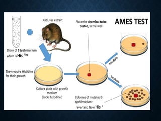
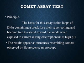
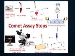
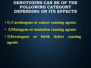
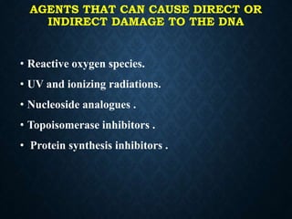
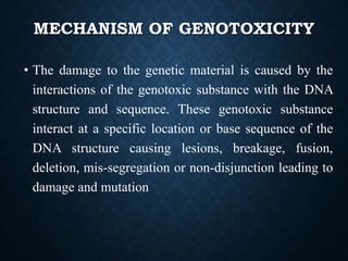
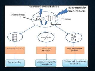
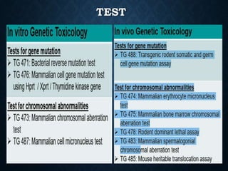
GENOTOXICITY
SUB.by- narsingh kashyap
m.f.Sc. , FishGenetic s and breeding
IFPGS, OMR Campus, CHENNAI
HISTORY & BACKGROUND
• Origin of genetic toxicology in 1900 , Genetic
toxicity independent branch of science started in
1927.
• OECD Genetic Toxicology TGs was first
published in 1987.
• : OECD ,ICH, SCHEDULE Y (D&C India).
MAJOR TEST GUIDELINES
• OECD (The Organization for Economic Cooperation
and Development) -1961
• ICH ( International Council for Harmonisation ) – 1990
• SCHEDULE Y of Drug and Cosmetic act -1940
INTRODUCTION
• Genotoxicity is a word used in genetics that describes the
possession of substance that has destructive effect on the
genetic material of the cell (DNA,RNA) , thus affecting
the integrity of the cell.
• Genotoxins are mutagens that can cause genotoxicity
leading to the damage of DNA or chromosomal
materials thus causing mutation.
• Genetic toxicology is the is the branch of science that
deal with study of agents or substance that can damage
the cells DNA chromosome.
IMPORTANCE OF GENOTOXICITY
STUDIES
• Genotoxicity studies can be defined as various in-vitro
and in-vivo tests designed to identify any substance or
compounds which may induce damage to genetic
material either directly or indirectly by various
mechanisms.
• These tests should enable the identification of hazard
with respect to DNA damage and fixation
GENOTOXINS CAN BE OF THE
FOLLOWING CATEGORY
DEPENDING ON ITS EFFECTS
• 1) Carcinogens or cancer causing agents
• 2)Mutagens or mutation causing agents
• 3)Teratogens or birth defect causing
agents
AGENTS THAT CAN CAUSE DIRECT OR
INDIRECT DAMAGE TO THE DNA
• Reactive oxygen species.
• UV and ionizing radiations.
• Nucleoside analogues .
• Topoisomerase inhibitors .
• Protein synthesis inhibitors .
MECHANISM OF GENOTOXICITY
• The damage to the genetic material is caused by the
interactions of the genotoxic substance with the DNA
structure and sequence. These genotoxic substance
interact at a specific location or base sequence of the
DNA structure causing lesions, breakage, fusion,
deletion, mis-segregation or non-disjunction leading to
damage and mutation
STANDARD TEST BATTERY FOR
GENOTOXICITY
• TG 471 Bacterial Reverse Mutation Test (Ames Test)
• TG 472 Genetic Toxicology: Escherichia coli, reverse assay
• TG 473 In-Vitro Mammalian Chromosome Aberration Test
• TG 474 Mammalian Erythrocyte Micronucleus Test
• TG 475 Mammalian Bone Marrow Chromosome Aberration Test
• TG 476 In-Vitro Mammalian Cell Gene Mutation Test
• TG 477 Genetic Toxicology: Sex-linked Recessive Lethal Test in
Drosophila melanogaster
• TG 478 Genetic Toxicology: Rodent Dominant Lethal Test
• TG 479 Genetic Toxicology: In-Vitro Sister Chromatid Exchange Assay
in Mammalian Cells
• TG 480 Genetic Toxicology: Saccharomyces cerevisiae, Gene Mutation
Assay
• TG 481 Genetic Toxicology: Saccharomyces cerevisiae, Mitotic
Recombination Assay
• TG 482 Genetic Toxicology: DNA Damage and Repair,
Unscheduled DNA Synthesis in Mammalian Cells In-Vitro
• TG 483 Mammalian Spermatogonial Chromosome Aberration
Test
• TG 484 Genetic Toxicology: Mouse Spot Test
• TG 485 Genetic Toxicology: Mouse Heritable Translocation
Assay
• TG 486 Unscheduled DNA Synthesis (UDS) Test with Mouse
Liver Cells In-Vitro
• TG 487 In-Vitro Mammalian Cell Micronucleus Test
TEST
TG 471 : AMES TEST (BACTERIAL
REVERSE
MUTATION TEST)
• Ames test was brought forward by Bruce Ames in
1970.
• • He is professor in university of California ,
• berkely . In department of biochemistry.
• • He developed this method because previous
methods were expensive and time consuming.
PLATE INCORPORATION METHOD
STEPS OF AMES TEST:
• Prepare the culture of Salmonella histidine auxotroph's (His-).
• Mix the bacterial cells and test substance in dilute molten top agar
with a small amount of histidine in one set, and control with
complete medium plus large amount of histidine .
• Pour the molten mixture on the top of minimal agar plates and
incubate at 37°C for 2-3 days.
• Until histidine is depleted all the His- cells will grow in the presence
of test mutagen.
• When the histidine is completely exhausted only the revertants will
grow on the plate.
• •High number of colonies represent the greater mutagenicity
• There are some chemicals that are non mutagenic , but they
become mutagenic when they come in contact in body
metabolism.
• Bacteria does not have metabolizing capacity, therefore, the
liver extract is added to this test, to promote transformation
• • And then the bacterial sample is inoculated. But from the
idea the negative test will not show any growth, but the
growth can occur due to spontaneous mutation, this is the only
limitation of this test.
C0NTI,,,
PRE-INCUBATION METHOD
• Pre-incubated with the test strain .
• 0.05-0.1ml (approx. 108 cells) & sterile buffer or the
metabolic activation system (s9 0.5 ml) usually for 20 min
@30-37°c [aeration+ shaker – 48 to 72hrs]
• Mix overlay agar (2ml) and pouring onto the surface of a
minimal agar plate
• • Report number of revertant colonies per plate
• • ( with +ve & -ve coloies nos)
• • Standard deviation
COMET ASSAY TEST
• Principle-
The basis for this assay is that loops of
DNA containing a break lose their super coiling and
become free to extend toward the anode when
exposed to current during electrophoresis at high pH.
• The results appear as structures resembling comets
observed by fluorescence microscopy
TG 474: MAMMALIAN
ERYTHROCYTE
MICRONUCLEUS TEST
• Animals are exposed to the test substance by an appropriate
route
• If bone marrow > the animals are sacrificed, bonemarrow
extracted, and preparations made and stained.
• If peripheral blood > the blood is collected at appropriate
times after treatment and smear preparations are made and
stained.
• Preparations are analysed for the presence of micronuclei
FISH AS A MODEL FOR THE AQUATIC
GENOTOXICITY
• Aquatic animal specially fishes are most susceptible to
genotoxic effect caused by the pollutants usually agricultural
wastes, chemicals heavy metal etc.
• The selection of fishes as a model in the eco-genotoxicalogical
studies could be made since fish is a very sensitive bio-
indicator of water quality and can highlight the potential danger
of new chemical introduced in the aquatic environment and
also respond in a manner similar to higher vertebrates.
• They have graater ability to metabolize xenbiotics and
accumulate polllutants.
CONTII,,,
• They capable of inhabiting practically all zones of the
aquatic habitat and have great Commercial and
recreational value.
• They play different roles in the tropic web such as
undergoing bio accumulation of environment pollutants
and biotransformation of xenobiotics through cytochrome
450- dependent oxidative metabolism like mammals,
besides they respond to mutagens at low concentration.
• In Addition as compared to mammalian cells, they have
been shown to be more sensitive for the induction of DNA
damage, there for they can used as sentinel organism for
bio-monitoring studies.
GENOTOXICITY DUE TO THE HEAVY
METALS
• Aquatic ecosystem receive a number of toxic substances,
among which heavy metals released from domestic
,industrial and other man-made activities are the significant
importance ,due to their toxicity their bioaccumulation
potential and their ability to induce damage in DNA.
• The studies carried out on various fishes have shown that
these metals alter the physiological activities and
biochemical parameters both in tissues and blood.
• The mercury toxicity for clarius batrachus resulted in marked
decrease in hemoglobin and Erythrocyte count.
GENOTOXICITY DUE TO THE
MICROBIAL TOXIN
• Ricardo et.al (2010) evaluated the toxicity and genotoxicity in
Astyanax bimaculatus ,as induced by an extract of
cyanobacterial microcystins ,using two administration routes
and different end points ,such as micronucleas and
Apoptosis-necrosis testing and comet assaying.
• The genotoxicity caused by the microcystins LR and LA
from a bloom collected in a eutrophic lake LC50 (72 h) was
determined as 242.81 µg L-1 and LD50 (72 h) as 49.19 µg kg-
1 bw. There observed a significant increase of DNA damage
in peripheral erythrocyteake LC50 was determine as 242.81
REFERENCE
• 1. Essential concepts in toxicology by prof.Dr.Gupta .
• 2. Shaily Umang Shah . Importance of Genotoxicity &
S2A guidelines for genotoxicity testing for
pharmaceuticals .IOSRJPBS.2012 1(2) 43-54 .
• 3. OECD website , www.oecd.org , genotoxicity test
guidelines :2016 OECD Test guidelines;
471,473,474,475,483,487 .
Genotoxicity

Genotoxicity

  • 1.
    GENOTOXICITY SUB.by- narsingh kashyap m.f.Sc., FishGenetic s and breeding IFPGS, OMR Campus, CHENNAI
  • 2.
    HISTORY & BACKGROUND •Origin of genetic toxicology in 1900 , Genetic toxicity independent branch of science started in 1927. • OECD Genetic Toxicology TGs was first published in 1987. • : OECD ,ICH, SCHEDULE Y (D&C India).
  • 3.
    MAJOR TEST GUIDELINES •OECD (The Organization for Economic Cooperation and Development) -1961 • ICH ( International Council for Harmonisation ) – 1990 • SCHEDULE Y of Drug and Cosmetic act -1940
  • 4.
    INTRODUCTION • Genotoxicity isa word used in genetics that describes the possession of substance that has destructive effect on the genetic material of the cell (DNA,RNA) , thus affecting the integrity of the cell. • Genotoxins are mutagens that can cause genotoxicity leading to the damage of DNA or chromosomal materials thus causing mutation. • Genetic toxicology is the is the branch of science that deal with study of agents or substance that can damage the cells DNA chromosome.
  • 6.
    IMPORTANCE OF GENOTOXICITY STUDIES •Genotoxicity studies can be defined as various in-vitro and in-vivo tests designed to identify any substance or compounds which may induce damage to genetic material either directly or indirectly by various mechanisms. • These tests should enable the identification of hazard with respect to DNA damage and fixation
  • 8.
    GENOTOXINS CAN BEOF THE FOLLOWING CATEGORY DEPENDING ON ITS EFFECTS • 1) Carcinogens or cancer causing agents • 2)Mutagens or mutation causing agents • 3)Teratogens or birth defect causing agents
  • 9.
    AGENTS THAT CANCAUSE DIRECT OR INDIRECT DAMAGE TO THE DNA • Reactive oxygen species. • UV and ionizing radiations. • Nucleoside analogues . • Topoisomerase inhibitors . • Protein synthesis inhibitors .
  • 10.
    MECHANISM OF GENOTOXICITY •The damage to the genetic material is caused by the interactions of the genotoxic substance with the DNA structure and sequence. These genotoxic substance interact at a specific location or base sequence of the DNA structure causing lesions, breakage, fusion, deletion, mis-segregation or non-disjunction leading to damage and mutation
  • 12.
    STANDARD TEST BATTERYFOR GENOTOXICITY • TG 471 Bacterial Reverse Mutation Test (Ames Test) • TG 472 Genetic Toxicology: Escherichia coli, reverse assay • TG 473 In-Vitro Mammalian Chromosome Aberration Test • TG 474 Mammalian Erythrocyte Micronucleus Test • TG 475 Mammalian Bone Marrow Chromosome Aberration Test • TG 476 In-Vitro Mammalian Cell Gene Mutation Test • TG 477 Genetic Toxicology: Sex-linked Recessive Lethal Test in Drosophila melanogaster • TG 478 Genetic Toxicology: Rodent Dominant Lethal Test • TG 479 Genetic Toxicology: In-Vitro Sister Chromatid Exchange Assay in Mammalian Cells • TG 480 Genetic Toxicology: Saccharomyces cerevisiae, Gene Mutation Assay
  • 13.
    • TG 481Genetic Toxicology: Saccharomyces cerevisiae, Mitotic Recombination Assay • TG 482 Genetic Toxicology: DNA Damage and Repair, Unscheduled DNA Synthesis in Mammalian Cells In-Vitro • TG 483 Mammalian Spermatogonial Chromosome Aberration Test • TG 484 Genetic Toxicology: Mouse Spot Test • TG 485 Genetic Toxicology: Mouse Heritable Translocation Assay • TG 486 Unscheduled DNA Synthesis (UDS) Test with Mouse Liver Cells In-Vitro • TG 487 In-Vitro Mammalian Cell Micronucleus Test
  • 14.
  • 15.
    TG 471 :AMES TEST (BACTERIAL REVERSE MUTATION TEST)
  • 16.
    • Ames testwas brought forward by Bruce Ames in 1970. • • He is professor in university of California , • berkely . In department of biochemistry. • • He developed this method because previous methods were expensive and time consuming.
  • 17.
    PLATE INCORPORATION METHOD STEPSOF AMES TEST: • Prepare the culture of Salmonella histidine auxotroph's (His-). • Mix the bacterial cells and test substance in dilute molten top agar with a small amount of histidine in one set, and control with complete medium plus large amount of histidine . • Pour the molten mixture on the top of minimal agar plates and incubate at 37°C for 2-3 days. • Until histidine is depleted all the His- cells will grow in the presence of test mutagen. • When the histidine is completely exhausted only the revertants will grow on the plate. • •High number of colonies represent the greater mutagenicity
  • 19.
    • There aresome chemicals that are non mutagenic , but they become mutagenic when they come in contact in body metabolism. • Bacteria does not have metabolizing capacity, therefore, the liver extract is added to this test, to promote transformation • • And then the bacterial sample is inoculated. But from the idea the negative test will not show any growth, but the growth can occur due to spontaneous mutation, this is the only limitation of this test. C0NTI,,,
  • 21.
    PRE-INCUBATION METHOD • Pre-incubatedwith the test strain . • 0.05-0.1ml (approx. 108 cells) & sterile buffer or the metabolic activation system (s9 0.5 ml) usually for 20 min @30-37°c [aeration+ shaker – 48 to 72hrs] • Mix overlay agar (2ml) and pouring onto the surface of a minimal agar plate • • Report number of revertant colonies per plate • • ( with +ve & -ve coloies nos) • • Standard deviation
  • 24.
    COMET ASSAY TEST •Principle- The basis for this assay is that loops of DNA containing a break lose their super coiling and become free to extend toward the anode when exposed to current during electrophoresis at high pH. • The results appear as structures resembling comets observed by fluorescence microscopy
  • 26.
    TG 474: MAMMALIAN ERYTHROCYTE MICRONUCLEUSTEST • Animals are exposed to the test substance by an appropriate route • If bone marrow > the animals are sacrificed, bonemarrow extracted, and preparations made and stained. • If peripheral blood > the blood is collected at appropriate times after treatment and smear preparations are made and stained. • Preparations are analysed for the presence of micronuclei
  • 29.
    FISH AS AMODEL FOR THE AQUATIC GENOTOXICITY • Aquatic animal specially fishes are most susceptible to genotoxic effect caused by the pollutants usually agricultural wastes, chemicals heavy metal etc. • The selection of fishes as a model in the eco-genotoxicalogical studies could be made since fish is a very sensitive bio- indicator of water quality and can highlight the potential danger of new chemical introduced in the aquatic environment and also respond in a manner similar to higher vertebrates. • They have graater ability to metabolize xenbiotics and accumulate polllutants.
  • 31.
    CONTII,,, • They capableof inhabiting practically all zones of the aquatic habitat and have great Commercial and recreational value. • They play different roles in the tropic web such as undergoing bio accumulation of environment pollutants and biotransformation of xenobiotics through cytochrome 450- dependent oxidative metabolism like mammals, besides they respond to mutagens at low concentration. • In Addition as compared to mammalian cells, they have been shown to be more sensitive for the induction of DNA damage, there for they can used as sentinel organism for bio-monitoring studies.
  • 32.
    GENOTOXICITY DUE TOTHE HEAVY METALS • Aquatic ecosystem receive a number of toxic substances, among which heavy metals released from domestic ,industrial and other man-made activities are the significant importance ,due to their toxicity their bioaccumulation potential and their ability to induce damage in DNA. • The studies carried out on various fishes have shown that these metals alter the physiological activities and biochemical parameters both in tissues and blood. • The mercury toxicity for clarius batrachus resulted in marked decrease in hemoglobin and Erythrocyte count.
  • 33.
    GENOTOXICITY DUE TOTHE MICROBIAL TOXIN • Ricardo et.al (2010) evaluated the toxicity and genotoxicity in Astyanax bimaculatus ,as induced by an extract of cyanobacterial microcystins ,using two administration routes and different end points ,such as micronucleas and Apoptosis-necrosis testing and comet assaying. • The genotoxicity caused by the microcystins LR and LA from a bloom collected in a eutrophic lake LC50 (72 h) was determined as 242.81 µg L-1 and LD50 (72 h) as 49.19 µg kg- 1 bw. There observed a significant increase of DNA damage in peripheral erythrocyteake LC50 was determine as 242.81
  • 35.
    REFERENCE • 1. Essentialconcepts in toxicology by prof.Dr.Gupta . • 2. Shaily Umang Shah . Importance of Genotoxicity & S2A guidelines for genotoxicity testing for pharmaceuticals .IOSRJPBS.2012 1(2) 43-54 . • 3. OECD website , www.oecd.org , genotoxicity test guidelines :2016 OECD Test guidelines; 471,473,474,475,483,487 .