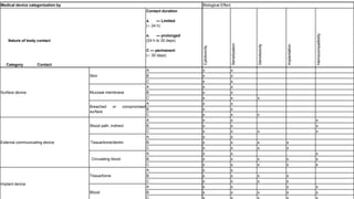
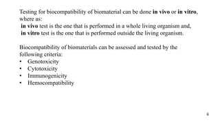
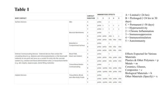
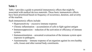
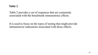
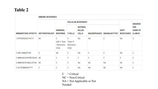
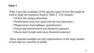
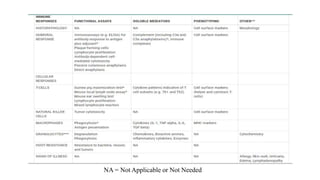
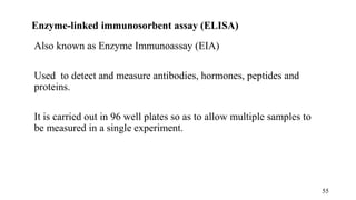
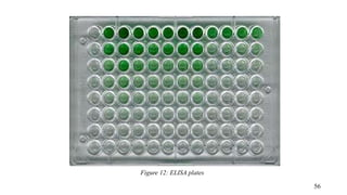
Bangalore Call Girls Hebbal Kempapura Number 7001035870 Meetin With Bangalor...
Biomaterials -Biocompatibility and characteristics
1. MUHIMBILI UNIVERSITY OF HEALTH AND ALLIED
SCIENCES (MUHAS)
SCHOOL OF MEDICINE
DEPARTMENT OF PHYSIOLOGY
BIOMEDICAL ENGINEERING UNIT
GROUPASSIGNMENT :BIOCOMPATIBILITY TESTING
FACULTY NAME: BACHELOR OF SCIENCE IN BIOMEDICAL
ENGINEERING
COURSE CODE: BME 304
2. Participants:
S/N Name Registration Number
1 Boniphace Aloyce 2020-04-14167
2 Anthony Katumba 2020-04-14114
3 Joseph Chengula 2020-04-14450
4 Mike Kabero 2020-04-14586
3. Objectives:
At the end of this presentation, one should be able to explain:-
• The concept of biocompatibility
• The concept of biocompatibility testing
• Cytotoxicity, genotoxicity, immunogenicity and hemocompability
as tests involved in biocompatibility testing
3
4. Content:
• Introduction to Biocompatibility Testing
• Genotoxicity
-Ames Test
-Comet Test
• Cytotoxicity
-Extract Test
-Direct Contact Procedure
• Immunogenicity
-ELISA
-Proliferation Assay
• Hemocompatibility
-Haemolysis Test
-Chandler Loop System 4
5. Medical device categorization by Biological Effect
Nature of body contact
Category Contact
Contact duration
A. — Limited
(< 24 h)
A. — prolonged
(24 h to 30 days)
C — permanent
(> 30 days)
Cytotoxicity
Sensitization
Genotoxicity
Implantation
Hemocompatibility
Surface device
Skin
A x x
B x x
C x x
Mucosal membrane
A x x
B x x
C x x x
Breached or compromised
surface
A x x
B x x
C x x x
External communicating device
Blood path, indirect
A x x x
B x x x
C x x x x
Tissue/bone/dentin
A x x
B x x x x
C x x x x
Circulating blood
A x x x
B x x x x x
C x x x x x
Implant device
Tissue/bone
A x x
B x x x x
C x x x x
Blood
A x x x x
B x x x x x
6. Testing for biocompatibility of biomaterial can be done in vivo or in vitro,
where as:
in vivo test is the one that is performed in a whole living organism and,
in vitro test is the one that is performed outside the living organism.
Biocompatibility of biomaterials can be assessed and tested by the
following criteria:
• Genotoxicity
• Cytotoxicity
• Immunogenicity
• Hemocompatibility
6
7. A Biomaterial is defined as a material used to make devices to
replace a part or function of the body in a safe, reliable, economic
and physiological acceptable manner.
Biocompatibility refers to the ability of a material to perform its
desired function with an appropriate host response in a specific
application
When we test whether a biomaterial is biocompatible or not, we do
biocompatibility testing.
Introduction to Biocompatibility Testing
7
8. The guidelines and standards for compatibility testing are defined by
International Standard Organization (ISO 10993). ISO 10993
provides the standards of results of biocompatibility tests that are
acceptable.
These standards guide the health professionals performing
biocompatibility tests to gauge their results and see whether the
biomaterial or medical device tested is compatible or non-
compatible.
The next slide shows a sample of medical device evaluation
categories and tests
Guidelines for Biocompatibility Testing
8
10. Genotoxicity is a genetic word used to describe the possession of
substance that has destructive effect on the genetic material of the
cell (DNA,RNA), thus affecting the integrity of the cell.
The study of genotoxicity is called Genetic Toxicology
Genotoxins are chemicals or agents that can cause DNA or
chromosomal damage. Depending on its effects, genotoxins can be
categorized as follows:
1. Mutagens; mutation-causing agent
2. Teratogens; birth defect-causing agent
3. Carcinogens; cancer causing-agent
10
11. Agents that cause direct or indirect damage to the DNA:
• UV and ionizing radiation
• Nucleoside analogues
• Reactive oxygen species
• Topoisomerase inhibitors
• Protein synthesis inhibitors
11
12. Figure 1: (a)Direct and indirect radiation damage to the DNA (b)schematic illustration of
damage to the DNA
source: https://www.researchgate.net/figure/
Mechanism of genotoxicity
12
13. Ames Test
It’s named after the developer Bruce Ames. It’s also known as the
bacterial reverse mutation assay.
Principle of the Ames Test
• The test uses several strains of bacteria (Salmonella,E.coli) that
carry a particular mutation.
• Point mutations are made in the histidine (Salmonella
typhimurium) or the tryptophan (Escherichia coli) operon,
rendering the bacteria incapable of producing the corresponding
amino acid.
• This mutations result in his- or trp- organism that cannot grow
unless histidine or tryptophan is supplied.
13
14. Procedure for the Ames Test
1. Isolate an auxotrophic strain of Salmonella Typhimurium for
histidine, His-ve
2. Prepare a test suspension of his- Salmonella Typhimurium in a
plain buffer with test biomaterial. Also add a small amount of
histidine.
3. Also prepare a control suspension of his- Salmonella
Typhimurium but without test biomaterial
4. Incubate the suspension at 37˚ C for 20 minutes
5. Prepare ager the two agar plates and spread the suspension on
ager plate.
6. Incubate the plates at for 48 hours, then count number of
colonies in each plate
14
15. Figure 2 :Procedure of Ames Test
source: https://microbiologyinfo.com/ames-test/
15
16. Results Interpretation
• The mutagenicity of biomaterial is proportional to the
number of colonies observed.
• Large number of colonies on the test plate in comparison
to control, then biomaterials are mutagens/non-
biocompatibility.
16
17. Advantage and Disadvantage of AMES test
• Advantages
Easy, inexpensive and high sensitivity
• Disadvantages
In case of reducing the number of strains, some mutagens
will be missed
17
18. Comet Test
• It’s a gel electrophoresis method, to visualize and
measure DNA strands breaks in individual cell
microscopy.
There are three procedures namely:
1. Encapsulation
2. Lysis
3. electrophoresis
18
19. 1.Encapsulation
• A sample of cells, after made in contact with the test biomaterial is
mixed with molten low-melting-point agarose at 37 °C.
• This mono-suspension is cast on a microscope slide;
Immobilization of cells on CometSlide
• The agarose forms a matrix of carbohydrate fibers that encapsulate
the cells, anchoring them in place.
19
20. 2.Lysis
• The slides with test cells are then treated with lysis solution
to remove membranes and histones from the DNA.
• The lysis solution often consists of a highly concentrated
aqueous salt (often, common table salt can be used) and a
detergent .
• The aqueous salt disrupts proteins and their bonding patterns
within the cell as well as disrupting the RNA content of the
cell.
• The detergent dissolves the cellular membrane.
• Only the DNA of the cell remains.
20
21. 3.Electrophoresis
• After lysis of the cells (typically 1 to 2 hours at 4 °C) the slides are
washed in distilled water to remove all salts and immersed in a
second solution – an electrophoresis solution.
• The slides are left for ~20 minutes in the electrophoresis solution
prior to an electric field being applied. In alkaline conditions the
DNA double helix is denatured and the nucleoid becomes single
stranded.
• An electric field is applied (typically 1 V/cm) for ~20 minutes. The
slides are then neutralized to pH 7, stained with a DNA-specific
fluorescent stain and analyzed using a microscope that is connected
to a computer with image analysis software
21
24. Interpretation
• The picture formed resembles to comet, the head
signifies nucleus and the tail signifies amount of DNA
damage.
• The extent of DNA liberated from the head from the
comet is directly proportional to the amount of DNA
damage.
24
25. Figure 4: Appearance of test cells under microscope connected to computer software
Source: https//hindawi.com/journals/bmri/2012/385245/fig1
25
27. Toxicity is defined as the quality a substance (for example, a drug)
has of being toxic or poisonous.
A biomaterial is said to be toxic when: -
• It alters local biological environment, causing general molecular
or cell dysfunction
• Interact specifically with the particular endogenous target
molecule
Whilst toxicity is a more general term for how harmful a substance is
to an organism; cytotoxicity is the term for how toxic a substance is
to cells.
27
28. Three types of cytotoxicity tests are stated in the International
Organization for Standardization 109993-5: Extract, direct contact
and indirect contact tests (specifically, the agar overlay test).
The extract test is suitable for detecting the toxicity of soluble
substances of medical devices, the direct contact assay is the most
sensitive for testing the cytotoxicity of the medical devices and the
agar overlay assay is suitable for the medical devices that have
large toxicity and bulk filtering.
28
29. 1. The Extract Test (MTT Assay)
This non-radioactive, colorimetric assay system using MTT was first
described by Tim Mosmann and improved in subsequent years by
several other investigators.
Figure 5: MTT Assay 29
30. By definition, the mitochondrial dehydrogenase performance
measurement, also known as the 3-(4,5-dimethyl-2-thiazolyl)-2,5-
diphenyl-2H-tetrazolium bromide (methyl thiazolyl tetrazolium;
MTT) assay, is a colorimetric assay to measure cell metabolism and
rapid assessment of cell proliferation and cytotoxicity.
Fig 6: Chemical formula of MTT 30
31. The main principle of the MTT assay is as follows:
Mitochondrial dehydrogenase in the cytochrome b and c sites of the
living cells can cleave the tetrazole ring, and the yellow, water-
soluble MTT is reduced to produce a purple crystalline formazan.
Fig 7: MTT Assay reaction
31
32. This substance (formazan) is soluble in dimethyl sulfoxide and other
organic solvents, but is insoluble in water. The amount of crystals
formed has a positive correlation to the number of cells and their
activity, and measuring the absorbance (optical density) colorimetric
value reflects the number of surviving cells and metabolic activity.
Fig 8: Color change in MTT Assay
32
33. The insoluble formazan crystals are dissolved using a solubilization
solution and the resulting colored solution is quantified by measuring
the absorbance at 500-600 nanometers using a multi-well
spectrophotometer. The darker the solution, the greater the number of
viable, metabolically active cells
Fig 9: A multiwell spectrophometer
33
34. Advantages of MTT Assay:
More accurate
MTT assay can be performed on 96-well microplates in a standard
reader (such as a Bio-Tek ELx808) allowing for fast screening of
multiple samples.
Fig 10: 96-well microplates and a Bio-Tek ELx808 reader 34
35. Limitations of MTT Assay
the MTT assay does not discriminate a specific cellular death
mechanism
it may underestimate cellular damage and only detect death at the
last stages of the cellular dying process
it is cumbersome and time-consuming
traditional tests use artificial methods, such as measuring platelets
to count the number of surviving cells, which can lead to errors
35
36. 2. The Direct Contact Assay
The direct contact method yields direct contact of the solid medical
devices with cultured mammalian cells in vitro.
This cytotoxicity test occurs by observing the morphological changes
and detecting the changes in the number of cells; it can directly
reflect the impact of testing the medical devices on the cells.
Although the method has high sensitivity, it is more demanding for
the medical devices, and suitable medical devices are limited.
36
37. • For the direct contact test’s sample preparation, sample pieces
with flat surfaces not less than 100 square millimeters (mm) are
prepared.
• Next, L-929 mammalian fibroblast cells are grown in a serum-
supplemented minimum essential medium (MEM), and a cell
suspension is prepared.
• Equal amounts of the L-929 cell suspension are added to culture
plates with a 35 mm diameter to create a single layer cell culture.
• After L-929 cells have been cultured to reach the appropriate
confluence, the cell culture medium is aspirated from the plates
and replaced with a fresh culture medium. 37
38. • Next, a single product sample, positive control, or negative
control is placed in each 35 mm culture dish.
• All samples and controls are cultured in duplicate or triplicate at
37 ± 1°C in a humidified incubator containing 5 ± 1% of carbon
dioxide.
Fig 11: Direct Contact procedure
38
39. • Following incubation, each sample, positive control, and negative
control are examined under a microscope. In some cases, cells are
stained with a cytochemical stain such as haematoxylin blue to
support assessing biological reactivity.
• The biological reactivity of the cells exposed to the sample or
sample extracts is rated on a scale of 0-4 (see the table in the next
slide).
• The biological reactivity is determined by assessing the non-lethal
injury of the cells (cellular degeneration) and any structural
defects (malformations) the cells have.
39
40.
41. • The direct contact test is valid if the observed responses to the
negative controls are grade 0 and the positive controls are all at
least grade 3
• The sample meets the requirements of the direct contact
cytotoxicity testing rules if the biological responses of the
samples are not greater than grade 2.
41
42. Advantages of the Direct Contact Assay
• The target cells come into contact with biomaterials.
• It has a high sensitivity
• It standardizes the test amount.
Limitations of the Direct Contact Assay
• High-density material has the potential to damage the cells when
the sample is placed on top of them
• Very low-density materials float in the cell growth media
• Cellular trauma if the biomaterial moves
42
44. Immunogenicity is defined as any adverse effect on the structure or
function of the immune system, or on other systems as a result of
immune system dysfunction.
An effect is considered adverse or immunotoxic if:
• it impairs humoral or cellular immunity needed by the host to
defend itself against infectious or neoplastic disease
(immunosuppression)
• it causes unnecessary tissue damage (autoimmunity,
hypersensitivity, or chronic inflammation).
44
45. Immunogenicity test format/guidance
• The guidance consists of a flow chart and three tables that follow
the ISO 10993 format.
• The flow chart is used for determining whether immunotoxicity
testing is likely to be needed, and includes the option of providing a
rationale for not performing the testing based upon available
published data or other sources of information on the same or like
materials.
45
47. The flow chart should be used first to determine whether
immunotoxicity testing may be needed to support the safety of the
device.
Immunotoxicity testing may not be needed if:
• the device material is the same as in a legally marketed device;
• has the same body contact, dose and duration; and
• there is either a long history of use without reported toxicity, or
scientific data in the public domain supporting lack of toxicity.
If immunotoxicity testing has been carried out as specified in ISO
10993 as part of overall safety evaluation, then it need not be
repeated.
47
48. Tables for Immunogenicity Testing
When the flow chart indicates that immunotoxicity testing is
recommended, Tables 1-3 are used sequentially to determine the
types of testing that might be used to help evaluate product safety
consistent with the intended use and indicated patient population and
expected risk vs. benefit.
48
49. Table 1
A = Limited (<24 hrs)
B = Prolonged (>24 hrs to 30
days)
C = Permanent (>30 days)
1 = Hypersensitivity
2 = Chronic Inflammation
3 = Immunosuppression
4 = Immunostimulation
5 = Autoimmunity
Effects Expected for Various
Materials:
Plastics & Other Polymers = p
Metals = m
Ceramics, Glasses,
Composites = c
Biological Materials = b
Other Materials (Specify) = x
50. Table 1
Table 1 provides a guide to potential immunotoxic effects that might be
associated with medical devices materials. These basic immunotoxic effects
have been prioritized based on frequency of occurrence, duration, and severity
of the reaction.
Such immunotoxic effects include:
• Hypersensitivity – excessive immune response
• Chronic inflammation – accumulation of cells to fight against antigen
• Immunosuppression – reduction of the activation or efficiency of immune
system.
• Immunostimulation – unwanted overreaction of the immune system upon
exposure to pathogens.
• Autoimmunity – immune response of an organism against its own healthy
cells, tissues and other normal body constituents.
50
51. Table 2
Table 2 provides a set of responses that are commonly
associated with the benchmark immunotoxic effects.
It is used to focus on the types of testing that might provide
immunotoxic indications associated with those effects.
51
52. Table 2
C = Critical
NC = Non-Critical
NA = Not Applicable or Not
Needed
53. Table 3
Table 3 provides examples of the specific types of tests that might be
used to study the responses listed in Table 2 . For example:
• ELISA (for antigen detection)
• Proliferation assay (for rapid cells division detection )
• Phagocytosis (macrophages, granulocytes)
• Guinea pig maximization test (humoral response)
• Mouse local lymph node assay (humoral response)
These selected examples are only representative of the large number
of tests that are currently available.
53
55. Enzyme-linked immunosorbent assay (ELISA)
Also known as Enzyme Immunoassay (EIA)
Used to detect and measure antibodies, hormones, peptides and
proteins.
It is carried out in 96 well plates so as to allow multiple samples to
be measured in a single experiment.
55
57. Principle of ELISA
ELISA works on the principle that specific antibodies bind the target
antigen and detect the presence and quantity of antigens binding.
In order to increase the sensitivity and precision of the assay, the
plate must be coated with antibodies with high affinity.
57
58. Procedures of ELISA
• The ELISA plate is coated with capture antibody, any
excess, unbound antibody is then washed from the plate. The
capture antibody is an antibody raised against the antigen of
interest.
• The sample is added. Any antigen found in the sample will
bind to the capture antibody already coating the plate.
• Detection antibody is added. This antibody is labelled with
an enzyme, usually horse radish peroxidase or alkaline
phosphatase. Detection antibody binds to any target antigen
already bound to the plate.
• Finally, the substrate is added. The substrate is converted by
the enzyme to form a colored product, which can be
measured by spectrophotometry or plate reader.
58
59. Proliferation assay
Objective:
The in vitro proliferation assay can be used to determine whether or
not cells are triggered to divide after exposure to a specific stimulus.
59
60. Procedures for Proliferation assay
• Cells in culture are given a specific stimulus.
• A radio-labelled nucleotide is then added to the culture media. The
most commonly used is 3H-thymidine. As the cells are stimulated
to divide, they utilize the radio-labelled nucleotide in the culture
media and incorporate it into their newly-synthesized DNA.
• After this incubation (generally 24-48 hours), the cells are removed
from the culture media by centrifugation and washed to remove any
free radio-labelled nucleotide that has not been incorporated into
the cells’ DNA. The total radioactivity is measured, and compared
against a control group of cells that did not receive the
proliferation-inducing stimulus.
60
62. Hemocompatibility or blood compatibility is the ability of a biomaterial
to not cause adverse effects when they come into contact with blood.
Hemocompatibility or blood compatibility is the property of a material or
device that permits it to function in contact with blood without inducing
adverse reaction (Ratner,2014).
62
63. Types of device in contact with blood (as categorized in ISO 10993-1)
I. External communicating device
External communicating devices that serve as an indirect blood path include
but are not limited to:
cannulae
extension sets
blood collection device
External communicating devices in contact with circulating blood include but
are not limited to:
atherectomy devices
blood monitors
catheters
Guidewires
intravascular endoscopes
intravascular ultrasound
intravascular laser systems, 63
64. II.Non-contact devices
An in-vitro diagnostic device is an example of a non-contact device.
III.Implant devices
Implant devices are placed largely or entirely within the vascular system.
Examples include but are not limited to:
annuloplasty rings
mechanical or tissue heart valves
prosthetic or tissue vascular grafts
circulatory support devices (ventricular-assist devices artificial hearts,
intra-aortic balloon pumps)
inferior vena cava filters
embolization devices,
64
65. Procedure for the Evaluation of the Hemocompatibility of
Biomaterials
The figure above shows the Schematic representation of the procedure
for the evaluation of the hemocompatibility of biomaterials:
65
66. Procedure for the Evaluation of the Hemocompatibility of
Biomaterials
i. Fresh human blood is collected and anticoagulated with low dose
heparin.
ii. The test material is incubated at 37°c using static, agitated, or
dynamic test models with the blood.
iii.The activation markers in the blood are analyzed before and after the
incubation with the test material.
iv. The surface of the biomaterial is analyzed to determine the
interaction of blood cells and proteins with the biomaterial
66
67. Test categories for evaluation of hemocompatibility
According to ISO 10993-4 Blood interactions can be classified into five
categories based on the primary process or system being measured.
Thrombosis
Coagulation
Platelets
Haematology
Immunology (complement system and leukocytes)
67
68. Test categories for evaluation of hemocompatibility
Test Category Method Comments
Thrombosis Light microscopy Can be replaced by electron
microscopy for more clarity or in case
of technical problems
Coagulation Partial thromboplastin time assay
Haematology Leukocyte count and differential
haemolysis (plasma haemoglobin)
Haemolysis is regarded as an especially
significant screening test to perform in
this category because of its
measurement of red blood cell
membrane fragility in contact with
materials and devices
Platelets Platelet count Flowcytometry
Immunology C3a, C5a, TCC, BB, iC3b, C4d,
SC5b-9
Can be conducted by immunology
analyser.
e.g. Architect I100se
68
69. Hemocompatibility Test Panel/model
According to ISO 10993-4 hemocompatibility Test are conducted in the
following model
Static
Blood coagulation Assay
Hemolysis Assay
Partial Thromboplastin Time(PPT)
Dynamic
Chandler loop system
Chronic shunting
69
70. Static hemocompatibility model:Haemolysis assay
In vitro tests are used to evaluate damage to erythrocytes. Direct
methods determine haemolysis due to physical and chemical
interactions with erythrocytes. Indirect methods determine haemolysis
due to extractables from test articles. The procedures for haemolysis
assay are:
Prepare three separate test tubes (N=3) containing red blood cells
dosed with the test compound dissolved in DMSO
Incubated at 37°C in a water bath with mild agitation
Sample each replicate at 0, 5, 15, 30, 60, 120 and 180 minutes
Centrifuge each sample
70
71. Measure the absorbance of the supernatant from each sample
using a UV/Visible spectrometer
Calculate percentage hemolysis at each sampling time point by
the formula
% ℎ𝑎𝑒𝑚𝑜𝑙𝑦𝑠𝑖𝑠
=
𝑎𝑏𝑠𝑜𝑟𝑏𝑎𝑛𝑐𝑒 𝑜𝑓 𝑠𝑎𝑚𝑝𝑙𝑒 − 𝑎𝑏𝑠𝑜𝑟𝑏𝑎𝑛𝑐𝑒 𝑜𝑓 𝑛𝑒𝑔𝑎𝑡𝑖𝑣𝑒 𝑐𝑜𝑛𝑡𝑟𝑜𝑙
𝑎𝑏𝑠𝑜𝑟𝑏𝑎𝑛𝑐𝑒 𝑜𝑓 𝑝𝑜𝑠𝑖𝑡𝑖𝑣𝑒 𝑐𝑜𝑛𝑡𝑟𝑜𝑙
× 100%
Conclusion:
• Over 5% hemolysis are classified as hemolytic
• Between 5% and 2% are slightly hemolytic
• Below 2% are non-hemolytic
71
72. Dynamic hemocompatibility model
Medical devices having contact to circulating blood should be examined
very thoroughly. For these devices the testing of single endpoints using
static systems may not be sufficient.
Testing should be preferably performed by using dynamic systems.
Physiochemically comparable materials can exhibit different effects on
hemocompatibility in clinical applications.
Therefore, appropriate in vitro models should offer the possibility of
dynamic testing in relation to high/ low shear stress and the use of human
whole blood with arbitrary anticoagulation.
72
73. Dynamic hemocompatibility model: Chandler loop system
The chandler loop system enables simulation of blood flow inside
the laboratory with arterial flow condition.
Procedures of chandler loop system to test hemocompatibility:
• Material to be tested is attached to the tubing by inserting it
inside tubing and it is covered by a tube with larger radius.
• Tubing is filled with 50 mills of prepared blood from testing
animals or human.
• The PVC tubes with blood and samples are attached to the
rotating shaft of electric motor and inserted into water bath with
water heated to 37 degrees temperature
73
74. Chandler system with testing material in operation (Buddy D. Ratner, 1996)
Source: https://www.nelsonlabs.com/testing/hemocompatibility/
74
75. References
•(International Organization for Standardization, 2. (2021, 12). Quality management
systems — Requirements. Retrieved from iso.org:
https://www.iso.org/standard/21823.html
•Celik, T. A. (2018). Cytotoxicity.
•Ethide. (2021). Ethide laboratories.https://ethidelabs.com/what-is-agar-diffusion-test-
for-cytotoxicity/
•Nelson Laboratories, L. (2021, 12). Hemocompatibility. Retrieved from Nelson
Laboratories, LLC: https://www.nelsonlabs.com/testing/hemocompatibility/
•STEMart. (2021, 12). Stemart. Retrieved from Cytotoxicity testing: https://www.ste-
mart.com/cytotoxicity-testing.htm
•Williams, D. (1999). the Williams dictionary of biomaterials.
Editor's Notes
His
Colorimetric analysis is a method of determining the concentration of a chemical element or chemical compound in a solution with the aid of a color reagent. It is applicable to both organic compounds and inorganic compounds and may be used with or without an enzymatic stage. The method is widely used in medical laboratories and for industrial purposes.
Confluency means the proportion of the surface which is covered by adherent cells
In cell culture biology, confluence refers to the percentage of the surface of a culture dish that is covered by adherent cells.
Replicate means any from a copies or repetitions, especially of a laboratory or research procedure.
DMSO short form of Dimethyl sulfoxide, (CH3)2SO.. Precipitation medication fluid.
Supernatant is the clear liquid that lies above the solid residue after centrifugation, precipitation, crystallization or settling.