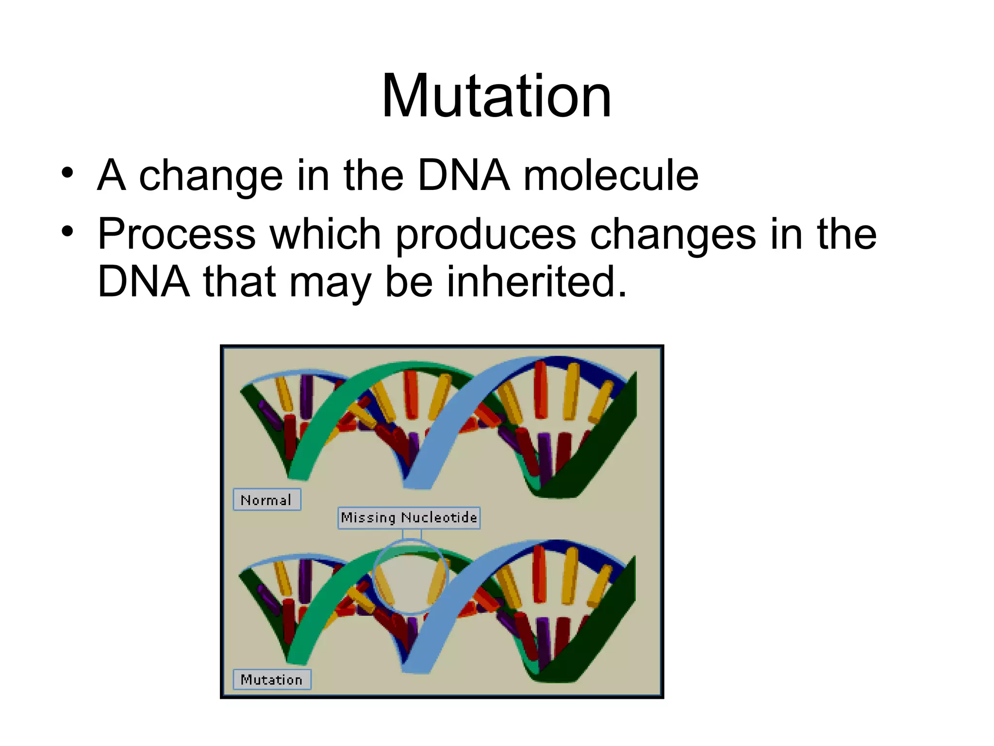
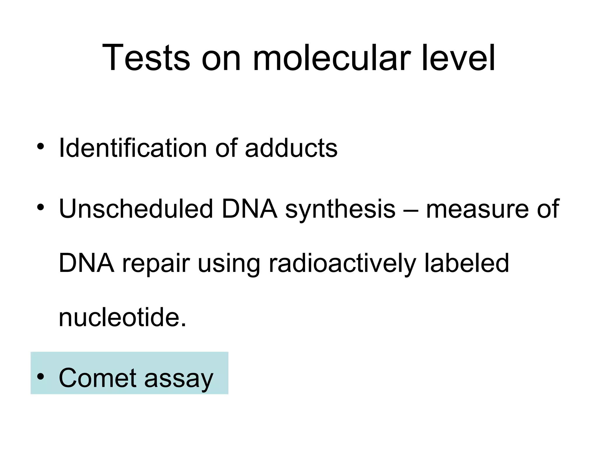
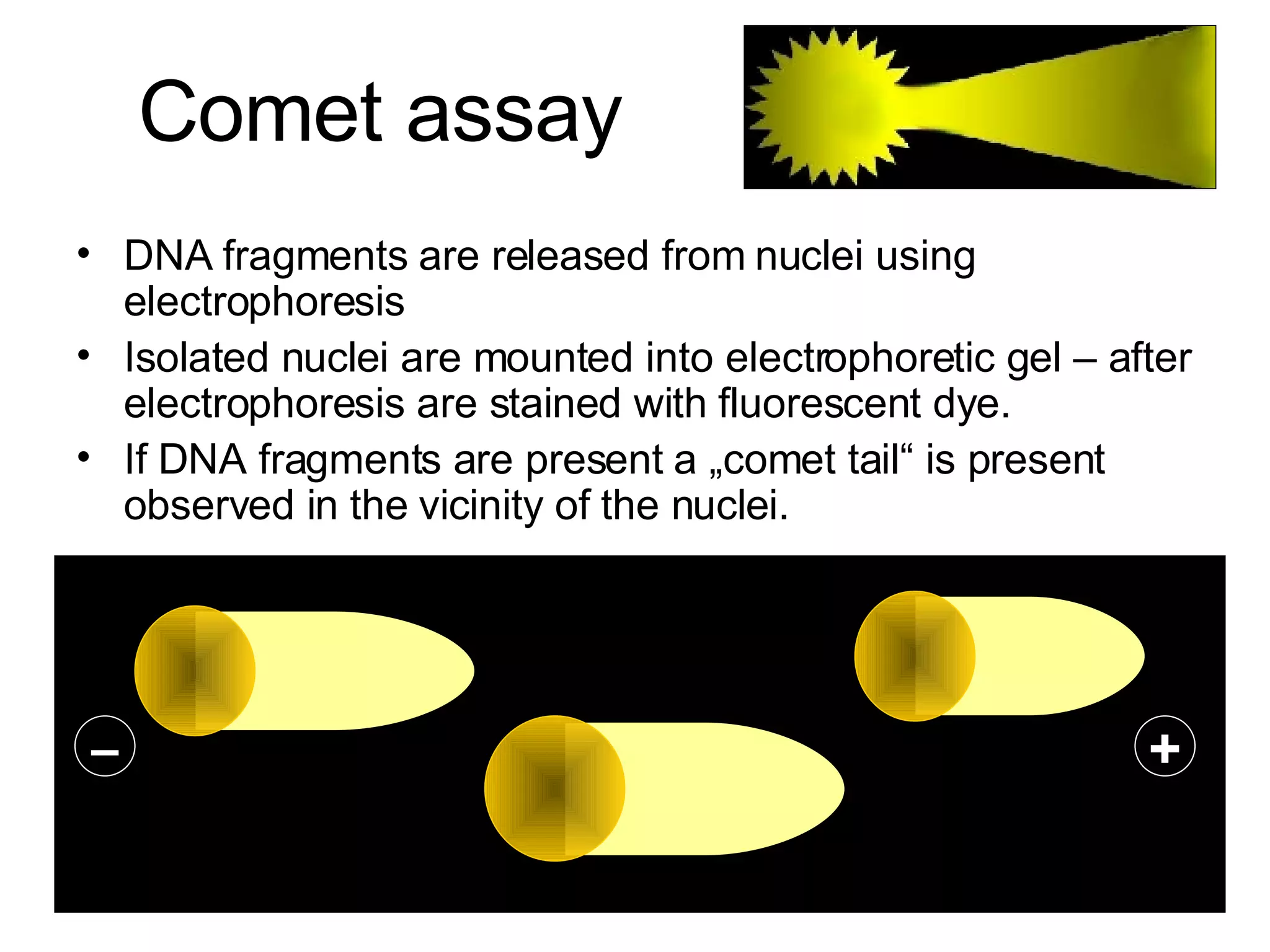
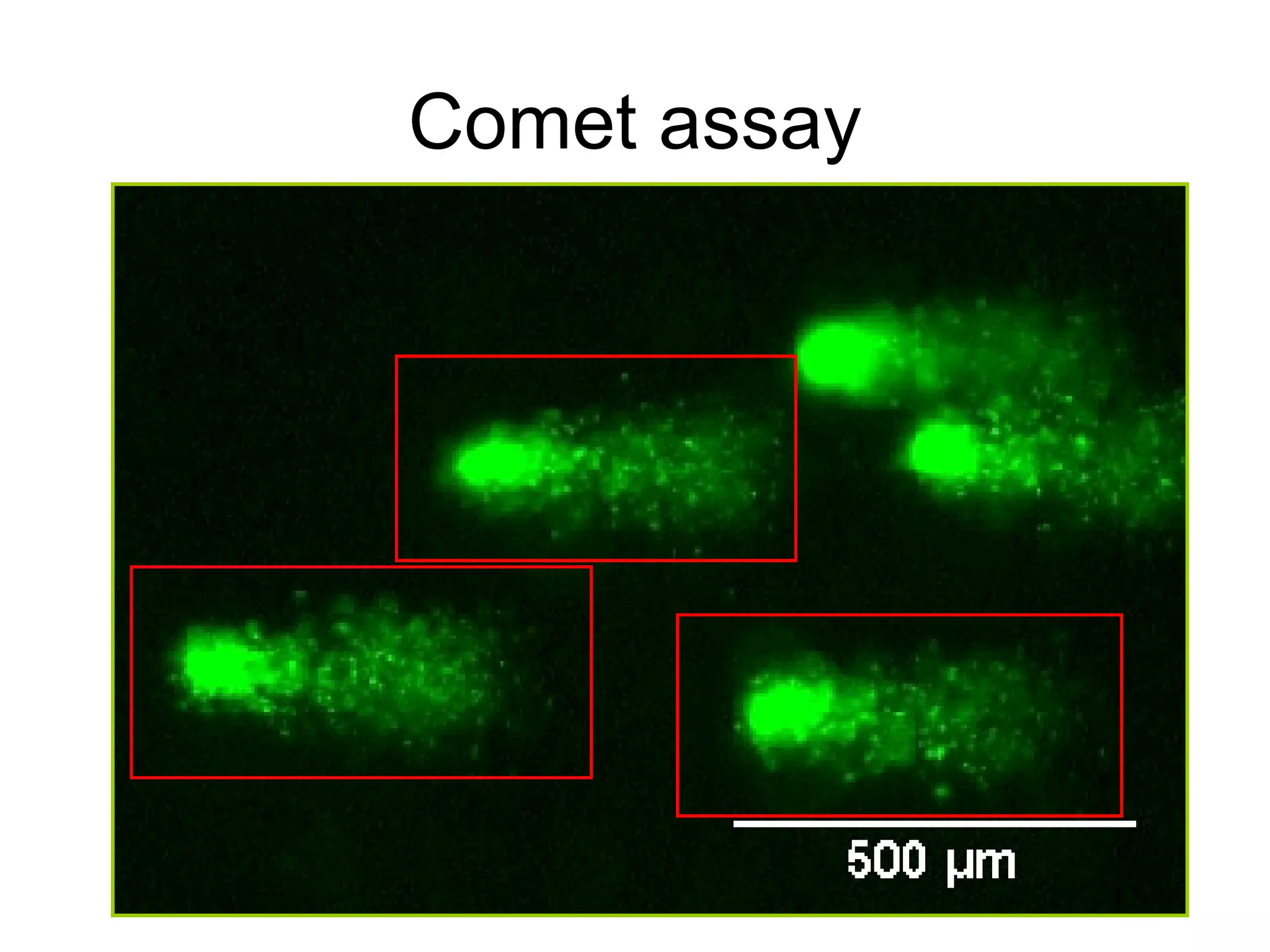
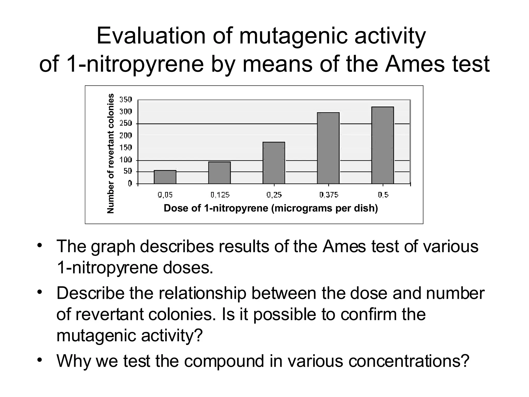
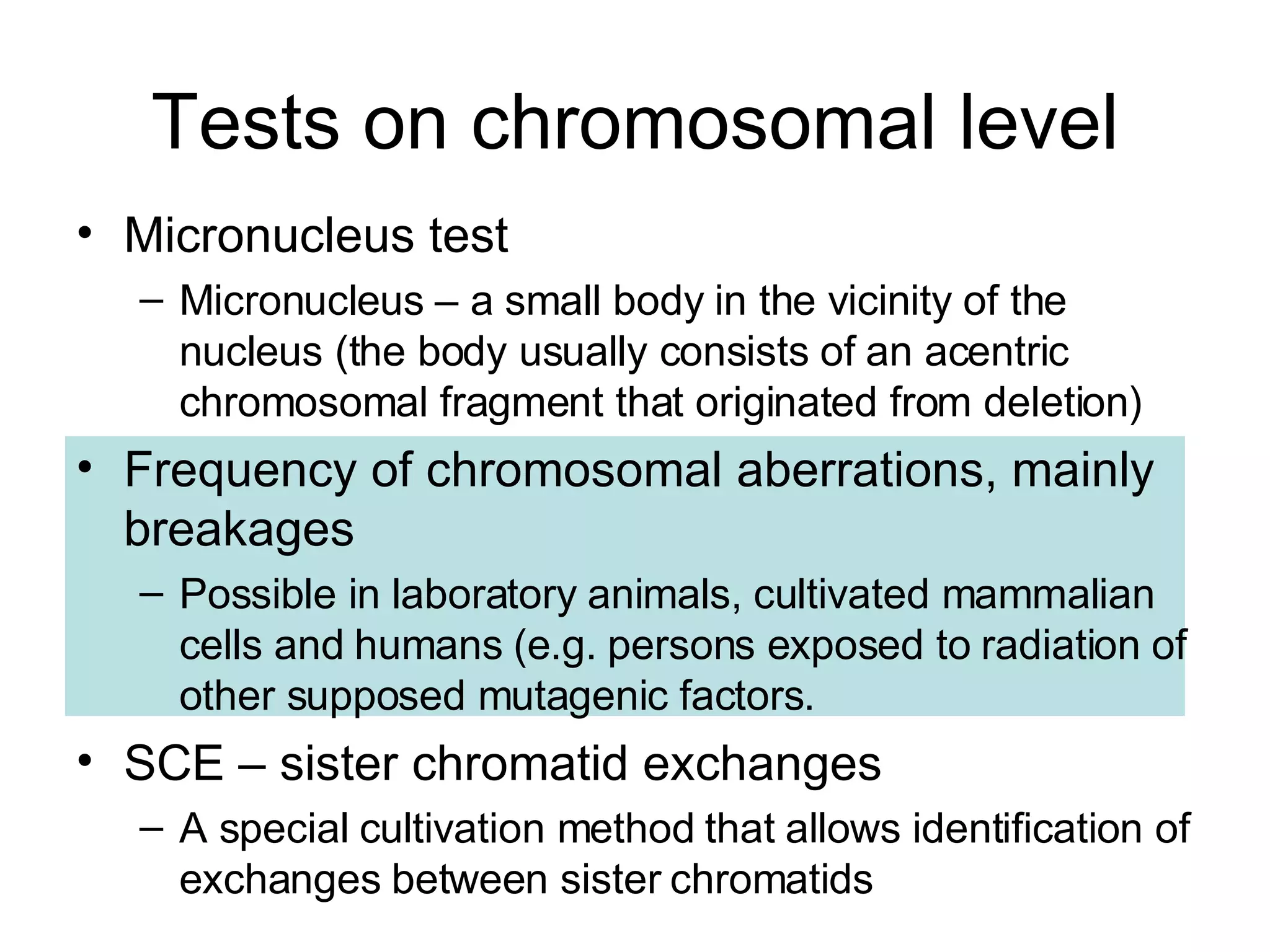
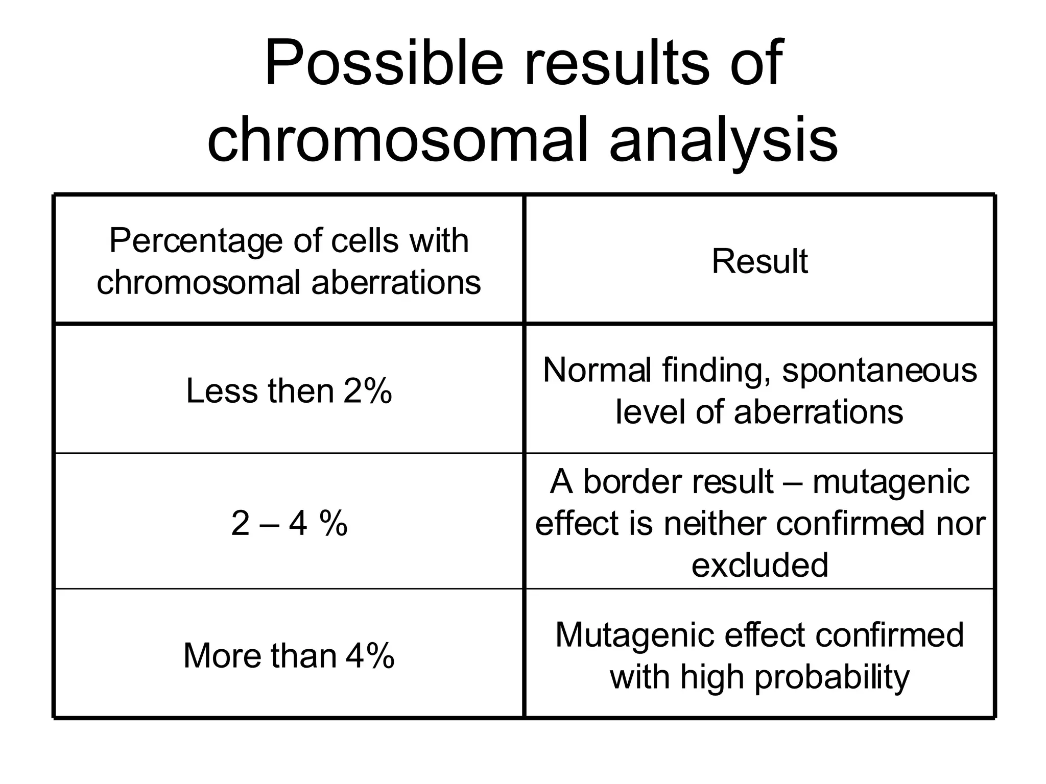
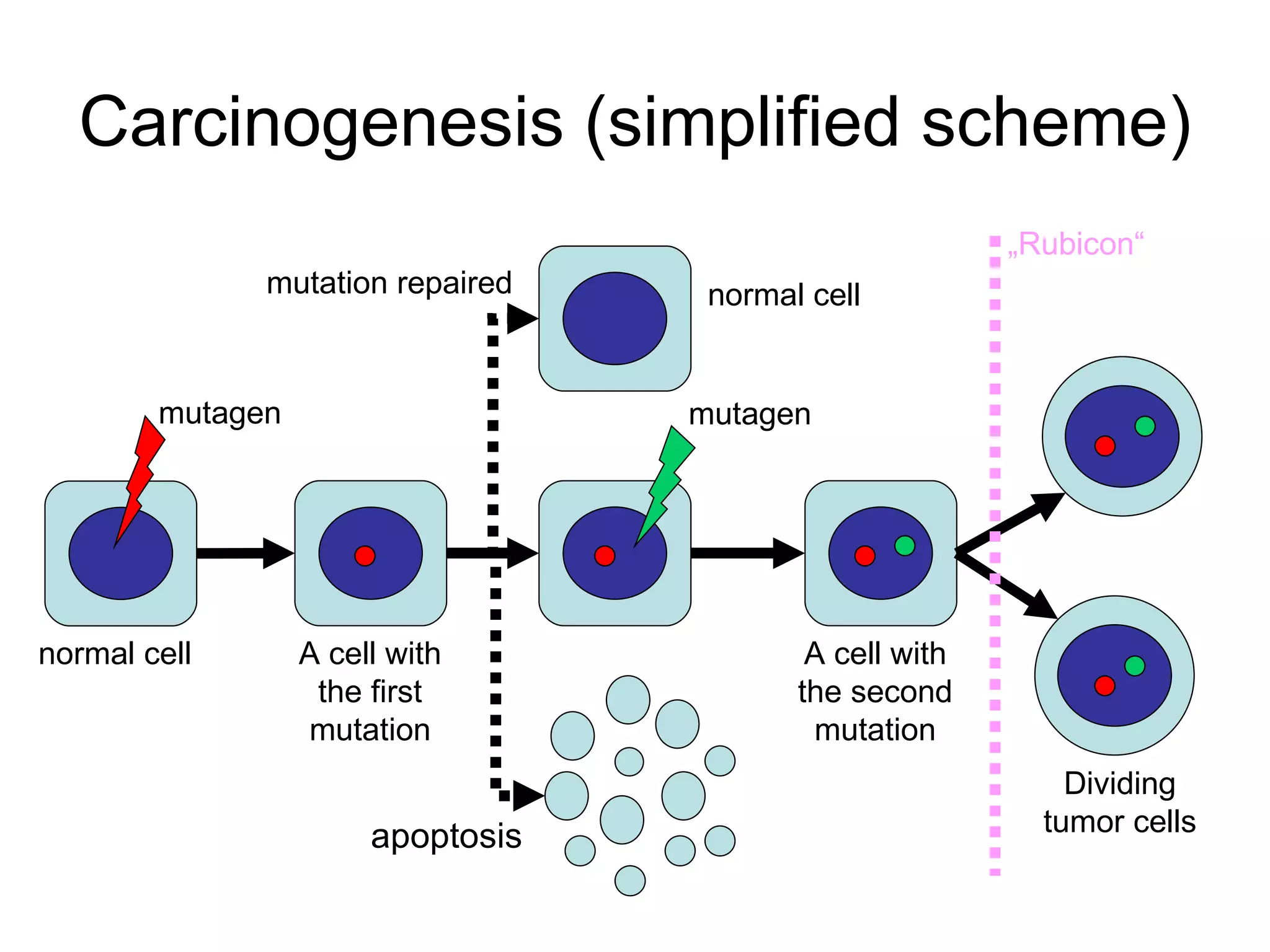
Mutagenicity and carcinogenicity of environmental factors
Next week A test in molecular biology (will be based on professor Goetz and professor Sedláček lectures) Don´t be late, please! Possibility of presentations (after negotiation with professor Mareš)
Find Kočárek´s presentations in: http://www.medik.cz/medik/kocarek/ or http://camelot2.lf2.cuni.cz/turnovec/ublg/vyuka/
Mutation A change in the DNA molecule Process which produces changes in the DNA that may be inherited .
Mutagenicity = mutagenic activity = genotoxicity (genotoxic activity) Mutagenic (genotoxic) compound = a compound that causes mutation Mutations in certain genes could lead to malignant transformation of the cell. Many mutagens are supposed to be carcinogenic.
Carcinogens Karkinos  (Greek)  =  crayfish Compounds or other factors that induce transformation of a normal cell into a tumor one
Classification of mutagens Chemical (different compounds) Alkylating agents Aromatic hydrocarbons  (e.g. benzo(a)pyrene) Intercalating agents ( e.g.  fluorescent dyes) Artificial derivatives of DNA bases Physical (ionizing radiation, UV-radiation) Biological (viruses, transposable elements)
According to metabolic conversion we distinguish: Direct acting mutagens (active without metabolic conversion) Promutagens (require metabolic activation)
Benzo(a)pyrene Polycyclic aromatic hydrocarbon Carcinogenic activity confirmed A product of combustion processes (e.g. tobacco smoking) A typical promutagenic agent Metabolic conversion (addition of epoxide group and two OH-groups). A metabolic product binds to DNA  ->  adduct (a compound that results from addition). Presence of adduct can cause mutation. Metabolic conversion binds to DNA BaP – guanin adduct
Metabolic activation of mutagens PROMUTAGEN ACTIVE (ULTIMATE) MUTAGEN DNA ADDUCT (OR OTHER CHEMICAL MODIFICATION OF DNA BASES) MUTATION enzymes metabolic activation TUMOR liver
Genotoxicity screening tests On molecular level On gene level On chromosomal level
Tests on molecular level Identification of adducts Unscheduled DNA synthesis – measure of DNA repair using radioactively labeled nucleotide. Comet assay
Comet assay  Single Cell Gel Electrophoresis (SCGE) Identification of small DNA fragments inside nuclei of affected cells  The fragments result from mutagenic affect.
Comet assay DNA fragments are released from nuclei using electrophoresis Isolated nuclei are mounted into electrophoretic gel – after electrophoresis are stained with fluorescent dye.  If DNA fragments are present a „comet tail“ is present observed in the vicinity of the nuclei. – +
Comet assay
Possible results of a comet assay Normal nucleus without fragments (DNA is not damaged – mutagenicity excluded) Two nuclei with DNA damage
Evaluation of a comet assay
Comet assay – computer analysis
Tests on gene level Induction of  in vitro  mammalian cells resistant to certain chemical compound  (e.g. 8-azaguanine, 6-thioguanin, ouabain) SOS/umu test – measurement of DNA repair in bacteria The bacteria is transformed by plasmide with mutator (repair) genes umuC and umuD fused with the gene for galactosidase. If the DNA is damaged by mutagen both umu mutator genes and gal gene are transcribed and galactosidase turns  a specific substrate into a color product. Ames test
Ames test Bruce Ames (born 1928)
Ames test Test system – auxotrophic strain of  Salmonella typhimurium  – survives only in medium with histidine (dies in normal medium without histidine) After treatment with mutagen some auxotrophic cells are turned into normal ones that synthesize histidine and survive in a normal medium. These cells are called revertants (due to reverse mutation).
Ames test His – bacteria Dies in a normal medium Medium  containing histidine Normal medium   His + bacteria Reverse mutation Mutagen
Ames test 0 Negative control Positive control A dish with  a  compound to be tested GENOTOXICITY CONFIRMED S PONTANEOUS  REVERTANTS IS USED FOR CONTROL OF THE TEST COMPOUND CONFIRME D  MUTAGENIC COMPOUND
Result of the Ames test
Ames test – automatic evaluation of results
Evaluation of mutagenic activity  of 1-nitropyren e  by means of the Ames test The graph describes results of the Ames test of various 1-nitropyren e  doses. Describe the relationship between the dose and number of revertant colonies. Is it possible to confirm the mutagenic activity?  Why we test the compound in various concentrations? Number of revertant colonies Dose of 1-nitropyrene (micrograms per dish)
Result of the task Number of colonies decreases due to toxic effects of the tested compound Number of revertant colonies Dose of 1-nitropyrene (micrograms per dish) Number of colonies increases with the dose – mutagenicity confirmed.
Tests on chromosomal level Micronucleus test Micronucleus – a small body in the vicinity of the nucleus (the body usually consists of an acentric chromosomal fragment that originated from deletion) Frequency of chromosomal aberrations, mainly breakages Possible in laboratory animals, cultivated mammalian cells and humans (e.g. persons exposed to radiation of other supposed mutagenic factors. SCE – sister chromatid exchanges  A special cultivation method that allows identification of exchanges between sister chromatids
Result of micronucleus test
Tests on chromosomal level Micronucleus test Micronucleus – a small body in the vicinity of the nucleus (the body usually consists of an acentric chromosomal fragment that originated from deletion) Frequency of chromosomal aberrations, mainly breakages Possible in laboratory animals, cultivated mammalian cells and humans (e.g. persons exposed to radiation of other supposed mutagenic factors. SCE – sister chromatid exchanges  A special cultivation method that allows identification of exchanges between sister chromatids
Possible results of chromosomal analysis Mutagenic effect confirmed with high probability More than 4% A border result – mutagenic effect is neither confirmed nor excluded 2 – 4 % Normal finding, spontaneous level of aberrations Less then 2% Result Percentage of cells with chromosomal aberrations
Tests on chromosomal level Micronucleus test Micronucleus – a small body in the vicinity of the nucleus (the body usually consists of an acentric chromosomal fragment that originated from deletion) Frequency of chromosomal aberrations, mainly breakages Possible in laboratory animals, cultivated mammalian cells and humans (e.g. persons exposed to radiation of other supposed mutagenic factors. SCE – sister chromatid exchanges  A special cultivation method that allows identification of exchanges between sister chromatids
Sister Chromatid Exchanges Normal chromosome A chromosome with SCE The higher SCE frequency – the higher mutagenic activity.
About the origin of a tumor cell
Carcinogenesis (simplified scheme) mutagen mutagen apoptosis mutation repaired A cell with the first mutation A cell with the second mutation Dividing tumor cells normal cell normal cell „ Rubicon“
Mutagenic X carcinogenic agent All mutagens are supposed to be carcinogenic but in some of them the carcinogenicity was not detected. Some carcinogenic agents are not mutagenic – they belong to the group of epigenetic carcinogens. Evaluation of carcinogenicity to humans is difficult and requires combination of various tests.
Carcinogenic agents Chemical (mostly mutagenic compounds) Physical (radiation, asbestos) Biological (oncogenic viruses – e.g. HTLV = human T-leukemia virus).
Direct evaluation of carcinogenicity Laboratory tests In animals In vitro  transformation of cultivated cells Epidemiologic studies of exposed human population – mostly reliable
IARC database  International Agency for Research of Cancer
IARC database  (International Agency for Research of Cancer) Direct connection to „Monographs“: http:// monographs.iarc.fr/index.php
IARC classification Group 1:  Compounds (mixtures) carcinogenic to humans Group 2A:  Probable human carcinogens Group 2B:  Possible human carcinogens Group 3:  Carcinogenicity not confirmed Group 4:  Compounds probably not carcinogenic to humans
See you after 3 weeks!

Ecology Practicals2

  • 1.
    Mutagenicity and carcinogenicityof environmental factors
  • 2.
    Next week Atest in molecular biology (will be based on professor Goetz and professor Sedláček lectures) Don´t be late, please! Possibility of presentations (after negotiation with professor Mareš)
  • 3.
    Find Kočárek´s presentationsin: http://www.medik.cz/medik/kocarek/ or http://camelot2.lf2.cuni.cz/turnovec/ublg/vyuka/
  • 4.
    Mutation A changein the DNA molecule Process which produces changes in the DNA that may be inherited .
  • 5.
    Mutagenicity = mutagenicactivity = genotoxicity (genotoxic activity) Mutagenic (genotoxic) compound = a compound that causes mutation Mutations in certain genes could lead to malignant transformation of the cell. Many mutagens are supposed to be carcinogenic.
  • 6.
    Carcinogens Karkinos (Greek) = crayfish Compounds or other factors that induce transformation of a normal cell into a tumor one
  • 7.
    Classification of mutagensChemical (different compounds) Alkylating agents Aromatic hydrocarbons (e.g. benzo(a)pyrene) Intercalating agents ( e.g. fluorescent dyes) Artificial derivatives of DNA bases Physical (ionizing radiation, UV-radiation) Biological (viruses, transposable elements)
  • 8.
    According to metabolicconversion we distinguish: Direct acting mutagens (active without metabolic conversion) Promutagens (require metabolic activation)
  • 9.
    Benzo(a)pyrene Polycyclic aromatichydrocarbon Carcinogenic activity confirmed A product of combustion processes (e.g. tobacco smoking) A typical promutagenic agent Metabolic conversion (addition of epoxide group and two OH-groups). A metabolic product binds to DNA -> adduct (a compound that results from addition). Presence of adduct can cause mutation. Metabolic conversion binds to DNA BaP – guanin adduct
  • 10.
    Metabolic activation ofmutagens PROMUTAGEN ACTIVE (ULTIMATE) MUTAGEN DNA ADDUCT (OR OTHER CHEMICAL MODIFICATION OF DNA BASES) MUTATION enzymes metabolic activation TUMOR liver
  • 11.
    Genotoxicity screening testsOn molecular level On gene level On chromosomal level
  • 12.
    Tests on molecularlevel Identification of adducts Unscheduled DNA synthesis – measure of DNA repair using radioactively labeled nucleotide. Comet assay
  • 13.
    Comet assay Single Cell Gel Electrophoresis (SCGE) Identification of small DNA fragments inside nuclei of affected cells The fragments result from mutagenic affect.
  • 14.
    Comet assay DNAfragments are released from nuclei using electrophoresis Isolated nuclei are mounted into electrophoretic gel – after electrophoresis are stained with fluorescent dye. If DNA fragments are present a „comet tail“ is present observed in the vicinity of the nuclei. – +
  • 15.
  • 16.
    Possible results ofa comet assay Normal nucleus without fragments (DNA is not damaged – mutagenicity excluded) Two nuclei with DNA damage
  • 17.
    Evaluation of acomet assay
  • 18.
    Comet assay –computer analysis
  • 19.
    Tests on genelevel Induction of in vitro mammalian cells resistant to certain chemical compound (e.g. 8-azaguanine, 6-thioguanin, ouabain) SOS/umu test – measurement of DNA repair in bacteria The bacteria is transformed by plasmide with mutator (repair) genes umuC and umuD fused with the gene for galactosidase. If the DNA is damaged by mutagen both umu mutator genes and gal gene are transcribed and galactosidase turns a specific substrate into a color product. Ames test
  • 20.
    Ames test BruceAmes (born 1928)
  • 21.
    Ames test Testsystem – auxotrophic strain of Salmonella typhimurium – survives only in medium with histidine (dies in normal medium without histidine) After treatment with mutagen some auxotrophic cells are turned into normal ones that synthesize histidine and survive in a normal medium. These cells are called revertants (due to reverse mutation).
  • 22.
    Ames test His– bacteria Dies in a normal medium Medium containing histidine Normal medium His + bacteria Reverse mutation Mutagen
  • 23.
    Ames test 0Negative control Positive control A dish with a compound to be tested GENOTOXICITY CONFIRMED S PONTANEOUS REVERTANTS IS USED FOR CONTROL OF THE TEST COMPOUND CONFIRME D MUTAGENIC COMPOUND
  • 24.
    Result of theAmes test
  • 25.
    Ames test –automatic evaluation of results
  • 26.
    Evaluation of mutagenicactivity of 1-nitropyren e by means of the Ames test The graph describes results of the Ames test of various 1-nitropyren e doses. Describe the relationship between the dose and number of revertant colonies. Is it possible to confirm the mutagenic activity? Why we test the compound in various concentrations? Number of revertant colonies Dose of 1-nitropyrene (micrograms per dish)
  • 27.
    Result of thetask Number of colonies decreases due to toxic effects of the tested compound Number of revertant colonies Dose of 1-nitropyrene (micrograms per dish) Number of colonies increases with the dose – mutagenicity confirmed.
  • 28.
    Tests on chromosomallevel Micronucleus test Micronucleus – a small body in the vicinity of the nucleus (the body usually consists of an acentric chromosomal fragment that originated from deletion) Frequency of chromosomal aberrations, mainly breakages Possible in laboratory animals, cultivated mammalian cells and humans (e.g. persons exposed to radiation of other supposed mutagenic factors. SCE – sister chromatid exchanges A special cultivation method that allows identification of exchanges between sister chromatids
  • 29.
  • 30.
    Tests on chromosomallevel Micronucleus test Micronucleus – a small body in the vicinity of the nucleus (the body usually consists of an acentric chromosomal fragment that originated from deletion) Frequency of chromosomal aberrations, mainly breakages Possible in laboratory animals, cultivated mammalian cells and humans (e.g. persons exposed to radiation of other supposed mutagenic factors. SCE – sister chromatid exchanges A special cultivation method that allows identification of exchanges between sister chromatids
  • 31.
    Possible results ofchromosomal analysis Mutagenic effect confirmed with high probability More than 4% A border result – mutagenic effect is neither confirmed nor excluded 2 – 4 % Normal finding, spontaneous level of aberrations Less then 2% Result Percentage of cells with chromosomal aberrations
  • 32.
    Tests on chromosomallevel Micronucleus test Micronucleus – a small body in the vicinity of the nucleus (the body usually consists of an acentric chromosomal fragment that originated from deletion) Frequency of chromosomal aberrations, mainly breakages Possible in laboratory animals, cultivated mammalian cells and humans (e.g. persons exposed to radiation of other supposed mutagenic factors. SCE – sister chromatid exchanges A special cultivation method that allows identification of exchanges between sister chromatids
  • 33.
    Sister Chromatid ExchangesNormal chromosome A chromosome with SCE The higher SCE frequency – the higher mutagenic activity.
  • 34.
    About the originof a tumor cell
  • 35.
    Carcinogenesis (simplified scheme)mutagen mutagen apoptosis mutation repaired A cell with the first mutation A cell with the second mutation Dividing tumor cells normal cell normal cell „ Rubicon“
  • 36.
    Mutagenic X carcinogenicagent All mutagens are supposed to be carcinogenic but in some of them the carcinogenicity was not detected. Some carcinogenic agents are not mutagenic – they belong to the group of epigenetic carcinogens. Evaluation of carcinogenicity to humans is difficult and requires combination of various tests.
  • 37.
    Carcinogenic agents Chemical(mostly mutagenic compounds) Physical (radiation, asbestos) Biological (oncogenic viruses – e.g. HTLV = human T-leukemia virus).
  • 38.
    Direct evaluation ofcarcinogenicity Laboratory tests In animals In vitro transformation of cultivated cells Epidemiologic studies of exposed human population – mostly reliable
  • 39.
    IARC database International Agency for Research of Cancer
  • 40.
    IARC database (International Agency for Research of Cancer) Direct connection to „Monographs“: http:// monographs.iarc.fr/index.php
  • 41.
    IARC classification Group1: Compounds (mixtures) carcinogenic to humans Group 2A: Probable human carcinogens Group 2B: Possible human carcinogens Group 3: Carcinogenicity not confirmed Group 4: Compounds probably not carcinogenic to humans
  • 42.
    See you after3 weeks!