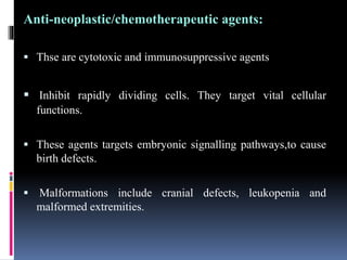
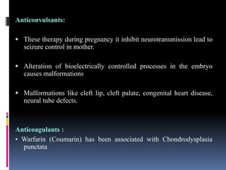
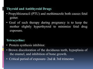
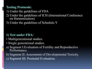
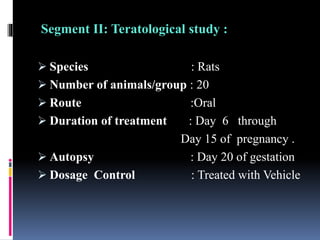
ADITYA INSTITUTE OF PHARMACY
EDUCATION & RESEARCH
(DEPARTMENT OF PHARMACOLOGY)
TOXICOLOGICAL SCREENING METHODS
TOPIC ON
TERATOGENICITY STUDIES(SEGMENT 2)
SUBMITTED TO :DR N VENKATESAN
SUBMITTED BY :RAJESH YADAV
Contents
 Introduction
 Mechanism of teratogenicity
 Principles
 Types of study
 Observation
 Data and reporting
 Evaluation of results
TERATOGENICITY
 The property or capability of producing congenital
malformations.
 Less than 2% of congenital malformations are caused by
drugs or chemicals.
 Teratogenic drugs should be avoided either during or
prior to conception.
 Women to avoid all medications in the first 8 weeks after
conception.
Principles of teratogenicity
1. Teratogenic susceptibility is determined by the
genotype of the conceptus .
2. Susceptibility to teratogenic agents depends on
the Developmental stage of the embryo or fetus
at the time of exposure .
3. These agents work by specific mechanisms on
developing cells and tissues to initiate pathogenesis .
4. Perturbations of developmental processes can result
in death, malformation, growth retardation.
Drugs can affect the foetus at 3 stages:
 Fertilization & implantation – conception to
17 days.
 Organogenesis- 18 to 55 days of gestation.
 Growth & development-56 days onwards.
Teratogenic mechanism
 1) Folate antagonism
 2) Neural crest cell disruption
 3) Endocrine disruption
 4) Oxidative stress
 5)Vascular disruption
 6) 5- HT receptors and transporters
 7) Enzyme mediated teratogenesis
Thalidomide:
 It binds to the GC promoter sites in transcription
process.
 Decrease in transcription efficiency of associated
genes.
 Decrease in angiogenesis,results in truncation of
limbs.
 Characteristic features include limb
abnormalities.
Anti-neoplastic/chemotherapeutic agents:
 Thse are cytotoxic and immunosuppressive agents
 Inhibit rapidly dividing cells. They target vital cellular
functions.
 These agents targets embryonic signalling pathways,to cause
birth defects.
 Malformations include cranial defects, leukopenia and
malformed extremities.
Anticonvulsants:
 These therapy during pregnancy it inhibit neurotransmission lead to
seizure control in mother.
 Alteration of bioelectrically controlled processes in the embryo
causes malformations
 Malformations like cleft lip, cleft palate, congenital heart disease,
neural tube defects.
Anticoagulants :
• Warfarin (Coumarin) has been associated with Chondrodysplasia
punctata
Thyroid and Antithyroid Drugs
• Propylthiouracil (PTU) and methimazole both causes fetal
goiter.
• Goal of such therapy during pregnancy is to keep the
mother slightly hyperthyroid to minimize fetal drug
exposure.
Tetracycline:
• Protein synthesis inhibitor.
• Brown discoloration of the deciduous teeth, hypoplasia of
the enamel, and inhibition of bone growth.
• Critical period of exposure- 2nd & 3rd trimester.
Testing Protocols:
1) Under the guidelines of FDA
2) Under the guidelines of ICH (International Conference
on Harmonisation)
3) Under the guidelines of Schedule-Y
1) Test under FDA:
• Multigenerational studies.
• Single generational studies.
a) Segment I:Evaluation of Fertility and Reproductive
Performance.
b) Segment II: Assessment of Developmental Toxicity.
c) Segment III: Postnatal Evaluation.
Single generation studies
 This Test Guideline(TG no 415) for reproduction
testing is designed to provide general information.
 Concerning the effects of a test substance on male
and female reproductive performance, such as
gonadal function, oestrous cycle, mating
behaviour, conception, parturition, lactation and
weaning.
Principle
 The test substance is administered in graduated
doses to several groups of males and females.
 Males should be dosed during growth.
 Females should be dosed for at least two complete
oestrous cycles.
 The animals are then mated.
 The test substance is administered to both sexes
during the mating period.
Description
Preparation
 Healthy young adult animals are randomised and
assigned to the treatment groups.
 The animals are kept in cages for at least five
days to allow for acclimatisation.
 Dosing should be on a seven-day per week basis.
Experimental animals
Selection of species
 This Test Guideline is designed for use with the
rat or mouse.
 The test animals should be characterised as to
species, strain, sex, weight and/or age.
 Healthy animals, not subjected to previous
experimental procedures, should be used.
Number and sex
 Each test and control group should contain a
sufficient number of animals to yield about 20
pregnant females.
 The objective is to produce enough
pregnancies and offspring.
Test conditions
a) Housing and feeding conditions
b) Dose levels
 At least three treatment and a control group should be
used.
1) Lowest dose level
2) Intermediate dose level
3) Highest dose level
Limit test
 If a dose of at least 1000 mg/kg produces no
evidence of interference with reproductive
performance, studies at other dose levels may not
be considered necessary.
 If a preliminary study at the high dose level, with
definite evidence of maternal toxicity, shows no
adverse effects on fertility, studies at other dose
levels may not be considered necessary.
Performance of the study
Experimental schedules
 Daily dosing of the parental (P) males should
begin when they are about five to nine weeks old.
 In rats dosing is continued for ten weeks prior to
the mating period .
 Males should be killed and examined either at the
end of the mating period or some time before the
end of the experiment
 For parental (P) females, dosing should begin
after at least five days of acclimatisation and
continue for at least two weeks prior to mating
 Daily dosing of the P females should continue
throughout the 3-week mating period, pregnancy
and up to the weaning of the F 1 offspring.
Mating procedure
 Based on 1: 1 mating, one female placed with the
same male until pregnancy occurs.
 Each morning the females should be examined
for presence of sperm or vaginal plugs.
 Day 0 of pregnancy is defined as the day a
vaginal plug or sperm are found.
 Those pairs that fail to mate should be evaluated
to determine the cause of the apparent infertility
Observations
 Each animal should be observed at least once
daily.
 Pertinent behavioural changes, signs of
difficult or prolonged parturition and all signs
of toxicity, including mortality, should be
recorded.
 During pre-mating and mating periods, food
consumption should be measured weekly.
 Each litter should be examined to establish the
number and sex of pups, stillbirths, live births
and the presence of gross anomalies.
 Dead pups and pups killed should be
preserved and studied for possible defects.
 Physical or behavioural abnormalities
observed in the dams or offspring should be
recorded.
Pathology
a) Gross necropsy
 At the time of sacrifice during the study the animals of the P
generation should be examined macroscopically for any structural
abnormalities with special attention paid to the organs of the
reproductive system.
 Dead or moribund pups should be examined for defects.
b) Histopathology
The ovaries, uterus, cervix, vagina, testes, epididymides, seminal
vesicles, prostate, coagulating gland, pituitary gland and target organ(s)
of all P animals should be preserved for microscopic examination.
Data and reporting
Treatment of results
 Data may be summarised in tabular form.
 Showing for each test group the number of
animals at the start of the test, the number of
fertile males, the number of pregnant females,
the types of changes and the percentage of
animals displaying each type of change.
Evaluation and interpretation of results
 This study should be evaluated in terms of the
observed effects, necropsy and microscopic
findings.
 Abnormalities including fertility, clinical
abnormalities, body weight changes, effects on
mortality and any other toxic effects.
Test report
 Species/strain used.
 Fertility, gestation, and viability indices.
 Time of death during the study or whether
animals survived to termination.
 Table presenting the weights of each litter, the
mean pup weights and the individual weights of
the pups at termination.
 Toxic or other effects on reproduction, offspring.
 The day of observation of each abnormal signs.
 Body weight data for P animals.
 Necropsy findings.
Segment II: Teratological study :
 Species : Rats
 Number of animals/group : 20
 Route :Oral
 Duration of treatment : Day 6 through
Day 15 of pregnancy .
 Autopsy : Day 20 of gestation
 Dosage Control : Treated with Vehicle
Procedure
 Rats are sacrificed on gestation day 20.
 Fetuses are removed by cesarean section after
noting the number of resorptions, implantations,
and normal fetuses.
 The size, weight, and any abnormality of each
fetus are noted along with Dams parameters
observation.
 Two thirds of the fetuses are eviscerated .
 then preserved in absolute alcohol for staining with
Alizarin Red S for skeletal assessment.
 The other one-third of the fetuses is fixed in Allen’s
modification of Bouin’s fluid for slicing with a razor
blade (Wilson’s Technique) to evaluate visceral
anomalies.
Invitro- study
 1) Whole embryo culture test
a) Rodent embryo culture
b) Zebrafish embryo
 2) Micromass teratogen test
 3) Embryonic stem cell test
 4) Dicto-stelium discoideum
…..THANK YOU…..

Teratogenicity studies

  • 1.
    ADITYA INSTITUTE OFPHARMACY EDUCATION & RESEARCH (DEPARTMENT OF PHARMACOLOGY) TOXICOLOGICAL SCREENING METHODS TOPIC ON TERATOGENICITY STUDIES(SEGMENT 2) SUBMITTED TO :DR N VENKATESAN SUBMITTED BY :RAJESH YADAV
  • 2.
    Contents  Introduction  Mechanismof teratogenicity  Principles  Types of study  Observation  Data and reporting  Evaluation of results
  • 3.
    TERATOGENICITY  The propertyor capability of producing congenital malformations.  Less than 2% of congenital malformations are caused by drugs or chemicals.  Teratogenic drugs should be avoided either during or prior to conception.  Women to avoid all medications in the first 8 weeks after conception.
  • 4.
    Principles of teratogenicity 1.Teratogenic susceptibility is determined by the genotype of the conceptus . 2. Susceptibility to teratogenic agents depends on the Developmental stage of the embryo or fetus at the time of exposure .
  • 5.
    3. These agentswork by specific mechanisms on developing cells and tissues to initiate pathogenesis . 4. Perturbations of developmental processes can result in death, malformation, growth retardation.
  • 6.
    Drugs can affectthe foetus at 3 stages:  Fertilization & implantation – conception to 17 days.  Organogenesis- 18 to 55 days of gestation.  Growth & development-56 days onwards.
  • 7.
    Teratogenic mechanism  1)Folate antagonism  2) Neural crest cell disruption  3) Endocrine disruption  4) Oxidative stress  5)Vascular disruption  6) 5- HT receptors and transporters  7) Enzyme mediated teratogenesis
  • 9.
    Thalidomide:  It bindsto the GC promoter sites in transcription process.  Decrease in transcription efficiency of associated genes.  Decrease in angiogenesis,results in truncation of limbs.  Characteristic features include limb abnormalities.
  • 10.
    Anti-neoplastic/chemotherapeutic agents:  Thseare cytotoxic and immunosuppressive agents  Inhibit rapidly dividing cells. They target vital cellular functions.  These agents targets embryonic signalling pathways,to cause birth defects.  Malformations include cranial defects, leukopenia and malformed extremities.
  • 11.
    Anticonvulsants:  These therapyduring pregnancy it inhibit neurotransmission lead to seizure control in mother.  Alteration of bioelectrically controlled processes in the embryo causes malformations  Malformations like cleft lip, cleft palate, congenital heart disease, neural tube defects. Anticoagulants : • Warfarin (Coumarin) has been associated with Chondrodysplasia punctata
  • 12.
    Thyroid and AntithyroidDrugs • Propylthiouracil (PTU) and methimazole both causes fetal goiter. • Goal of such therapy during pregnancy is to keep the mother slightly hyperthyroid to minimize fetal drug exposure. Tetracycline: • Protein synthesis inhibitor. • Brown discoloration of the deciduous teeth, hypoplasia of the enamel, and inhibition of bone growth. • Critical period of exposure- 2nd & 3rd trimester.
  • 13.
    Testing Protocols: 1) Underthe guidelines of FDA 2) Under the guidelines of ICH (International Conference on Harmonisation) 3) Under the guidelines of Schedule-Y 1) Test under FDA: • Multigenerational studies. • Single generational studies. a) Segment I:Evaluation of Fertility and Reproductive Performance. b) Segment II: Assessment of Developmental Toxicity. c) Segment III: Postnatal Evaluation.
  • 14.
    Single generation studies This Test Guideline(TG no 415) for reproduction testing is designed to provide general information.  Concerning the effects of a test substance on male and female reproductive performance, such as gonadal function, oestrous cycle, mating behaviour, conception, parturition, lactation and weaning.
  • 15.
    Principle  The testsubstance is administered in graduated doses to several groups of males and females.  Males should be dosed during growth.  Females should be dosed for at least two complete oestrous cycles.  The animals are then mated.  The test substance is administered to both sexes during the mating period.
  • 16.
    Description Preparation  Healthy youngadult animals are randomised and assigned to the treatment groups.  The animals are kept in cages for at least five days to allow for acclimatisation.  Dosing should be on a seven-day per week basis.
  • 17.
    Experimental animals Selection ofspecies  This Test Guideline is designed for use with the rat or mouse.  The test animals should be characterised as to species, strain, sex, weight and/or age.  Healthy animals, not subjected to previous experimental procedures, should be used.
  • 18.
    Number and sex Each test and control group should contain a sufficient number of animals to yield about 20 pregnant females.  The objective is to produce enough pregnancies and offspring.
  • 19.
    Test conditions a) Housingand feeding conditions b) Dose levels  At least three treatment and a control group should be used. 1) Lowest dose level 2) Intermediate dose level 3) Highest dose level
  • 20.
    Limit test  Ifa dose of at least 1000 mg/kg produces no evidence of interference with reproductive performance, studies at other dose levels may not be considered necessary.  If a preliminary study at the high dose level, with definite evidence of maternal toxicity, shows no adverse effects on fertility, studies at other dose levels may not be considered necessary.
  • 21.
    Performance of thestudy Experimental schedules  Daily dosing of the parental (P) males should begin when they are about five to nine weeks old.  In rats dosing is continued for ten weeks prior to the mating period .  Males should be killed and examined either at the end of the mating period or some time before the end of the experiment
  • 22.
     For parental(P) females, dosing should begin after at least five days of acclimatisation and continue for at least two weeks prior to mating  Daily dosing of the P females should continue throughout the 3-week mating period, pregnancy and up to the weaning of the F 1 offspring.
  • 23.
    Mating procedure  Basedon 1: 1 mating, one female placed with the same male until pregnancy occurs.  Each morning the females should be examined for presence of sperm or vaginal plugs.  Day 0 of pregnancy is defined as the day a vaginal plug or sperm are found.  Those pairs that fail to mate should be evaluated to determine the cause of the apparent infertility
  • 24.
    Observations  Each animalshould be observed at least once daily.  Pertinent behavioural changes, signs of difficult or prolonged parturition and all signs of toxicity, including mortality, should be recorded.  During pre-mating and mating periods, food consumption should be measured weekly.
  • 25.
     Each littershould be examined to establish the number and sex of pups, stillbirths, live births and the presence of gross anomalies.  Dead pups and pups killed should be preserved and studied for possible defects.  Physical or behavioural abnormalities observed in the dams or offspring should be recorded.
  • 26.
    Pathology a) Gross necropsy At the time of sacrifice during the study the animals of the P generation should be examined macroscopically for any structural abnormalities with special attention paid to the organs of the reproductive system.  Dead or moribund pups should be examined for defects. b) Histopathology The ovaries, uterus, cervix, vagina, testes, epididymides, seminal vesicles, prostate, coagulating gland, pituitary gland and target organ(s) of all P animals should be preserved for microscopic examination.
  • 27.
    Data and reporting Treatmentof results  Data may be summarised in tabular form.  Showing for each test group the number of animals at the start of the test, the number of fertile males, the number of pregnant females, the types of changes and the percentage of animals displaying each type of change.
  • 28.
    Evaluation and interpretationof results  This study should be evaluated in terms of the observed effects, necropsy and microscopic findings.  Abnormalities including fertility, clinical abnormalities, body weight changes, effects on mortality and any other toxic effects.
  • 29.
    Test report  Species/strainused.  Fertility, gestation, and viability indices.  Time of death during the study or whether animals survived to termination.  Table presenting the weights of each litter, the mean pup weights and the individual weights of the pups at termination.  Toxic or other effects on reproduction, offspring.  The day of observation of each abnormal signs.  Body weight data for P animals.  Necropsy findings.
  • 30.
    Segment II: Teratologicalstudy :  Species : Rats  Number of animals/group : 20  Route :Oral  Duration of treatment : Day 6 through Day 15 of pregnancy .  Autopsy : Day 20 of gestation  Dosage Control : Treated with Vehicle
  • 31.
    Procedure  Rats aresacrificed on gestation day 20.  Fetuses are removed by cesarean section after noting the number of resorptions, implantations, and normal fetuses.  The size, weight, and any abnormality of each fetus are noted along with Dams parameters observation.
  • 32.
     Two thirdsof the fetuses are eviscerated .  then preserved in absolute alcohol for staining with Alizarin Red S for skeletal assessment.  The other one-third of the fetuses is fixed in Allen’s modification of Bouin’s fluid for slicing with a razor blade (Wilson’s Technique) to evaluate visceral anomalies.
  • 33.
    Invitro- study  1)Whole embryo culture test a) Rodent embryo culture b) Zebrafish embryo  2) Micromass teratogen test  3) Embryonic stem cell test  4) Dicto-stelium discoideum
  • 34.