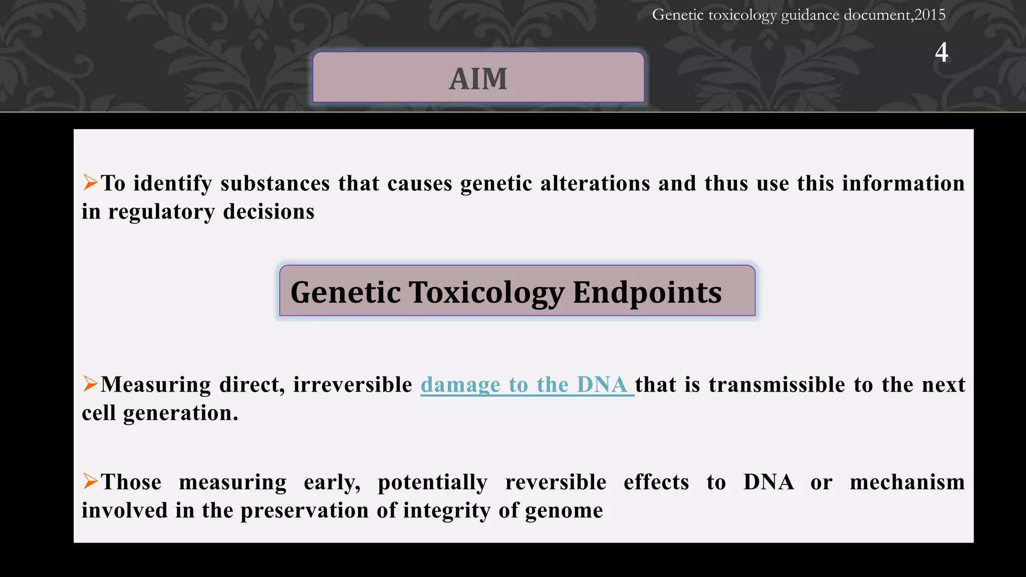
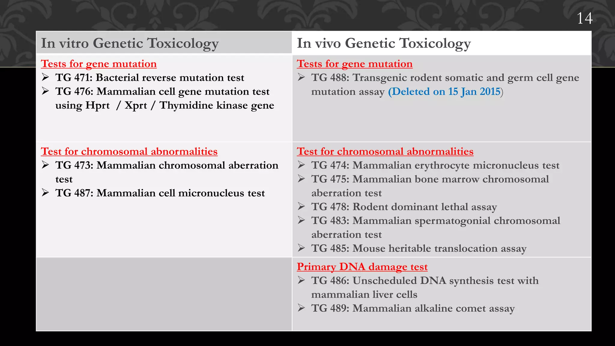
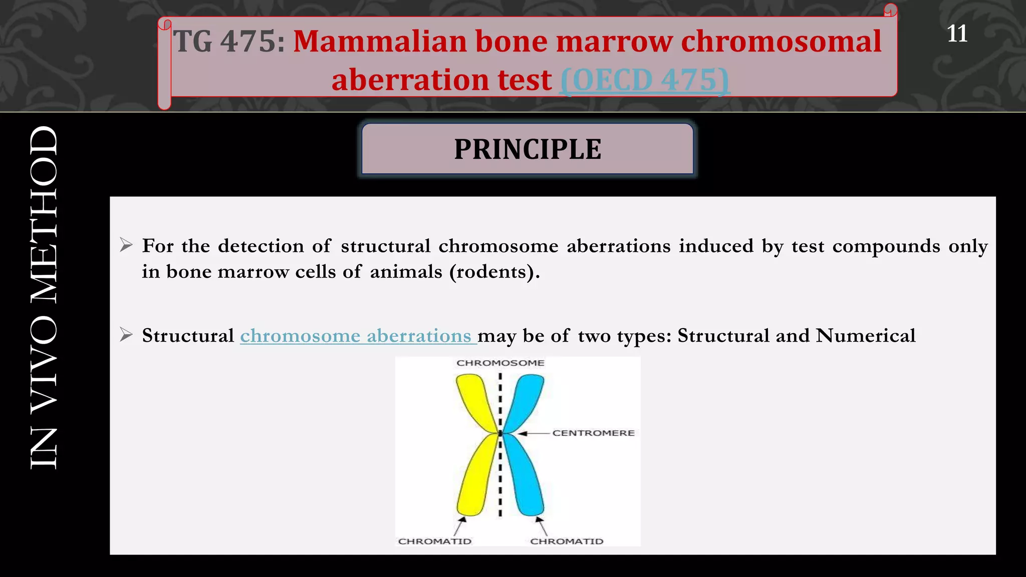
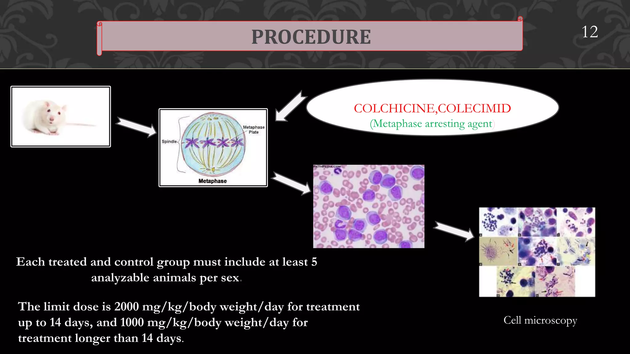
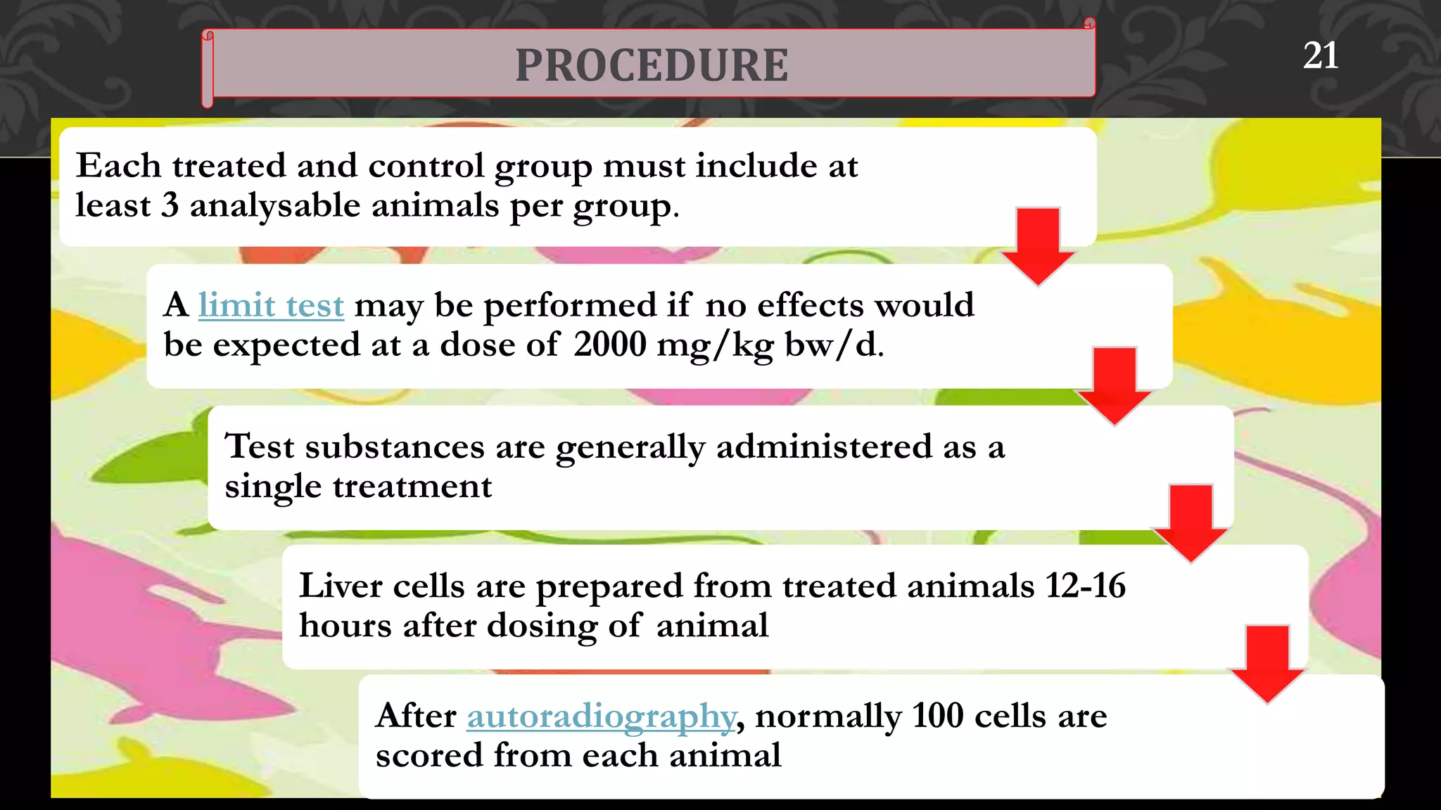
1
Presentation by :
Ashwini Patil
Department Of
Pharmacology
M.Pharm -2nd –Sem
R.C.P.I.P.E.R,Shirpur.
1
Presentation by:- Ashwini Patil
Department Of
Pharmacology
M.Pharm -2nd Sem
R.C.P.I.P.E.R,Shirpur.
HISTORY OF TOXICITY STUDIES
GENERAL CLASSIFICATION OF GUIDELINES
GENETIC TOXICITY GUIDELINES
CONTENT
2
PHASES OF CELL CYCLE
TEST GUIDELINES ON GENOTOXICITYTEST GUIDELINES
REFERNCES
Paracelsus (Father of Toxicology)
"All substances are poisons; there is none which is not a poison.
The right dose differentiates a poison and a remedy”.
Determined specific chemicals responsible for the toxicity of plants
and animals (dose-response relationship).
Mathieu Orfila, determined the relationship between poisons and their
biological. He is referred to as the father of modern toxicology
3 / 1 / 2 0 1 7 3
History of Toxicity Studies
Paracelsus
(1493-1541)
Mathieu Orfila
(24 April 1787 – 12
March 1853)
To identify substances that causes genetic alterations and thus use this information
in regulatory decisions
Measuring direct, irreversible damage to the DNA that is transmissible to the next
cell generation.
Those measuring early, potentially reversible effects to DNA or mechanism
involved in the preservation of integrity of genome
4
AIM
Genetic Toxicology Endpoints
Genetic toxicology guidance document,2015
5
In vitro Genetic Toxicology In vivo Genetic Toxicology
Tests for gene mutation
 TG 471: Bacterial reverse mutation test
 TG 476: Mammalian cell gene mutation test
using Hprt / Xprt / Thymidine kinase gene
Tests for gene mutation
 TG 488: Transgenic rodent somatic and germ cell gene
mutation assay (Deleted on 15 Jan 2015)
Test for chromosomal abnormalities
 TG 473: Mammalian chromosomal aberration
test
 TG 487: Mammalian cell micronucleus test
Test for chromosomal abnormalities
 TG 474: Mammalian erythrocyte micronucleus test
 TG 475: Mammalian bone marrow chromosomal
aberration test
 TG 478: Rodent dominant lethal assay
 TG 483: Mammalian spermatogonial chromosomal
aberration test
 TG 485: Mouse heritable translocation assay
Primary DNA damage test
 TG 486: Unscheduled DNA synthesis test with
mammalian liver cells
 TG 489: Mammalian alkaline comet assay
14
PHASES OF CELL CYCLE 14
 For the detection of damage induced by the test substance to the chromosomes.
 Identifies micronuclei containing lagging chromosome fragments or whole chromosomes.
 An increase in the frequency of micronucleated polychromatic erythrocytes in treated animals
is an indication of induced chromosome damage because they lack main nucleus
3 / 1 / 2 0 1 7
7TG 474: Mammalian erythrocyte micronucleus
Test(OECD 474)
INVIVOMETHOD
PRINCIPLE
8
9PROCEDURE
Each treated and control group must include at least 5
analyzable animals per sex.
Administration of the treatments consists of a single
dose or two daily doses (or more).
The limit dose is 2000 mg/kg/body weight/day for
treatment up to 14 days, and 1000 mg/kg/body
weight/day for treatment longer than 14 days.
v
10
PARAMETER
1. Automated system that measures
micronucleated erythrocyte
frequencies
1. Cells must be proliferated before
tissue collection and must be
properly sampled
2. Restricts only for detection of
organ specific genotoxic substances
10
 For the detection of structural chromosome aberrations induced by test compounds only
in bone marrow cells of animals (rodents).
 Structural chromosome aberrations may be of two types: Structural and Numerical
3 / 1 / 2 0 1 7 11TG 475: Mammalian bone marrow chromosomal
aberration test (OECD 475)
INVIVOMETHOD
PRINCIPLE
PROCEDURE 12
COLCHICINE,COLECIMID
(Metaphase arresting agent)
Each treated and control group must include at least 5
analyzable animals per sex.
The limit dose is 2000 mg/kg/body weight/day for treatment
up to 14 days, and 1000 mg/kg/body weight/day for
treatment longer than 14 days.
Cell microscopy
various types of chromosomal aberrations is visualized in individual cell using microscopy.
13
PARAMETER
1. Increased incidences of polyploidy can
be seen
1. Standard design is not optimized for
detection of aneuploidy.
2. Long exposure decreases the sensitivity
of this test.
FISH Staining technique have been used to increase the senstivity of CA
,allowing each chromosome to be differentially stained.
Dominant lethal (DL) effects cause embryonic or fetal death.
 Induction of a dominant lethal event after exposure to a test substance indicates that the
substance has affected germinal tissue of the test species.
Dominant lethals are generally accepted to be the result of chromosomal aberrations
(structural and numerical anomalies)
Chemicals that causes Dominant lethality also causes F1 congenital malformations.
3 / 1 / 2 0 1 7
14TG 478: Rodent dominant lethal assay) (OECD 478)INVIVOMETHOD
PRINCIPLE
PROCEDURE 15
Normally, three dose levels should be used.
The various germ cell stages can be tested separately
by the use of sequential mating intervals.
The females are sacrificed after an appropriate period
of time
 The calculation of the dominant lethal effect is based on comparison of the live implants
per female in the treated group to the live implants per female in the control group
 Chemicals that are positive in Dominant lethal test also are +ve in translocation test and
Specific locus test
16
PARAMETER
Detects structural and numerical chromosome changes in mammalian germ cells
 The types of chromosome changes detected in this test system are reciprocal
translocations.
 Carriers of translocations and XO-females show reduced fertility which is used to
select first generation progeny for cytogenetic analysis.
 Translocations are cytogenetically observed in meiotic cells at diakinesis metaphase I
3 / 1 / 2 0 1 7 17TG 485: Mouse heritable translocation assay
(OECD 485)
INVIVOMETHOD
PRINCIPLE
The test is usually performed by
analysis of male first generation
progeny.
Translocation are cytogenetically observed as
quadrivalent which are compromised of two
sets of homologous chromosome in meiotic
cells at diakinesis of meiosis of F1 male
progeny
A single administration of the test substance
or the administration of the test substance on
7 days/week for 35 days, are possible.
One dose level is tested, usually the
highest dose associated with the
production of minimal toxic effects, and
administered by oral intubation or
intraperitoneal injection
About 500 first generation
males per dose level are
required.
PROCEDURE
18
Monitoring of litter size of F1 generation indicates that Dominant lethality is
occuring.
Requires large number of animals and rarely used.
19
LIMITATION
PARAMETER
 To identify substances that induce DNA repair after excision and removal of a stretch of
DNA containing a region of damage induced by chemical substances or physical agents in
the liver.
 The test is usually based on the incorporation of tritium-labelled thymidine, 3H-TdR,
(during 3-8 hours) into the DNA of liver cells
 The uptake of 3H-TdR is usually determined by autoradiography
 `
3 / 1 / 2 0 1 7 20TG 486: Unscheduled DNA synthesis (UDS) test
with mammalian liver cells (OECD 486)
INVIVOMETHOD
PRINCIPLE
21
Each treated and control group must include at
least 3 analysable animals per group.
A limit test may be performed if no effects would
be expected at a dose of 2000 mg/kg bw/d.
Test substances are generally administered as a
single treatment.
Liver cells are prepared from treated animals 12-16
hours after dosing of animal.
After autoradiography, normally 100 cells are
scored from each animal
PROCEDURE
Response is dependent on number of DNA bases excised and replaced at the site of origin.
A positive result indicates that a substance induces DNA damage in mammalian liver cells in
vivo that can be repaired by unscheduled DNA synthesis in vitro.
This test is not considered as surrogate mutation test, thus less reliable than other primary
DNA damage test.
22
LIMITATION
PARAMETER
Measures the DNA strand breaks in eukaryotic cells.
These strand breaks may be: 1) repaired, 2) lethal to the cell, 3) fixed as mutation resulting in
permanent heritable change.
Alternate name: Alkaline single-cell gel electrophoresis assay.
Under alkaline condition (>13) this assay can detect single and double strand breaks.
For eg: direct interactions with DNA alkali liable sites or consequences of transient DNA
strand discontinuities resulting from DNA excision repair.
3 / 1 / 2 0 1 7
23TG 489: In vivo Mammalian alkaline comet assay
(OECD 489)
INVIVOMETHOD
PRINCIPLE
Administration of the treatment consists of
daily doses over duration of 2 days or more,
ensuring the test chemical reaches the target
tissue
.
24
PARAMETER
Based on their size DNA fragments migrate away from the head to the tail, and
the intensity of the comet tail relative to the total intensity (head plus tail)
reflects the amount of DNA breakage.
28
PARAMETER
26
CASPLAB SOFTWARE
.
27
1. Cell division is not required.
2. Detecting exposure to target tissue
3. Small amount of test substances.
4. Less time consuming.
1. Does not detect aneuploidy.
2. Structural chromosomal damage or
mutation is not detected directly.
3. Long exposure: decreased sensitivity.
4. Difficult to distinguish between genotoxicity
and apoptosis after electrophoresis.
5. Not able to detect small DNA fragment
Sengupta , Alokparna (2012) Toxicity testing in India: An animal welfare perspective. Federation of Indian
Animal Protection Organizations and Humane Society International April 2012
Combes RD, Gaunt, I, Balls M (2004). A Scientific and Animal Welfare Assessment of the OECD Health Effects
Test Guidelines for the Safety Testing of Chemicals under the European Union REACH System. ATLA 32, 163-
208.
NRC (2007). Toxicity Testing in the 21st Century: A Vision and a Strategy. Washington, DC: The
National Academies Press.
www.oecd.org
Genetic toxicology guidance document,2015
Hacettepe University, Faculty of Pharmacy, Department of Toxicology, Ankara
Department of Environmental Biotechnology, University of Warmia and Mazury in Olsztyn, ul. Sloneczna 45G,
10–712 Olsztyn, Poland
28REFERENCES
29

Ashwini presentation

  • 1.
    1 Presentation by : AshwiniPatil Department Of Pharmacology M.Pharm -2nd –Sem R.C.P.I.P.E.R,Shirpur. 1 Presentation by:- Ashwini Patil Department Of Pharmacology M.Pharm -2nd Sem R.C.P.I.P.E.R,Shirpur.
  • 2.
    HISTORY OF TOXICITYSTUDIES GENERAL CLASSIFICATION OF GUIDELINES GENETIC TOXICITY GUIDELINES CONTENT 2 PHASES OF CELL CYCLE TEST GUIDELINES ON GENOTOXICITYTEST GUIDELINES REFERNCES
  • 3.
    Paracelsus (Father ofToxicology) "All substances are poisons; there is none which is not a poison. The right dose differentiates a poison and a remedy”. Determined specific chemicals responsible for the toxicity of plants and animals (dose-response relationship). Mathieu Orfila, determined the relationship between poisons and their biological. He is referred to as the father of modern toxicology 3 / 1 / 2 0 1 7 3 History of Toxicity Studies Paracelsus (1493-1541) Mathieu Orfila (24 April 1787 – 12 March 1853)
  • 4.
    To identify substancesthat causes genetic alterations and thus use this information in regulatory decisions Measuring direct, irreversible damage to the DNA that is transmissible to the next cell generation. Those measuring early, potentially reversible effects to DNA or mechanism involved in the preservation of integrity of genome 4 AIM Genetic Toxicology Endpoints Genetic toxicology guidance document,2015
  • 5.
    5 In vitro GeneticToxicology In vivo Genetic Toxicology Tests for gene mutation  TG 471: Bacterial reverse mutation test  TG 476: Mammalian cell gene mutation test using Hprt / Xprt / Thymidine kinase gene Tests for gene mutation  TG 488: Transgenic rodent somatic and germ cell gene mutation assay (Deleted on 15 Jan 2015) Test for chromosomal abnormalities  TG 473: Mammalian chromosomal aberration test  TG 487: Mammalian cell micronucleus test Test for chromosomal abnormalities  TG 474: Mammalian erythrocyte micronucleus test  TG 475: Mammalian bone marrow chromosomal aberration test  TG 478: Rodent dominant lethal assay  TG 483: Mammalian spermatogonial chromosomal aberration test  TG 485: Mouse heritable translocation assay Primary DNA damage test  TG 486: Unscheduled DNA synthesis test with mammalian liver cells  TG 489: Mammalian alkaline comet assay 14
  • 6.
  • 7.
     For thedetection of damage induced by the test substance to the chromosomes.  Identifies micronuclei containing lagging chromosome fragments or whole chromosomes.  An increase in the frequency of micronucleated polychromatic erythrocytes in treated animals is an indication of induced chromosome damage because they lack main nucleus 3 / 1 / 2 0 1 7 7TG 474: Mammalian erythrocyte micronucleus Test(OECD 474) INVIVOMETHOD PRINCIPLE
  • 8.
  • 9.
    9PROCEDURE Each treated andcontrol group must include at least 5 analyzable animals per sex. Administration of the treatments consists of a single dose or two daily doses (or more). The limit dose is 2000 mg/kg/body weight/day for treatment up to 14 days, and 1000 mg/kg/body weight/day for treatment longer than 14 days. v
  • 10.
    10 PARAMETER 1. Automated systemthat measures micronucleated erythrocyte frequencies 1. Cells must be proliferated before tissue collection and must be properly sampled 2. Restricts only for detection of organ specific genotoxic substances 10
  • 11.
     For thedetection of structural chromosome aberrations induced by test compounds only in bone marrow cells of animals (rodents).  Structural chromosome aberrations may be of two types: Structural and Numerical 3 / 1 / 2 0 1 7 11TG 475: Mammalian bone marrow chromosomal aberration test (OECD 475) INVIVOMETHOD PRINCIPLE
  • 12.
    PROCEDURE 12 COLCHICINE,COLECIMID (Metaphase arrestingagent) Each treated and control group must include at least 5 analyzable animals per sex. The limit dose is 2000 mg/kg/body weight/day for treatment up to 14 days, and 1000 mg/kg/body weight/day for treatment longer than 14 days. Cell microscopy
  • 13.
    various types ofchromosomal aberrations is visualized in individual cell using microscopy. 13 PARAMETER 1. Increased incidences of polyploidy can be seen 1. Standard design is not optimized for detection of aneuploidy. 2. Long exposure decreases the sensitivity of this test. FISH Staining technique have been used to increase the senstivity of CA ,allowing each chromosome to be differentially stained.
  • 14.
    Dominant lethal (DL)effects cause embryonic or fetal death.  Induction of a dominant lethal event after exposure to a test substance indicates that the substance has affected germinal tissue of the test species. Dominant lethals are generally accepted to be the result of chromosomal aberrations (structural and numerical anomalies) Chemicals that causes Dominant lethality also causes F1 congenital malformations. 3 / 1 / 2 0 1 7 14TG 478: Rodent dominant lethal assay) (OECD 478)INVIVOMETHOD PRINCIPLE
  • 15.
    PROCEDURE 15 Normally, threedose levels should be used. The various germ cell stages can be tested separately by the use of sequential mating intervals. The females are sacrificed after an appropriate period of time
  • 16.
     The calculationof the dominant lethal effect is based on comparison of the live implants per female in the treated group to the live implants per female in the control group  Chemicals that are positive in Dominant lethal test also are +ve in translocation test and Specific locus test 16 PARAMETER
  • 17.
    Detects structural andnumerical chromosome changes in mammalian germ cells  The types of chromosome changes detected in this test system are reciprocal translocations.  Carriers of translocations and XO-females show reduced fertility which is used to select first generation progeny for cytogenetic analysis.  Translocations are cytogenetically observed in meiotic cells at diakinesis metaphase I 3 / 1 / 2 0 1 7 17TG 485: Mouse heritable translocation assay (OECD 485) INVIVOMETHOD PRINCIPLE
  • 18.
    The test isusually performed by analysis of male first generation progeny. Translocation are cytogenetically observed as quadrivalent which are compromised of two sets of homologous chromosome in meiotic cells at diakinesis of meiosis of F1 male progeny A single administration of the test substance or the administration of the test substance on 7 days/week for 35 days, are possible. One dose level is tested, usually the highest dose associated with the production of minimal toxic effects, and administered by oral intubation or intraperitoneal injection About 500 first generation males per dose level are required. PROCEDURE 18
  • 19.
    Monitoring of littersize of F1 generation indicates that Dominant lethality is occuring. Requires large number of animals and rarely used. 19 LIMITATION PARAMETER
  • 20.
     To identifysubstances that induce DNA repair after excision and removal of a stretch of DNA containing a region of damage induced by chemical substances or physical agents in the liver.  The test is usually based on the incorporation of tritium-labelled thymidine, 3H-TdR, (during 3-8 hours) into the DNA of liver cells  The uptake of 3H-TdR is usually determined by autoradiography  ` 3 / 1 / 2 0 1 7 20TG 486: Unscheduled DNA synthesis (UDS) test with mammalian liver cells (OECD 486) INVIVOMETHOD PRINCIPLE
  • 21.
    21 Each treated andcontrol group must include at least 3 analysable animals per group. A limit test may be performed if no effects would be expected at a dose of 2000 mg/kg bw/d. Test substances are generally administered as a single treatment. Liver cells are prepared from treated animals 12-16 hours after dosing of animal. After autoradiography, normally 100 cells are scored from each animal PROCEDURE
  • 22.
    Response is dependenton number of DNA bases excised and replaced at the site of origin. A positive result indicates that a substance induces DNA damage in mammalian liver cells in vivo that can be repaired by unscheduled DNA synthesis in vitro. This test is not considered as surrogate mutation test, thus less reliable than other primary DNA damage test. 22 LIMITATION PARAMETER
  • 23.
    Measures the DNAstrand breaks in eukaryotic cells. These strand breaks may be: 1) repaired, 2) lethal to the cell, 3) fixed as mutation resulting in permanent heritable change. Alternate name: Alkaline single-cell gel electrophoresis assay. Under alkaline condition (>13) this assay can detect single and double strand breaks. For eg: direct interactions with DNA alkali liable sites or consequences of transient DNA strand discontinuities resulting from DNA excision repair. 3 / 1 / 2 0 1 7 23TG 489: In vivo Mammalian alkaline comet assay (OECD 489) INVIVOMETHOD PRINCIPLE
  • 24.
    Administration of thetreatment consists of daily doses over duration of 2 days or more, ensuring the test chemical reaches the target tissue . 24
  • 25.
    PARAMETER Based on theirsize DNA fragments migrate away from the head to the tail, and the intensity of the comet tail relative to the total intensity (head plus tail) reflects the amount of DNA breakage. 28 PARAMETER
  • 26.
  • 27.
    . 27 1. Cell divisionis not required. 2. Detecting exposure to target tissue 3. Small amount of test substances. 4. Less time consuming. 1. Does not detect aneuploidy. 2. Structural chromosomal damage or mutation is not detected directly. 3. Long exposure: decreased sensitivity. 4. Difficult to distinguish between genotoxicity and apoptosis after electrophoresis. 5. Not able to detect small DNA fragment
  • 28.
    Sengupta , Alokparna(2012) Toxicity testing in India: An animal welfare perspective. Federation of Indian Animal Protection Organizations and Humane Society International April 2012 Combes RD, Gaunt, I, Balls M (2004). A Scientific and Animal Welfare Assessment of the OECD Health Effects Test Guidelines for the Safety Testing of Chemicals under the European Union REACH System. ATLA 32, 163- 208. NRC (2007). Toxicity Testing in the 21st Century: A Vision and a Strategy. Washington, DC: The National Academies Press. www.oecd.org Genetic toxicology guidance document,2015 Hacettepe University, Faculty of Pharmacy, Department of Toxicology, Ankara Department of Environmental Biotechnology, University of Warmia and Mazury in Olsztyn, ul. Sloneczna 45G, 10–712 Olsztyn, Poland 28REFERENCES
  • 29.